Manuscript version: Author’s Accepted Manuscript
The version presented in WRAP is the author’s accepted manuscript and may differ from the published version or Version of Record.
Persistent WRAP URL:
http://wrap.warwick.ac.uk/127117 How to cite:
Please refer to published version for the most recent bibliographic citation information. If a published version is known of, the repository item page linked to above, will contain details on accessing it.
Copyright and reuse:
The Warwick Research Archive Portal (WRAP) makes this work by researchers of the University of Warwick available open access under the following conditions.
Copyright © and all moral rights to the version of the paper presented here belong to the individual author(s) and/or other copyright owners. To the extent reasonable and
practicable the material made available in WRAP has been checked for eligibility before being made available.
Copies of full items can be used for personal research or study, educational, or not-for-profit purposes without prior permission or charge. Provided that the authors, title and full
bibliographic details are credited, a hyperlink and/or URL is given for the original metadata page and the content is not changed in any way.
Publisher’s statement:
Please refer to the repository item page, publisher’s statement section, for further information.
Development of a routinely applicable
imaging protocol for fast and precise
middle cerebral artery occlusion
assessment and perfusion deficit
measure in an ovine stroke model: a
case study
Giorgio Cattaneo1, Andrea M. Herrmann2, 3, Sebastian A. Eiden3, Manuela Wieser3, Elias Kellner4, Christoph Maurer5, Jörg Haberstroh6, Wolf-Dirk Niesen7, Horst Urbach3, Johannes Boltze8, 9, Stephan Meckel3*, Mukesch Shah10
1Other, Germany, 2Institute of Veterinary Anatomy, Histology and Embryology, Faculty of Veterinary
Medicine, University of Leipzig, Germany, 3Department of Neuroradiology, University Hospital Freiburg, Germany, 4Department of Radiology and Medical Physics, University Hospital Freiburg, Germany, 5Department for Diagnostic and Interventional Radiology and Neuroradiology, Augsburg University Hospital, Germany, 6Center for Experimental Models and Transgenic Service, University Hospital Freiburg, Germany, 7Department of Neurology, University of Freiburg, Germany, 8Institute of Medical and Marine Biotechnology, University of Lübeck, Germany, 9School of Life Sciences, Faculty of Science, University of Warwick, United Kingdom, 10Department of Neurosurgery, University Hospital Freiburg, Germany
Submitted to Journal:
Frontiers in Neurology
Specialty Section:
Stroke
Article type:
Original Research Article
Manuscript ID:
465499
Received on:
11 Apr 2019
Revised on:
23 Sep 2019
Frontiers website link:
www.frontiersin.org
Conflict of interest statement
The authors declare a potential conflict of interest and state it below
Giorgio Cattaneo was an employee of Acandis GmbH during the duration of this study.
The other authors declare that the research was conducted in the absence of any commercial or financial relationships that could be construed as a potential conflict of interest
Author contribution statement
C. G: significant contribution to study design, article drafting, critical review of intellectual contents H. AM: significant contribution to data acquisition, article drafting, critical review of intellectual contents
E. S: significant contribution to data acquisition, analysis, interpretation of data, critical review of intellectual contents W. M: significant contribution to data acquisition, analysis of data, critical review of intellectual contents
K. E: significant contribution to data acquisition, analysis of data, critical review of intellectual contents M. C: significant contribution to data acquisition, analysis of data, critical review of intellectual contents H. J: significant contribution to concept, study design, data acquisition, critical review of intellectual contents N. W-D: significant contribution to data acquisition, critical review of intellectual contents
U. H: significant contribution to concept, study design, critical review of intellectual contents
B. J: significant contribution to concept, study design, data interpretation, article drafting, critical review of intellectual contents M. S: significant contribution to experimental design image analysis, data acquisition, analysis, interpretation of data, article drafting, critical review of intellectual contents
S. MJ: significant contribution to experimental design, article drafting, critical review of intellectual contents all authors: approval of final version
Keywords
MCAO, sheep stroke model, Reperfusion, CT perfusion, DSA = digital subtraction angiography
Abstract
Word count: 201
Temporary middle cerebral artery occlusion (MCAO) in sheep allows modelling of acute large vessel occlusion stroke and subsequent vessel recanalization. However, rapid and precise imaging-based assessment of vessel occlusion and the resulting perfusion deficit during MCAO still represents an experimental challenge. Here, we tested feasibility and suitability of a strategy for MCAO verification and perfusion deficit assessment. We also compared the extent of the initial perfusion deficit and subsequent lesion size for different MCAO durations.
The rete mirabile prevents reliable vascular imaging investigation of middle cerebral artery filling status. Hence, computed tomography perfusion imaging was chosen for indirect confirmation of MCAO. Follow-up infarct size evaluation by diffusion-weighted magnetic resonance imaging revealed fluctuating results, with no apparent relationship of lesion size with MCAO at occlusion times below 4 hours, potentially related to the variable collateralization of the MCA territory. This underlines the need for intra-ischemic perfusion assessment and future studies focusing on the correlation between perfusion deficit, MCAO duration, and final infarct volume.
Temporary MCAO and intra-ischemic perfusion imaging nevertheless has the potential to be applied for the simulation of novel recanalization therapies, particularly those that aim for a fast reperfusion effect in combination with mechanical thrombectomy in a clinically realistic scenario.
Contribution to the field
Recent clinical trials have shown that endovascular mechanical thrombectomy is beneficial for patients with acute ischemic stroke due to large artery occlusion. However, there is a lack of animal stroke models to study the effects of vessel recanalization and reperfusion, as standard rodent models are not suitable to simulate complex interventional procedures. Large animal models may help overcoming these limitations. However, inter-individual collateral extent and capacity may cause significant variations in final lesion volume. Thus, rapid and precise imaging-based assessment of vessel occlusion and the resulting perfusion deficit during middle cerebral artery occlusion (MCAO) still represents an experimental challenge. Here, we tested feasibility and suitability of different imaging strategies for MCAO verification and perfusion deficit assessment in an ovine stroke model using permanent and transient MCAO. We applied a realistic protocol offering only a short imaging time window between vessel occlusion and
Funding statement
This work was supported by Federal Ministry of Education and Research, Germany (grant 13GW0015B)
Ethics statements
(Authors are required to state the ethical considerations of their study in the manuscript, including for cases where the study was exempt from ethical approval procedures)
Does the study presented in the manuscript involve human or animal subjects: Yes
Please provide the complete ethics statement for your manuscript. Note that the statement will be directly added to the manuscript file for peer-review, and should include the following information:
Full name of the ethics committee that approved the study
Consent procedure used for human participants or for animal owners
Any additional considerations of the study in cases where vulnerable populations were involved, for example minors, persons with disabilities or endangered animal species
As per the Frontiers authors guidelines, you are required to use the following format for statements involving human subjects: This study was carried out in accordance with the recommendations of [name of guidelines], [name of committee]. The protocol was approved by the [name of committee]. All subjects gave written informed consent in accordance with the Declaration of Helsinki.
For statements involving animal subjects, please use:
This study was carried out in accordance with the recommendations of 'name of guidelines, name of committee'. The protocol was approved by the 'name of committee'.
If the study was exempt from one or more of the above requirements, please provide a statement with the reason for the exemption(s).
Ensure that your statement is phrased in a complete way, with clear and concise sentences.
no exempt from one or more of the above requirements
Data availability statement
Development of a routinely applicable imaging protocol for fast and
precise middle cerebral artery occlusion assessment and perfusion
deficit measure in an ovine stroke model: a case study
1
Andrea Maria Herrmann1,2#Giorgio Franco Maria Cattaneo3#, Sebastian Alexander Eiden1, 2
Manuela Wieser1, Elias Kellner4, Christoph Maurer5, Jörg Haberstroh6, Christoph Mülling2, 3
Wolf-Dirk Niesen7, Horst Urbach2, Johannes Boltze8,9, Stephan Meckel2#,*, Mukesch Johannes 4
Shah10# 5
#
authors contributed equally to this work 6
1
Neuroradiology, 4MR Physics, 6Experimantal Surgery, CEMT-FR, 7Neurology and 10Neurosurgery, 7
Medical Center – University of Freiburg, Faculty of Medicine, University of Freiburg, Germany 8
2
Faculty of Veterinary Medicine, Institute of Veterinary Anatomy, Histology and Embryology, 9
Leipzig University, Leipzig, Germany 10
3
Acandis GmbH, Pforzheim, Germany 11
5
Department of Diagnostic and Interventional Neuroradiology, University Hospital Augsburg, 12
Germany 13
8
Fraunhofer Research Institution for Marine Biotechnology and Institute for Medical and Marine 14
Biotechnology, University of Lübeck, Germany 15
9
School of Life Sciences, University of Warwick, Coventry, CV4 7AL, UK 16
17
*Correspondence: 18
Corresponding Author 19
Stephan Meckel 20
Department of Neuroradiology, Medical Center –University of Freiburg, Faculty of Medicine, 21
University of Freiburg, Germany 22
Breisacher Straße 64 23
79106 Freiburg, Germany 24
Email: [email protected] 25
26
This work was supported by the German Ministry of Research and Education [grant number
27
13GW0015A].
28
Running headline: Novel imaging protocol for temporary MCAO in sheep
29 30
Keywords: CT perfusion, DSA, MCAO, Reperfusion, Sheep stroke model 31
32
Abstract 33
Temporary middle cerebral artery occlusion (MCAO) in sheep allows modelling of acute large vessel 34
occlusion stroke and subsequent vessel recanalization. However, rapid and precise imaging-based 35
assessment of vessel occlusion and the resulting perfusion deficit during MCAO still represents an 36
experimental challenge. Here, we tested feasibility and suitability of a strategy for MCAO 37
verification and perfusion deficit assessment. We also compared the extent of the initial perfusion 38
deficit and subsequent lesion size for different MCAO durations. 39
The rete mirabile prevents reliable vascular imaging investigation of middle cerebral artery filling 40
status. Hence, computed tomography perfusion imaging was chosen for indirect confirmation of 41
MCAO. Follow-up infarct size evaluation by diffusion-weighted magnetic resonance imaging 42
revealed fluctuating results, with no apparent relationship of lesion size with MCAO at occlusion 43
times below 4 hours, potentially related to the variable collateralization of the MCA territory. This 44
underlines the need for intra-ischemic perfusion assessment and future studies focusing on the 45
correlation between perfusion deficit, MCAO duration, and final infarct volume. 46
Temporary MCAO and intra-ischemic perfusion imaging nevertheless has the potential to be applied 47
for the simulation of novel recanalization therapies, particularly those that aim for a fast reperfusion 48
effect in combination with mechanical thrombectomy in a clinically realistic scenario. 49
50
1 Introduction 51
Several recent randomized-controlled trials have shown that endovascular mechanical thrombectomy 52
is highly beneficial for patients with acute ischemic stroke and large vessel occlusion (LVO) (Goyal 53
et al., 2016). This breakthrough in acute stroke treatment has led to steadily increasing numbers of 54
patients undergoing endovascular treatment with recanalization, providing options for novel 55
combined treatment strategies. For instance, companion neuroprotective therapies are believed to 56
augment the beneficial impact of recanalization therapies in future settings (Linfante and Cipolla, 57
2016; Savitz et al., 2017). 58
Although ischemia/reperfusion rodent models exist, these models have limitations in simulating 59
endovascular approaches under conditions which are similar to a clinical intervention in humans. The 60
major limitation are the much smaller vessels which, for instance, would not allow to test 61
intravascular test devices used for or to support thrombectomy. Large animal models can fill this gap 62
by providing a suitable vascular anatomy and size for preclinical evaluation of new endovascular or 63
combination treatment concepts for LVO stroke (Herrmann et al., 2019). Non-human primate and 64
canine stroke models are restricted by ethical concerns and high mortality in the acute and subacute 65
stages after stroke, preventing long-term assessment of functional outcome and final lesion size as 66
the most important clinical endpoints. Alternatively applied porcine and ovine models are more 67
suitable to monitor long-term impact of an intervention, but exhibit a rete mirabile which does not 68
allow direct endovascular access to the middle cerebral artery (MCA) for occlusion (MCAO). Stroke 69
models using these species therefore require surgical access to the MCA. Recently, ovine permanent 70
and transient MCAO stroke models were established (Boltze et al., 2008; Wells et al., 2012). 71
Effective occlusion of the MCA main trunk or its branches was reported to depend on the qualitative 72
visual assessment of the operating surgeon, but this may only be predictive in permanent occlusion 73
studies. In reperfusion studies, the individual extent and capacity of collaterals can cause significant 74
variations in final lesion volume similarly to the situation in human LVO stroke. Thus, a reliable, 75
rapid and unbiased estimation of the perfusion deficit during MCAO is an important prerequisite for 76
acute and long-term MCA recanalization studies. Investigating how the initial diffusion deficit 77
corresponds to final infarct size is another important aspect awaiting clarification. 78
In this feasibility study, we tested several imaging modalities for application in acute ovine MCAO 79
modelling human LVO stroke. We specifically aimed to assess (i) the reliability to confirm 80
successful transient MCAO, (ii) MCA territory hypoperfusion, and (iii) feasibility of the imaging 81
strategy in an experimental MCAO setting only offering a short imaging time window between 82
vessel occlusion and reopening. This work is also intended to report pitfalls and challenges we faced 83
during this development. We finally want to share the experience we have gained with other groups 84
in the field, or trying to access it. 85
86
2 Material and Methods 87
This study was carried out in accordance with the recommendations of the German animal protection 88
law and the animal care guidelines of the European Community (2010/63/EU). The protocol was 89
approved by the local ethics committee (Regierungspräsidium Freiburg, Germany; reference numbers 90
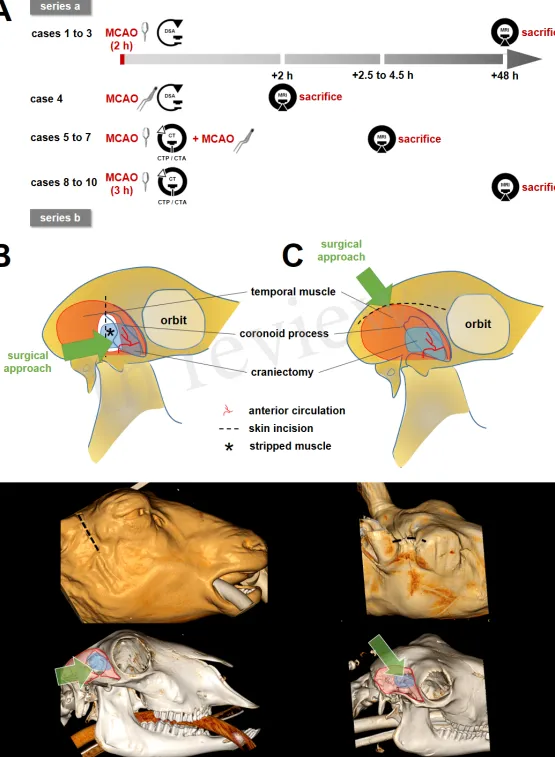
#35-9185.81/G-14/85 and #39-9185.81/G-15/38). Study design is illustrated in Figure 1A. ARRIVE 91
guidelines were followed as applicable for a pilot study. 92
93
2.1 Animal baseline data
94
The study involved ten merino sheep half breed (age, 1-3 years; weight, 80.2±7.4 kg), kept in the 95
CEMT-FR (Center for Experimental Models and Transgenic Service, Freiburg, Germany) under 96
following conditions: straw boxes, daily grazing, water and hay ad libitum, concentrated feed pellets 97
as reward and to foster human familiarization. 98
99
[Figure 1 around here] 100
101
2.2 Anesthesia
102
Anesthesia was prepared by intramuscular injection of midazolam (0.5 mg/kg bodyweight (BW)) and 103
ketamine hydrochloride (20 mg/kg BW), and was induced by intravenous propofol administration 104
(2–4 mg/kg BW). Following endotracheal intubation, 12–15 breaths/min were provided by a volume-105
controlled ventilator at a 10–15 mL/kg BW tidal volume and 5-mbar positive end-expiratory 106
pressure. Settings were adjusted to normalize oxygen and carbon dioxide tension, and pH values. 107
Anesthesia for surgical and endovascular procedures was maintained by isoflurane in oxygen/air 108
(FiO2 > 0.4), intravenous ketamine (10 mg/kg BW/h) and fentanyl (2–3 μg/kg BW/h) administration. 109
For CT perfusion and CT angiography as well as brain MRI and angiography anesthesia was 110
maintained by intravenous propofol (15-18 mg/kg/h). 111
Fluid homeostasis was maintained by intravenous infusion of Ringer solution (10 mg/kg BW/h). 112
Electrocardiogram and blood pressure were monitored continuously. A postsurgical antibiotic 113
(dihydrostreptomycin sulfate 12.9 mg/kg, benzylpenicillin-procaine 8 mg/kg) and analgesic 114
(carprofen 4 mg/kg) treatment was performed. 115
116
2.3 Surgical MCA preparation, occlusion and recanalization 117
Sheep were placed in the supine position slightly elevating the right shoulder. The head was then 118
tilted to the left by ninety degrees. The wool between the ear and eye was shorn, and sterile draping 119
was applied to cover the surgical field. 120
Two different approaches to the MCA were performed. MCAO surgery in the first series of 121
experiments (series a, cases 1-3) was carried out as described by Wells et al. (Wells et al., 2012), with 122
the following modifications (Figure 1B). A 5 cm vertical incision was made, terminating at the 123
zygomatic arch. Temporal and other mastication muscles were divided and stripped from the 124
coronoid process of the mandible. Partial removal of the coronoid process was omitted when 125
accessing the proximal MCA. The remaining masticators were then divided and stripped from the 126
outer table as far rostral as the fibrous ring attaching the posterior orbit to the concave border of the 127
parietal bone. Thereafter, a small craniectomy over the junction of the parietal and squamous 128
temporal bones was performed using an electric high-speed drill (microspeed, Aesculap, Tuttlingen, 129
Germany) to access the floor of the middle cranial fossa directly behind the orbita. The dura was then 130
opened carefully. A 3 Head VM-900 surgical microscope (Möller-Wedel, Wedel, Germany) was 131
used for surgical preparation of the proximal MCA and terminal ICA. The proximal MCA was 132
occluded by a Yasargil temporary titanium clip (Aesculap) for 2 hours. 133
Surgery in the second series (series b, cases 4-10) was carried out as described by Boltze et al. 134
(Figure 1C) (Boltze et al., 2008). The skin between the eye and ear was incised at 5 to 7 cm along the 135
superior temporal fossa. The fascia of the temporal muscle was opened and the muscle was stripped 136
away in lateral manner to expose the temporal fossa. During this maneuver, the coronoid process was 137
lateralized and thereafter kept laterally with a self-holding spreader. The remaining masticators were 138
then stripped from the outer table of the cranium as far rostral as the fibrous ring attaching the 139
posterior orbit to the concave border of the parietal bone. 140
Craniectomy was performed as described for series a. The distal branches of the MCA were followed 141
proximally until the optic nerve and the terminal internal carotid artery (ICA) had been identified. 142
The MCA was permanently occluded using an electrocoagulation device (KLS Martin, Mühlheim, 143
Germany) in case 4. This was performed to control for the influence of the exact occlusion site. In 144
cases 5-7, a clip was placed on the MCA and left in place during CT imaging. The clip was then 145
removed and the vessel was immediately electrocoagulated at the same location (Figure 1A). MCAO 146
varied between 2.5 and 4.5h depending on the particular research question to be addressed in each 147
case. In cases 8-10, the clip was placed on the MCA and removed after 3.0 h without subsequent 148
electrocoagulation (Figure 1A). 149
150
2.4 Endovascular procedure
151
MCAO was immediately followed by surgical cut down of the femoral artery for introduction of a 152
12F sheath by an experienced veterinarian (J.H.). An 8F 90-cm sheath (Flexor Shuttle Guiding 153
Sheath; Cook, Bloomington, Indiana, USA) was then inserted into the right common carotid artery 154
(CCA) using a coaxial 125-cm 5F vertebral or Simmons-2 shaped inner catheter for vessel selection 155
by an experienced interventional neuroradiologist (S.M., C.M.). Selective digital subtraction 156
angiography (DSA) with injections of contrast media (Solutrast 300, Bracco Imaging Deutschland, 157
Konstanz, Germany) into the right CCA that was performed using a C-arm monoplanar angiography 158
system (XA BV300, Philips Health Systems, Hamburg, Germany). Angiographic imaging for 159
visualization of the clip-occluded right MCA was performed in variable angulations. 160
161
2.5 Brain MRI and MR Angiography
162
Magnetic resonance imaging (MRI) was performed on a 3T MRI Scanner (Trio, Siemens, Erlangen 163
Germany) using a combined 12-channel head/neck coil. The MRI protocol included sequences as 164
shown in Table 1. 165
Volumetric analysis of ischemic volume (on coronal DWI) and volume of edema (ischemic area plus 166
surrounding edema on coronal T2w) was based on manual segmentations using the medical imaging 167
platform NORA (www.nora-imaging.org). Ischemic areas were classified as such after correlation 168
with generated ADC maps. Image evaluation and infarct localization was performed by an 169
experienced neuroradiologist (S.M., C.M.) on a PACS station. 170
171
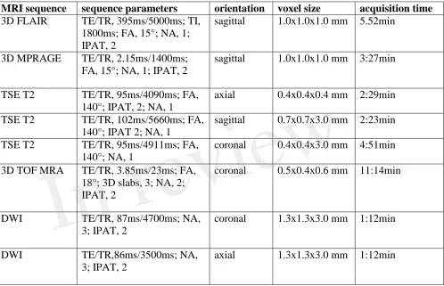
[Table 1 around here] 172
173
2.6 CT perfusion and CT angiography
174
Cases 5 to 10 in series b were transferred to a 16-slice computed tomography (CT) scanner 175
(Somatom Sensation 16, Siemens) immediately after surgical clip placement. Plain CT of the brain 176
was performed in coronary plane sequential acquisition (5-mm slice thickness) to localize the 177
surgical clip and to rule out intracranial hemorrhage. Then, a CT perfusion (CTP) scan was 178
performed covering a 2.4 cm slab of the sheep brain which was centered on the tips of the MCAO 179
clip within the MCA territory (4 slices; 6-mm slice thickness). Post-processing of standard perfusion 180
maps (CBV, CBF, and Tmax) was conducted using a dedicated commercial software package 181
(SyngoVia, Siemens). These perfusion maps were rated by an experienced neuroradiologist (S.M.) 182
for presence and degree of MCA territory hypoperfusion using the following semiquantitative score: 183
0 = no lesion visible on Tmax/CBF/CBV, 1 = lesion visible on Tmax only, 2 = lesion visible on 184
Tmax and partially visible on CBF/CBV, 3 = lesion visible on Tmax/CBF and partially on CBV. 185
Finally, thin-section CT angiography of the craniocervical arterial vasculature (slice thickness; 186
0.75mm) was performed with arterial bolus tracking. Assessment of CTA 3D datasets was conducted 187
by an experienced neuroradiologist (S.M., C.M.) on a PACS station. 188
189
2.7 End of experiments
190
Sheep were killed in deep anesthesia by an intravenous potassium chloride overdose at the end of 191
each experiment (after MRI acquisition on day 2 in cases 1-3 and 8-10 and on day 0 in cases 4-7). 192
Death by cardiac arrest was certified by an independent veterinarian. 193
3 Results 194
All procedures were performed without major complications. No sheep suffered from any clinical 195
complications except for neurological deficits after MCAO. Physiological parameters were 196
continuously monitored before and directly after MCAO, and were in normal ranges throughout the 197
experiments. Mean arterial blood pressure (MAP, median [IQR]) was 93 [82.25-103.75] mmHg / 92 198
[82-107] mmHg, and pulse rate was 78 [71-99] beats/min / 81 [73-97] beats/min before / after 199
MCAO, respectively. Both parameters did not differ significantly between pre- and intraischemic 200
measurements (p = 0.903 for MAP and p = 0.451 for pulse rate). Imaging results from all 201
experiments are summarized in Table 2. 202
203
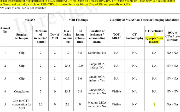
[Table 2 around here] 204
205
3.1 Results from series a 206
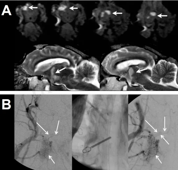
3.1.1 Case 1 207
DWI and T2w MRI on day 2 after MCAO showed a small ischemic lesion (1.7 mL; Figure 2A) in 208
right thalamic and midbrain regions after 2h of transient clip MCAO. The midbrain ischemia 209
suggested an erroneous confusion of the MCA main trunk (M1 segment) with the terminal ICA, 210
resulting in occlusion of terminal ICA and thus of perforating and choroidal artery branches with 211
mesencephalic supply. This appears likely since the proximal segment of the MCA trunk forms a 212
steep 180° curvature with an almost parallel course to the terminal ICA at the anterior skull base of 213
sheep (please also see case 2, Figure 2B; and case 4, Figure 4). 214
215
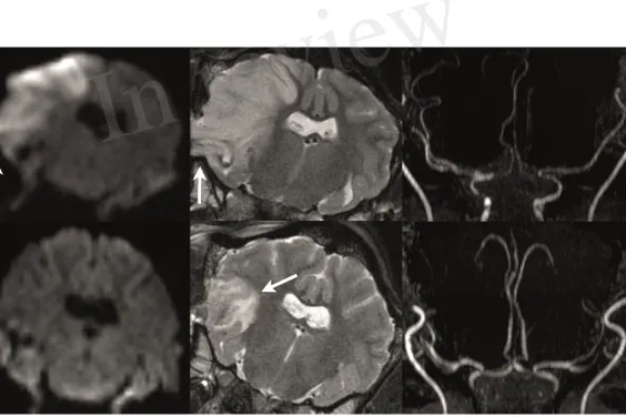
3.1.2 Case 2 216
Transient clip MCAO was performed for 2h. Selective DSA of the CCA could not unequivocally 217
demonstrate MCA main trunk occlusion despite variable angulations of the DSA images during 218
angiography (Figure 2B). MRI on day 2 showed a large-sized MCA territory infarct (Figure 3,upper 219
panels) with recanalized MCA on 3D TOF MRA. 220
221
[Figure 2 around here] 222
223
3.1.3 Case 3 224
Transient clip MCAO was performed for 2h. Selective DSA of CCA during MCAO again failed to 225
demonstrate MCA main trunk occlusion despite variable angulations of the DSA images during 226
angiography. MRI on day 2 showed no relevant ischemia on DWI (DWI lesion volume, 0.5 ml). A 227
small area of vasogenic edema with scattered and small hemorrhagic foci was found in the area of the 228
surgical access to the MCAO (Figure 3, lower panels). The MCA showed a normal flow signal on 3D 229
TOF-MRA images at day 2 after temporary clip occlusion. The neuro-deficit of the animal was light. 230
231
[Figure 3 around here] 232
233
The chosen approach in series a (cases 1-3) resulted in a highly variable infarct configuration for two 234
potential reasons. First, the vessel location for the surgical clip placement was inappropriate in case 1 235
(resulting in mid brain infarcts). Second and similar to the human situation, there might be a variable 236
extent of MCA vessel collateral flow resulting in highly different infarct sizes between cases 2 and 3. 237
Thus, we decided to modify the surgical approach in series b. We further tested whether the chosen 238
MCAO location was correct by using an optimized imaging algorithm during the ischemia phase. 239
240
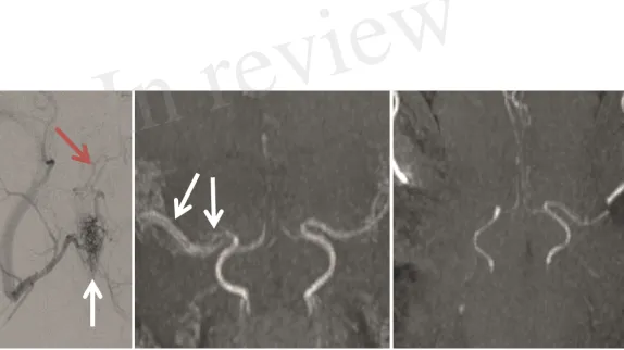
3.2 Results from series b 241
3.2.1 Case 4 242
In this case, we tested whether DSA of the CCA with additional superselective views from injection 243
of the right rete mirabile is capable of proofing MCAO. For immediate comparative assessment of 244
the vessel status after MCAO on 3D TOF MRA, the MCA main trunk was electrocoagulated to avoid 245
MRI artifacts emerging from the clip. A 0.021 inch microcatheter (Prowler Select Plus, Codman & 246
Shurtleff, Inc., Raynham, USA) was introduced into the largest inferior arterial branch supplying the 247
rete mirabile via long sheath endovascular access to right CCA directly after MCAO. Despite 248
multiple angulated vessel views on superselective DSA (Figure 4, left panel), MCAO could not be 249
correctly visualized. Further distal microcatheter navigation towards the rete mirabile led to 250
subsequent vasospasm with impaired demonstration of downstream vasculature. MRI was performed 251
directly at 2 hours following vessel occlusion. MRA visualized the MCAO site at the MCA main 252
trunk (Figure 4, right panel). The resulting early MCA territory infarct was visible on DWI images 253
(DWI lesion volume 13.3 ml) with beginning edematous change on T2w images (T2 lesion volume 254
5.8 ml). 255
256
[Figure 4 around here] 257
258
3.2.2 Case 5 259
Since DSA (including superselective views used in case 4) failed to demonstrate adequate vessel 260
occlusion, we decided to further amend the imaging protocol by introducing CTA with CTP imaging 261
in cases 5 to 7. Since electrocoagulation is not a feasible technique for transient MCAO, we decided 262
to first perform MCAO with a clip followed by immediate transfer to CTA/CTP imaging. Thereafter, 263
the clip was removed and the vessel was occluded in the same location by electrocoagulation in order 264
to perform subsequent MRI without clip-borne artifacts. Thus, CTP findings could be correlated with 265
the results of MRI simulating a temporary MCAO with ischemia duration of 2.5h (time interval from 266
initial vessel occlusion to MRI acquisition). 267
On CTP, a large area of right MCA territory hypoperfusion was seen on Tmax, whereas CBF and 268
CBV maps showed no areas hypoperfusion (perfusion score 1; Figure 5). Missing flow signal of the 269
MCA main trunk was seen on 3D TOF MRA. Visualization of the MCAO at the main trunk was not 270
possible on CTA images due to beam hardening artifacts originating from skull bone and the clip. On 271
DWI, signs of a small infarct in the MCA territory were detected (lesion volume 2.2 ml) without 272
edematous change on T2w images. In this case, evidence of correct temporary MCAO at the main 273
trunk by visualization of CTP hypoperfusion during the time window of clip occlusion was first 274
demonstrated with good correspondence to findings in immediate MRI. Hence, duration of MCAO 275
for 2.5h may still have been too short to detect a fully evolved infarct. 276
277
[Figure 5 around here] 278
279
3.2.3 Case 6 280
MCAO and imaging procedures were performed as described in case 5 except for the longer (4.5h) 281
duration of ischemia at the time of the MRI measurements in order to avoid a small final infarct due 282
to premature recanalization. Perfusion in the right hemisphere could not be evaluated on CTP due to 283
major streak artifacts from extensive jugular venous contrast media reflux. Correct MCA main trunk 284
occlusion could be reliably demonstrated using 3D TOF MRA, but CTA again failed to do so due to 285
beam hardening artifacts. Four and a half hours of ischemia led to a rather large-sized MCA infarct 286
that was seen on DWI MRI (lesion volume 14.5 ml) with resulting early edematous changes on T2w 287
images. 288
289
3.2.4 Case 7 290
MCAO was performed as described in case 5 and 6 except for a modification in the positioning of the 291
animal during CTP acquisition in order to avoid streak artifacts originating from contrast media 292
reflux into the jugular veins. To this end, the animal was placed in left anterior-lateral position on the 293
CT scanner table to relieve paunch-related increase in central venous pressure. DSA was also added 294
directly after CTP and before removal of the clip, and subsequent permanent electrocogaluation of 295
the MCA. However, as in the previous cases, DSA images could not clearly demonstrate correct 296
vessel occlusion. MRI was performed at 4.0 hours after MCAO. MRA was able to correctly visualize 297
MCA main trunk occlusion. On CTP, a large area of MCA territory hypoperfusion was seen on and 298
on CBF maps with minimal hypoperfusion also visible on CBV maps (perfusion score 3). There were 299
no major artifacts on CTP images. However, MCAO was not visible on CTA images due to beam 300
hardening artifacts similar to cases 5 and 6. Likewise, a rather large-sized MCA territory infarct was 301
seen on MRI (DWI volume, 16.8 ml) without significant early edematous change on T2w images 302
after 4h of ischemia. 303
304
3.2.4 Cases 8 to 10 305
During an interim summary of cases 5-7, the utilization of CT perfusion for demonstrating MCA 306
territory hypoperfusion as an indicator of correct MCAO was found successful except for extensive 307
beam hardening artifacts in case 6, caused by jugular venous reflux. Thus, we planned to gain further 308
experience with this CT perfusion protocol (applied with modified animal positioning as described in 309
case 7) in combination with the modified surgical approach of series b. However, we decided to 310
continue by performing a transient clip MCAO only (omitting electrocoagulation) and infarct size 311
measurement by MRI on day 2. The latter modifications were chosen in order to establish an 312
imaging-based MCAO model which is designed for testing novel combined endovascular approaches 313
of LVO stroke therapy in the future. Such transient MCAO stroke model should not only allow for 314
ultra-early MRI but also for delayed imaging assessment of final infarct evolution and clinical 315
follow-up as additional outcome measures. 316
The clip was removed after an ischemic period of 3.0 hours. CTP imaging was performed directly 317
after clip placement with modified animal positioning on the scanner table as described in case 7. 318
Ultra-early MRI scanning was skipped and animals were allowed to wake-up and recover from the 319
procedure. Infarct size measurement was performed on MRI at day 2 after MCAO in all three cases. 320
On CTP, MCA territory hypoperfusion was visible on Tmax in all three cases. In addition, CBF 321
reduction 9 and mild CBV reduction within the MCA territory was found in case 9 (perfusion score 322
3). In case 10, there were streak artifacts within the MCA territory from clip placement which, 323
however, did not severely impair visibility of hypoperfusion (perfusion score 2). The MCAO was 324
again not visible at all on DSA of the CCA in cases 8 and 9 after clip placement, and only poorly 325
visible in case 10. On MRI at day 2, medium-sized MCA territory infarcts were evident on both DWI 326
and T2w images (DWI volume, 6-8.5 ml) in cases 8 and 9. In contrast, the MCA territory infarction 327
was rather small-sized (DWI volume, 0.9 ml) despite proved MCA territory hypoperfusion on CTP in 328
case 10. This surprising result was explained by MRA on day 2 showing an early duplication of the 329
MCA vessels as a normal variation in this case (Figure 4, mid panel). This variant may be a source 330
for strongly improved collateralization within the MCA territory in some individuals. Identification 331
and occlusion of the duplicate MCA main trunk can be challenging as it could be located deeply 332
within a cerebral sulcus or the brain parenchyma. 333
334
4 Discussion 335
The aim of this case study was to establish a feasible imaging modality for MCA territory 336
hypoperfusion assessment in an ovine transient MCAO model, and to document our experience 337
collected on the way towards this aim. Final infarct size on MRI represents a meaningful efficacy 338
surrogate in experiments on acute stroke therapeutic interventions. However, in studies using 339
transient MCAO this is only valid when the extent of brain hypoperfusion and thus the expected final 340
lesion size without reperfusion or therapeutic intervention is known in order to compare it to the final 341
lesion volume with recanalization and/or accompanying therapeutic intervention. 342
We evaluated different imaging protocols for both intra-ischemic and post-ischemic perfusion and 343
infarct assessment, and performed a step-wise amendment of the imaging procedures and protocols. 344
The finally resulting imaging strategy was feasible to demonstrate temporary MCA territory 345
hypoperfusion during clip occlusion prior to vessel reopening in the intra-ischemic phase of 346
temporary MCAO. Furthermore, we performed a “two-step” occlusion by MCA clipping prior to 347
CTP, and electrocoagulation after CTP at the exact same vessel location to validate the results by 348
means of TOF MRA without the risk of clip-derived artifacts. 349
We also determined CTP using standard post-processed image maps (Tmax, CBF and CBV). This 350
imaging technique was feasible to confirm hypoperfusion and thus the correct clip placement during 351
MCAO. Such confirmation is an important quality assurance method when later removing the clip to 352
model successful recanalization. Although derived from a relatively small number of animals 353
undergoing CTP, our results indicate that final infarct size may be highly variable at least within a 354
time window of 3-4.5 hours of MCAO. These results are in-line with previous experiments done by 355
Wells et al. (Wells et al., 2015) that demonstrated DWI volumes ranging from 7 to 15% of whole 356
brain tissue after 2 hours of ischemia with proximal clip MCAO in 6 animals. In principle, this 357
variability may arise from incomplete MCA occlusion or a variable extent of collateral circulation to 358
the MCA territory. Although a definite conclusion is hard to make, we argue for the latter as the most 359
likely explanation due to numerous reasons. First, the Yasargil clips used in our experiments are also 360
used in humans and exhibit closing forces (>150g, 1.47N) that should be absolutely sufficient to 361
occlude the ovine MCA. Complete vessel coverage by the clip was confirmed after thorough visual 362
inspection by the surgeon (M.J.S.). Of note, the ovine MCA is smaller than the vessels Yarsagil clips 363
are usually placed on, so it is not difficult to cover it entirely. Second, we report cases of considerable 364
infarcts (e.g. cases 2, 8, and 9). This points at a factor being different between individual subjects 365
rather than a technical failure. Indeed, the extent of collateral circulation determines the extent of the 366
core infarct size very early after onset of human LVO stroke of the MCA (Wheeler et al., 2015; 367
Maurer et al., 2016), and the situation can be similar in sheep. If this assumption was right, it would 368
underpin the translational value of the model described herein, but also calls for pretest assessment of 369
collateral status to exclude extreme outcomes. In the sheep model, collateral circulation of the MCA 370
may further be enhanced by dedicated variants of the ovine cerebral arteries such as a duplicated 371
MCA main trunk (see case 10). Permanent MCAO by electrocoagulation as employed in our study 372
was previously reported to result in reproducible infarct volumes throughout a 7 week surveillance 373
period, starting 24h after MCAO (Boltze et al. 2008). Similar findings were reported for swine (Imai 374
et al., 2006). This might come in line with our assumption, as the initially „tissue-preserving“ effects 375
of collateralization will become less prominent over time in case a critical hypoperfusion/complete 376
blood flow disruption is present. During the acute stage, however, individual differences in 377
collateralization capacity would result in much more variable lesion volumes. 378
The volume of hypoperfused brain tissue at early time points of vessel occlusion may be later 379
correlated to the final lesion size. In our series, some cases that demonstrated profound 380
hypoperfusion at the time of vessel occlusion (score 3) showed rather large-sized infarct volumes on 381
follow-up DWI MRI. However, owing to the small number of cases with well-evaluable CTP images 382
(n=4) we were not able to clearly prove a suggested association between the extent of hypoperfusion 383
early after clip application and final infarct on DWI by using a semiquantitative analysis of perfusion 384
deficits. In order to provide a robust estimation of final infarcts, more data from CTP before clip 385
removal (simulating the endovascular recanalization) should be compared to final infarct size on MRI 386
or infarct histology in future studies. 387
388
4.1 Surgical approach
389
Reproducible and reliable infarcts could not be induced in series a, and effective occlusion depended 390
on qualitative visual assessment by the surgeon. Due to the basal approach, the proximal MCA and 391
terminal ICA could be reached easily. However, the narrow loop between the terminal ICA and the 392
proximal MCA may have led to erroneous terminal ICA occlusion, resulting in brain stem infarct 393
presumably from associated choroidal vessel occlusion with the absence of any MCA territory 394
ischemia (case 2). Surgical knowledge of this dedicated anatomy being different to human basal brain 395
arteries is crucial to avoid such complication. Moreover, duplication of MCA main trunk (M1 396
segment, see case 10 and Figure 4) represents a relatively frequently observed anatomical variant in 397
sheep, and is also supposed to be the source for of a high degree of collateralization within the MCA 398
territory. Such duplication may not be entirely visible during neurosurgical exposure. 399
According to the impression from our experienced vascular neurosurgeon (M.J.S.), the surgical 400
approach chosen in series b was technically more suitable for MCAO, as long as possible early 401
duplication of the MCA was ruled out and the proximal MCA (vascular loop near the optic nerve) 402
was clearly identified. 403
404
4.2 MCAO imaging
405
DSA, superselective DSA of vessels supplying the rete mirabile, and CTA were not suitable to 406
confirm correct MCAO. This was presumably due to the tiny caliber of intracranial arteries distal to 407
the rete mirabile on superselective DSA and CTA, and many overlapping large-sized extracranial 408
branches of the carotid artery on non-selective DSA, respectively. 3D rotational DSA might be an 409
alternative option to visualize the occlusion of the MCA main trunk which was however not possible 410
due to limited technical capabilities of our experimental angiography suite. Imaging of 411
leptomeningeal collateral status in MCAO may be of interest as it could serve as an estimate for 412
clinical outcome and final infarct size. However, the same methodological limitations of DSA and 413
CTA as in the confirmation of correct MCAO may also account for the poor visualization of the tiny 414
pial vessels in the sheep brain that impair a sufficient analysis of collaterals. However, in analogy to 415
human large vessel occlusion stroke, CT perfusion penumbral imaging may also provide indirect 416
information on the presence or absence of collaterals (Vagal et al., 2016; Lu et al., 2019). Non-human 417
primate (NHP) models of ischemic stroke (for review see Herrmann et al., 2019) might be 418
advantageous when assessing small-caliber intracerebral vessels and the cranial anatomy in NHPs is 419
even closer to the human situation. However, the use of NHPs is ethically restricted in many 420
contains, and costs related to using the species by far exceed those of other large animal stroke 421
models. In turn, this restricts sample sizes and often severely limits endpoints that can be addressed 422
quantitatively. 423
Beam-hardening metal artifacts were visible on CTA during temporary MCAO induced by titanium 424
clip application. 3D TOF MRA at 3T reliably showed MCAO, but required (permanent) occlusion by 425
electrocoagulation to avoid clip-borne artifacts (Steiger and van Loon, 1999). This issue might be 426
mitigated by the use of newly developed and improved, but extremely expensive MRI-compatible 427
clips. These are made of special titanium based alloys or Phynox, an alloy composed of cobalt, 428
chrome, nickel, and molybdenum (e.g., Aesculap Yasargil mini clips) and cause only minimal 429
artifacts. However, in transient MCAO, this imaging modality may be less efficient and also difficult 430
to apply during a short ischemic time window between two neurosurgical procedures for clip 431
placement and subsequent removal. This is particularly relevant for experiments that add additional 432
time for endovascular procedures, e.g. for intra-arterial neuroprotective therapy, which necessitate 433
additional navigation and placement of a catheter into the brain supplying arteries. Nevertheless, 434
MRI-compatible clips might allow perfusion-weighted imaging sequences to assess the perfusion 435
deficit during occlusion. 436
Positron emission tomography (PET) has been reported as a gold standard for experimental perfusion 437
deficit assessment and is applicable in sheep (Terpolilli et al., 2012; Werner et al., 2015). Moreover, 438
PET imaging is hardly susceptible to metal artifacts from a placed clip. However, PET imaging 439
requires a dedicated infrastructure while its application and in particular full data analysis may be too 440
time-consuming to be applied in acute experimental settings during a short time window of 441
temporary vessel occlusion. 442
We found that CT perfusion reliably provided indirect evidence of MCAO by demonstrating MCA 443
territory hypoperfusion already in a small number of cases. Importantly, it was the most feasible 444
modality that could be applied in a time-efficient manner during temporary neurosurgical clip 445
MCAO among the tested imaging techniques. In our experience, streak artifacts related to venous 446
reflux into the internal jugular vein may be reduced by placing the animal in a left anterior-lateral 447
position on the CT scanner table to relieve paunch-related venous pressure. This finally resulted in 448
good diagnostic quality of the CTP images. 449
450
4.3 Study limitations 451
In this pilot study with small number of consecutive animals, no complete blinding or randomization 452
could be performed which may potentially bias the analysis of study outcomes. However, the 453
investigators who performed the volumetric analysis of infarct volume on MRI images were blinded 454
to the respective animals’ treatment protocols. Detailed information derived on correlation of infarct 455
size with hypoperfusion volume could not be obtained due to variable imaging protocols and the 456
small number of animals that finally underwent CTP, warranting additional research on this aspect. 457
Consequently, further studies with a fixed CTP imaging protocol and ischemic time window of 458
temporary MCAO are necessary. 459
460
5 Conflict of Interest
461
Giorgio Cattaneo was an employee of Acandis GmbH during the duration of this study. 462
The other authors declare that the research was conducted in the absence of any commercial or
463
financial relationships that could be construed as a potential conflict of interest.
464 465
6 Author Contributions
466
Herrmann AM: significant contribution to study design, to data acquisition, analysis of data, to article 467
drafting, critical review of intellectual contents and approval of final version 468
Cattaneo G: significant contribution to study design, to article drafting, critical review of intellectual 469
contents and approval of final version 470
Eiden S: significant contribution to data acquisition, analysis and interpretation of data, and to critical 471
review of intellectual contents and approval of final version 472
Wieser M: significant contribution to data acquisition, analysis of data, and to critical review of 473
intellectual contents and approval of final version 474
Kellner E: significant contribution to data acquisition, analysis of data, and to critical review of 475
intellectual contents and approval of final version 476
Maurer C: significant contribution to data acquisition, analysis of data, and to critical review of 477
intellectual contents and approval of final version 478
Haberstroh J: significant contribution to concept and study design, to data acquisition, and to critical 479
review of intellectual contents and approval of final version 480
Mülling C.: significant contribution to concept and study design, and to critical review of intellectual 481
contents and approval of final version 482
Niesen W-D: significant contribution to data acquisition, and to critical review of intellectual 483
contents and approval of final version 484
Urbach H: significant contribution to concept and study design, and to critical review of intellectual 485
contents and approval of final version 486
Boltze J: significant contribution to concept and study design, to data interpretation, and to article 487
drafting, critical review of intellectual contents and approval of final version 488
Meckel S: significant contribution to experimental design image analysis, data acquisition and 489
analysis and interpretation of data, article drafting, critical review of intellectual contents and 490
approval of final version 491
Shah MJ: significant contribution to experimental design, to article drafting, critical review of 492
intellectual contents and approval of final version 493
494
7 Funding
495
This work was supported by Federal Ministry of Education and Research, Germany (grant 496
13GW0015B). 497
498
8 Acknowledgments
499
We thank our research technician Hansjörg Mast for valuable support with the MRI measurements of 500
all animals. 501
502
9 Data Availability Statement
503
The datasets generated and/or analyzed during the current study are available from the corresponding 504
author on request. 505
10 REFERENCES 507
Boltze, J., Förschler, A., Nitzsche, B., Waldmin, D., Hoffmann, A., Boltze, C. M., et al. (2008). 508
Permanent middle cerebral artery occlusion in sheep: a novel large animal model of focal cerebral 509
ischemia. Journal of cerebral blood flow and metabolism : official journal of the International
510
Society of Cerebral Blood Flow and Metabolism 28, 1951–1964. doi: 10.1038/jcbfm.2008.89
511
Goyal, M., Menon, B. K., van Zwam, W. H., Dippel, D. W. J., Mitchell, P. J., Demchuk, A. M., et al. 512
(2016). Endovascular thrombectomy after large-vessel ischaemic stroke: a meta-analysis of 513
individual patient data from five randomised trials. The Lancet 387, 1723–1731. doi: 514
10.1016/S0140-6736(16)00163-X 515
Herrmann, A. M., Meckel, S., Gounis, M. J., Kringe, L., Motschall, E., Mülling, C., et al. (2019). 516
Large animals in neurointerventional research: A systematic review on models, techniques and 517
their application in endovascular procedures for stroke, aneurysms and vascular malformations. 518
Journal of cerebral blood flow and metabolism : official journal of the International Society of
519
Cerebral Blood Flow and Metabolism, 271678X19827446. doi: 10.1177/0271678X19827446
520
Imai, H., Konno, K., Nakamura, M., Shimizu, T., Kubota, C., Seki, K., et al. (2006). A new model of 521
focal cerebral ischemia in the miniature pig. Journal of neurosurgery 104, 123–132. doi: 522
10.3171/ped.2006.104.2.123 523
Linfante, I., and Cipolla, M. J. (2016). Improving Reperfusion Therapies in the Era of Mechanical 524
Thrombectomy. Translational stroke research 7, 294–302. doi: 10.1007/s12975-016-0469-3 525
Lu, S.-S., Zhang, X., Xu, X.-Q., Cao, Y.-Z., Zhao, L. B., Liu, Q.-H., et al. (2019). Comparison of CT 526
angiography collaterals for predicting target perfusion profile and clinical outcome in patients with 527
acute ischemic stroke. European radiology. doi: 10.1007/s00330-019-06027-9 528
Maurer, C. J., Egger, K., Dempfle, A.-K., Reinhard, M., Meckel, S., and Urbach, H. (2016). Facing 529
the Time Window in Acute Ischemic Stroke: The Infarct Core. Clinical neuroradiology 26, 153– 530
158. doi: 10.1007/s00062-016-0501-8 531
Savitz, S. I., Baron, J.-C., Yenari, M. A., Sanossian, N., and Fisher, M. (2017). Reconsidering 532
Neuroprotection in the Reperfusion Era. Stroke 48, 3413–3419. doi: 533
10.1161/STROKEAHA.117.017283 534
Steiger, H. J., and van Loon, J. J. (1999). Virtues and drawbacks of titanium alloy aneurysm clips. 535
Acta neurochirurgica. Supplement 72, 81–88.
536
Terpolilli, N. A., Kim, S.-W., Thal, S. C., Kataoka, H., Zeisig, V., Nitzsche, B., et al. (2012). 537
Inhalation of nitric oxide prevents ischemic brain damage in experimental stroke by selective 538
dilatation of collateral arterioles. Circulation research 110, 727–738. doi: 539
10.1161/CIRCRESAHA.111.253419 540
Vagal, A., Menon, B. K., Foster, L. D., Livorine, A., Yeatts, S. D., Qazi, E., et al. (2016). 541
Association Between CT Angiogram Collaterals and CT Perfusion in the Interventional 542
Management of Stroke III Trial. Stroke 47, 535–538. doi: 10.1161/STROKEAHA.115.011461 543
Wells, A. J., Vink, R., Blumbergs, P. C., Brophy, B. P., Helps, S. C., Knox, S. J., et al. (2012). A 544
surgical model of permanent and transient middle cerebral artery stroke in the sheep. PloS one 7, 545
e42157. doi: 10.1371/journal.pone.0042157 546
Wells, A. J., Vink, R., Helps, S. C., Knox, S. J., Blumbergs, P. C., and Turner, R. J. (2015). Elevated 547
Intracranial Pressure and Cerebral Edema following Permanent MCA Occlusion in an Ovine 548
Model. PloS one 10, e0130512. doi: 10.1371/journal.pone.0130512 549
Werner, P., Saur, D., Zeisig, V., Ettrich, B., Patt, M., Sattler, B., et al. (2015). Simultaneous 550
PET/MRI in stroke: a case series. Journal of cerebral blood flow and metabolism : official journal
551
of the International Society of Cerebral Blood Flow and Metabolism 35, 1421–1425. doi:
552
10.1038/jcbfm.2015.158 553
Wheeler, H. M., Mlynash, M., Inoue, M., Tipirnini, A., Liggins, J., Bammer, R., et al. (2015). The 554
growth rate of early DWI lesions is highly variable and associated with penumbral salvage and 555
clinical outcomes following endovascular reperfusion. International journal of stroke : official
556
journal of the International Stroke Society 10, 723–729. doi: 10.1111/ijs.12436
557 558 559
Figure legends 560
561
Figure 1. Study design and surgical approaches 562
(A) Overview on study design with two experimental series. Clip pictograms indicate (transient) 563
MCAO by vessel clipping whereas the forceps indicate MCAO by electrocoagulation. (B) and (C): 564
Schematic illustration of the surgical approaches (top), 3D-Reconstruction showing the skin incision 565
(middle), 3D-bony-reconstruction with muscle (red) and craniectomy (blue) overlays (bottom). (B) 566
The surgical approach according to Wells et al. (series a). The proximal MCA and terminal ICA were 567
reached easily. Bony CT reconstruction shows the partial removal of the coronoid process. (C) 568
Approach according to Boltze et al. (series b), in which the distal branches of the MCA were 569
followed proximally until the optic nerve and the terminal internal carotid artery (ICA) had been 570
identified. A partial resection of the coronoid process was not necessary (CT reconstruction). 571
Dotted lines: skin incision; blue areas: craniectomy; *: muscle dissection (series a only). The green 572
arrows describes the surgeon's approach and the approximate line of vision. 573
574
Figure 2: Results from case 1 575
(A) MRI images on day 2 after MCAO from case 1. Upper panels showDWI images in coronal view 576
with ischemic lesions of the midbrain tegmental area within the right crus cerebri and right thalamus 577
(white arrows).Lower panelsshowconsecutive edema on T2w images in mid-sagittal views.
578
(B) CCA DSA images before and after clip MCAO. After clip MCAO, no clear cut-off of MCA main 579
trunk was visible with possible faint MCA filling (DSA image in left panel) at the clip level 580
(unsubtracted image in mid panel). After clip removal (DSA image in right panel), filling of the main 581
MCA trunk was visible. However, the distal MCA branch vasculature was not seen due to vessel 582
overlap from rete mirabile and larger extracranial arteries. 583
584
Figure 3: Results from cases 2 and 3 585
MRI images of case 2 (upper panels) and case 3 (lower panels) on day 2 after 2h of transient MCAO. 586
DWI and T2w images show a large right MCA territory infarct lesion (DWI lesion volume, 25.6 ml) 587
with swelling and mild herniation through the craniectomy site (arrows in upper left and mid panels). 588
MRA (upper right panel) demonstrates adequate MCA recanalization after right temporary MCAO. 589
In case 3, no relevant MCA territory ischemia is seen on DWI images (lower left panels; DWI lesion 590
volume, 0.5ml), whereas T2w images exhibit vasogenic edema in the area of the craniectomy with 591
mild hemorrhagic foci (arrow in lower middle panel). Again, MRA demonstrates adequate MCA 592
vessel recanalization after right temporary MCAO (lower left panel). 593
594
Figure 4: Results from case 4 595
DSA image (left panel) showing superselective rete mirabile injection (red arrow) after permanent 596
MCAO (animal 4) without clear evidence of MCA main trunk occlusion (red arrow). 3T TOF MRA 597
showing duplicated right MCA main trunk post temporary MCAO (mid panel, arrows) and clear 598
evidence of right MCA occlusion after permanent MCAO (right panel). 599
600
Figure 5: Results from case 5 601
CTP images from case 5. Directly after clip MCAO profound hypoperfusion is visible on Tmax maps 602
(upper panels) without reduction in CBV (lower panels). False color scale indicates Tmax from 0 603
(purple) to 2.5s (red) in upper panels and CBV from 0 ml/100g (purple) to 6 ml/100g (red) in lower 604
panels, respectively. 605
606
Table 1: Summary of 3 Tesla MRI sequence parameters 607
DWI - diffusion weighted imaging; FA - flip angle; FLAIR - Fluid-attenuated inversion recovery; 608
IPAT - integrated parallel imaging techniques; MPRAGE - Magnetization Prepared Rapid 609
Acquisition GRE (gradient echo); NA - number of averages; TE - echo time; TOF MRA - time-of-610
flight MR angiography; TR - repetition time; TSE - turbo spin echo 611
612
MRI sequence sequence parameters orientation voxel size acquisition time
3D FLAIR TE/TR, 395ms/5000ms; TI,
1800ms; FA, 15°; NA, 1; IPAT, 2
sagittal 1.0x1.0x1.0 mm 5.52min
3D MPRAGE TE/TR, 2.15ms/1400ms;
FA, 15°; NA, 1; IPAT, 2
sagittal 1.0x1.0x1.0 mm 3:27min
TSE T2 TE/TR, 95ms/4090ms; FA,
140°; IPAT, 2; NA, 1
axial 0.4x0.4x0.4 mm 2:29min
TSE T2 TE/TR, 102ms/5660ms; FA,
140°; IPAT 2; NA, 1
sagittal 0.7x0.7x3.0 mm 2:23min
TSE T2 TE/TR, 95ms/4911ms; FA,
140°; NA, 1
coronal 0.4x0.4x3.0 mm 4:51min
3D TOF MRA TE/TR, 3.85ms/23ms; FA, 18°; 3D slabs, 3; NA, 2; IPAT, 2
coronal 0.5x0.4x0.6 mm 11:14min
DWI TE/TR, 87ms/4700ms; NA,
3; IPAT, 2
coronal 1.3x1.3x3.0 mm 1:12min
DWI TE/TR,86ms/3500ms; NA,
3; IPAT, 2
axial 1.3x1.3x3.0 mm 1:12min
613
Table 2: Summary of MCAO technique, MRI findings and visualization of MCAO with various imaging modalities 614
*refers to time interval from start of MCAO until MRI DWI was performed in cases 4-7 (vessel coagulation). § refers to semiquantitative 615
visual assessment of hypoperfusion in MCA territory: 0 = no lesion on Tmax/CBF/CBV, 1 = lesion visible on Tmax only, 2 = lesion visible 616
on Tmax and partially visible on CBF/CBV, 3 = lesion fully visible on Tmax/CBF and partially on CBV 617
NV = not visible, NA = not available 618
619
Animal No.
MCAO MRI Findings Visibility of MCAO on Vascular Imaging Modalities
Surgical technique
Duration of ischemia*
[hours]
Day of MRI
DWI lesion volume
[ml]
T2 lesion volume
[ml]
Location of ischemia / surrounding
edema
TOF
MRA#
CT Angiography
CT Perfusion –
hypoperfusio n score§
DSA of CCA / rete
mirabile
1 Clip 2 2 1.7 4.8 Midbrain / No NA NA NA NA / NA
2 Clip 2 2 25.6 17.0 Large MCA
infarct / No NA NA NA NV / NA
3 Clip 2 2 0.5 3.6 Small MCA
infarct / Yes NA NA NA NV / NA
4 Coagulation 2 0 13.3 5.8 Large MCA
ischemia / No Visible NA NA NV / NV
5
Clip for CTP - coagulation for
MRI
2.5 0 2.2 0 Medium MCA
ischemia / No Visible NV 1 NA / NA
6
Clip for CTP - coagulation for
MRI
4.5 0 14.5 11.3 Large MCA
ischemia / No Visible NV NV - Artifacts NA / NA
7
Clip for CTP - coagulation for
MRI
4.0 0 16.8 0.2 Large MCA
ischemia / No Visible NV 3 NV / NA
8 Clip 3.0 2 6.0 5.4 Medium MCA
ischemia / No NA NV 1 NV / NA
9 Clip 3.0 2 8.5 9.2
Medium MCA ischemia /
minimal
NA NV 3 NV / NA
10 Clip 3.0 2 0.9 3.9 Small MCA
infarct / Yes NA NV
2 - clip Artifacts
Partially visible /
NA
620