STUDY ON ANGULAR LEAF SPOT CAUSING BACTERIA OF PUMPKIN (
C. MAXIMA
1
Sabina Eyesmin Sumi,
1Afrin Akter,
3
Asadul Islam
1
MS Student, Professor Joarder DNA and Chromosome Research Lab., Department of Genetic Engineering and
Biotechnology, Faculty of Life and Earth Sciences, University of Rajshahi, Rajs
2
Assistant Professor, Professor Joarder DNA and Chromosome Research Lab., Department of Genetic
Engineering and Biotechnology, Faculty of Life and Earth Sciences, University of Rajshahi,
3
Professor Joarder DNA and Chromosome Research Lab., Department of Genetic Engineering and
Biotechnology, Faculty of Life and Earth Sciences, University of Rajshahi, Rajshahi
ARTICLE INFO ABSTRACT
The present inquisition was conveyed to isolation, characterization of Lachrymans
control.
performed gram negative, rod shaped and pink color in gram staining test. It showed positive result in Catalase, Indole, Simmon’s citrate, MacConkey agar, Potassium hydroxide, Methyl Red test and negative
fermented the carbohydrates method.The highest 22
30µg/disc concentration against the isolated bacterium.
antimicrobial activity with inhibition zone 12.1±0.2mm that means the plant extract had antimicrobial activity against the isolated bacteri
this devastating disease.
Copyright © 2017, Sabina Eyesmin Sumi. This is an open use, distribution, and reproduction in any medium, provided
INTRODUCTION
Pumpkin (Cucurbita pepo, C. maxima) is an important vegetable crop, grown all over the world. It’s locally known as misty kumra which belongs to the family of Cucurbitaceae. color arise from orange carotenoid pigments, such as cryptoxanthin, alpha and beta carotene, all of which are provitamin A compounds converted to vitamin A
(Provesi et al., 2011). Pumpkin is used for a variety of purposes ranging from agricultural (e.g., animal feed) to ornamental sales. Nowadays, pumpkins are grown in many countries like China, India, Russia, Iran, and the United States. Pumpkins have a great nutritional value. Pu
vegetable, that are incredibly rich in vital antioxidants, and vitamin.
*Corresponding author: Sabina Eyesmin Sumi,
MS Student, Professor Joarder DNA and Chromosome Research Lab., Department of Genetic Engineering and Biotechnology, Faculty of Life and Earth Sciences, University of Rajshahi, Rajshahi-6205, Bangladesh
ISSN: 0975-833X
Vol.
Article History:
Received 16th September, 2017
Received in revised form 21st October, 2017
Accepted 08th November, 2017
Published online 27th December, 2017
Citation: Sabina Eyesmin Sumi, Afrin Akter, Roushan Ali, Rizwoana Sharmin Lia, Faruk Hasan, Asadul Islam and Biswanath Sikdar on angular leaf spot causing bacteria of pumpkin (
Research, 9, (12), 62162-62168.
Key words:
Pumpkin,Angular leaf spot disease,
Biochemical test, Antibiotic test, Antibacterial test.
RESEARCH ARTICLE
STUDY ON ANGULAR LEAF SPOT CAUSING BACTERIA OF PUMPKIN (
C. MAXIMA
) AND EVALUATION OF ITS SENSITIVITY
Afrin Akter,
1Roushan Ali,
1Rizwoana Sharmin Lia,
Asadul Islam and *
,3Biswanath Sikdar
MS Student, Professor Joarder DNA and Chromosome Research Lab., Department of Genetic Engineering and
Biotechnology, Faculty of Life and Earth Sciences, University of Rajshahi, Rajshahi
Assistant Professor, Professor Joarder DNA and Chromosome Research Lab., Department of Genetic
Engineering and Biotechnology, Faculty of Life and Earth Sciences, University of Rajshahi,
Rajshahi-6205, Bangladesh
NA and Chromosome Research Lab., Department of Genetic Engineering and
Biotechnology, Faculty of Life and Earth Sciences, University of Rajshahi, Rajshahi
ABSTRACT
The present inquisition was conveyed to isolation, characterization of
Lachrymans bacterium from angular leaf spot disease of pumpkin and evaluation of its biological control. The isolated bacterium was characterized by different biochemical test.
performed gram negative, rod shaped and pink color in gram staining test. It showed positive result in Catalase, Indole, Simmon’s citrate, MacConkey agar, Potassium hydroxide, Methyl Red test and negative to Motility and Urease test. In Triple Sugar Iron and Kligler Iron Agar test,
fermented the carbohydrates. Antibiotic and antimicrobial activities were screened by disc diffusion method.The highest 22±0.0mm diameter of zone of inhibition was
30µg/disc concentration against the isolated bacterium. Adhatoda vasica
antimicrobial activity with inhibition zone 12.1±0.2mm that means the plant extract had antimicrobial activity against the isolated bacterium. The present research could be helpful for biological control of this devastating disease.
open access article distributed under the Creative Commons Attribution provided the original work is properly cited.
) is an important vegetable crop, grown all over the world. It’s locally known as misty kumra which belongs to the family of Cucurbitaceae. Its pigments, such as
beta-, all of which are min A in the body Pumpkin is used for a variety of purposes ranging from agricultural (e.g., animal feed) to ornamental sales. Nowadays, pumpkins are grown in many countries like China, India, Russia, Iran, and the United States. Pumpkins have a great nutritional value. Pumpkins, a vegetable, that are incredibly rich in vital antioxidants, and
DNA and Chromosome Research Lab., Department of Genetic Engineering and Biotechnology, Faculty of Life and Earth Sciences, University of Rajshahi,
This vegetable is effective against some cancers and cardiovascular disease and the seeds are often eaten roasted and salted, also contain a plenty of mineral e.g. potassium. As far we know, pumpkin play a vital role in our economy and agriculture. Angular leaf spot disease of pumpkin, a bacterial disease, caused by Pseudomonas syrin
bacterium. Generally angular leaf spot of cucurbits is caused by Pseudomonas syringae pv.
Zitter et. al., 1996). It is distributed worldwide. The disease is emerged sporadically under humid and wet weather con (Bhat et. al., 2010; Riffaud et. al.,
Angular leaf spot of pumpkin is a devastating disease that results yield loss and fruit of poor quality. The disease may notice after a period of warm weather accompanied by rain. Leaf spots are variable in size and may be angular in shape because leaf veins limit enlargement of spots. The symptoms may vary depending on the host and environmental conditions, and are initially observed as circular necrotic lesions. As far our knowledge goes, there is no suitable report on isolation, characterization and their biological control measurement. International Journal of Current Research
Vol. 9, Issue, 12, pp.62162-62168, December, 2017
Sabina Eyesmin Sumi, Afrin Akter, Roushan Ali, Rizwoana Sharmin Lia, Faruk Hasan, Asadul Islam and Biswanath Sikdar spot causing bacteria of pumpkin (Cucurbita pepo, C. maxima) and evaluation of its sensitivity
STUDY ON ANGULAR LEAF SPOT CAUSING BACTERIA OF PUMPKIN (
CUCURBITA PEPO,
) AND EVALUATION OF ITS SENSITIVITY
Rizwoana Sharmin Lia,
2Faruk Hasan,
MS Student, Professor Joarder DNA and Chromosome Research Lab., Department of Genetic Engineering and
hahi-6205, Bangladesh
Assistant Professor, Professor Joarder DNA and Chromosome Research Lab., Department of Genetic
Engineering and Biotechnology, Faculty of Life and Earth Sciences, University of Rajshahi,
NA and Chromosome Research Lab., Department of Genetic Engineering and
Biotechnology, Faculty of Life and Earth Sciences, University of Rajshahi, Rajshahi-6205, Bangladesh
The present inquisition was conveyed to isolation, characterization of Pseodomonas syringae pv. bacterium from angular leaf spot disease of pumpkin and evaluation of its biological
terized by different biochemical test. The isolate performed gram negative, rod shaped and pink color in gram staining test. It showed positive result in Catalase, Indole, Simmon’s citrate, MacConkey agar, Potassium hydroxide, Methyl Red test and In Triple Sugar Iron and Kligler Iron Agar test, the bacterium Antibiotic and antimicrobial activities were screened by disc diffusion ±0.0mm diameter of zone of inhibition was observed by Cefotaxime in Adhatoda vasica showed the highest antimicrobial activity with inhibition zone 12.1±0.2mm that means the plant extract had antimicrobial um. The present research could be helpful for biological control of
ribution License, which permits unrestricted
This vegetable is effective against some cancers and the seeds are often eaten roasted and salted, also contain a plenty of mineral e.g. potassium. As far we know, pumpkin play a vital role in our economy and Angular leaf spot disease of pumpkin, a bacterial
Pseudomonas syringae pv. lachrymans
bacterium. Generally angular leaf spot of cucurbits is caused pv. lachrymans (Bradbury 1986; . It is distributed worldwide. The disease is emerged sporadically under humid and wet weather condition
et. al., 2003; Zitter et. al., 1996). Angular leaf spot of pumpkin is a devastating disease that results yield loss and fruit of poor quality. The disease may notice after a period of warm weather accompanied by rain. Leaf spots are variable in size and may be angular in shape e leaf veins limit enlargement of spots. The symptoms may vary depending on the host and environmental conditions, and are initially observed as circular necrotic lesions. As far our knowledge goes, there is no suitable report on isolation, n and their biological control measurement.
INTERNATIONAL JOURNAL OF CURRENT RESEARCH
Therefore, the present investigation was conducted to isolate the pathogenic bacteria from angular leaf spot of pumpkin, characterization by different biochemical test and evaluation of its biological control measurement.
MATERIALS AND METHODS
Plant Material
In the present research, disease infected pumpkin plant leaves were collected from the graveyard area of Rajshahi University, Rajshahi, Bangladesh and were identified by Bangladesh Fruit Research Station, Binodpur, Rajshahi. Angular leaf spot disease infected leaves of pumpkin were selected as plant material.
Isolation of bacteria
The infected leaves were surface disinfected using a dilute sodium hypochlorite (NaOCl) solution (10%) and rinsed thoroughly. Infected leaf portions were cut and grinded using a sterile mortar and pestle. The extract was poured into Luria-Bertani (LB) liquid medium and incubator at 37ºC for 16 hours. The next day, a sterile loop was used to streak the bacteria onto a solid agar medium very carefully. The bacteria were allowed to grow for at least 12 hours at 37ºC temperature and the colony were observed.
Morphological and Biochemical characterization
Gram staining
Gram staining test was performed according to Vincent and Humphrey, 1970. Crystal violet, iodine, ethanol and safranin were used. At first the isolated bacterial culture was heat fixed onto a glass slide. Then crystal violet was added to the bacterial sample and incubated for 1 min. After washing the slide, iodine was added in the medium. Then safranin was used to counterstaining (Jacquelyn, 1993). After all these steps the slide was used to observe under the light microscope at 100X using oil immersion.
SIM-medium test (Sulphide-Indole-Motility medium)
SIM medium is a multi-test agar medium that is used to test for indole production at the same time determining other characteristics of the bacterium (Harley, 2005). First of all in a conical flask 50 ml distilled water and 1.5 gm ready-made media was taken. Then the medium, two test tube, conical flask, cotton were autoclaved at 121°C. In the laminar air flow, 25 ml media was taken in two test tubes. After Cooling, a single bacterial colony and inoculated at 37°C temperature for 24 hours. It will react with the sodium thiosulfate in the medium and the indicator, ferric ammonium citrate, to produce ferrous sulfide which falls out of solution as a blackish precipitate. The indole portion of the test was performed by adding Kovac's reagent to the inoculated medium.
Simmon’s citrate test
The citrate test was used to check the ability to utilize citrate as its carbon and energy source (MacFaddin, 2000). In the laminar air flow, medium was poured in two test tubes and kept in slant condition. When cooled a single pure isolated colony was picked with a needle and the slant surface of one
test tube was lightly streaked. The incubation was done at 37°C for 18 hours. Citrate-positive result was interpreted on the basis of intense Prussian blue and in case of citrate -negative no color change occurred.
Catalase test
Catalase test is used to check whether the microbes produce catalase or not (Facklam and Elliott, 1995). A single colony was taken on a clean slide and hydrogen per oxide was added, smeared carefully. Catalase positive result was interpreted on the basis of production of oxygen bubbles.
MacConkey agar test
The differentiation of lactose fermenting bacteria, MacConkey medium was used (MacConkey, 1905). At first components were taken in conical flask and pH was adjusted to 7.0 and boiled to dissolved agar and sterilized at 15Ibs and 121°C for 20 minutes. MacConkey agar plates were prepared by pouring this medium on petridish and allowed to cool. The plates were streaking with desired pure colonies and were incubated at 37°C for 48 hours.
Kligler Iron Agar test
For Kligler Iron Agar test, 55gm of the medium was suspended in one liter of distilled or deionized water, mixed well and heated with frequent agitation. Volumes of 3 ml in tubes of 13 x 100 mm were dispensed and autoclaved at 121°C (15lbs. of pressure) for 20 minutes. The tubes were cooled in a slanted position to obtain a butt of 1.5 - 2.0 cm depth. Then the medium was inoculated with the colony in study by stabbing the butt and streaking the surface of the tube. After that the tubes were incubated at 37 °C for 24 hours and the results were recorded.
Triple Sugar Iron Agar (TSI) test
Triple Sugar Iron Agar is a differential medium that contains lactose, sucrose, a small amount of glucose (dextrose), ferrous sulfate, and the pH indicator phenol red. In a conical flask appropriate amount of readymade TSI medium were taken in 50 ml distilled water. Two test tube, and the medium was autoclaved, then taken in laminar air flow. Then autoclaved medium was poured into two test tubes and kept in a rack in slant condition. After cooling, a bacterial colony was taken. Then a sterilized inoculating loop was used for streaking back and forth along the surface of the slant. After capping the tubes were incubated at 37°C for 24 hours.
Potassium Hydroxide test
At first, a clean slide was taken. One or two large colony was placed on the slide from unknown stock. Then 1 drop of KOH (.3M) was added on top of the cells and kept for 1 minute. After waiting for a minute, the mixture was smeared by a tooth pick. After a moment positive result was confirmed by viscous
appearance of the bacteria.
Urease test
taken. Then a sterilized inoculating loop was used for streaking back and forth along the surface of the slant. Streak the surface of a urea agar slant with a portion of a well-isolated colony or inoculate slant with 1 to 2 drops from an overnight brain-heart infusion broth culture and incubate at 35-37°C in ambient air for 3 to 7 days and observed for the development of a pink color.
Methyl Red (MR) test
The methyl red solution was prepared and the medium was allowed to equilibrate at room temperature. Bacteria were inoculated into the MR broth medium in test tubes and incubated at 30°C or 37° C for 16-18 hours with observation. After inoculation, 1/3 of the suspension was poured into a clean non sterile tube then run the MR Test with the tube with 2/3. Then methyl red solution was added 2-3 drops to aliquot. A distinct red color showed positive result and yellow color showed negative result.
Antibiotic sensitivity test of isolated bacteria
Antibiotic sensitivity test was performed by moderate disc diffusion method (Hasan and Sikdar, 2016). The selected bacterial strains were grown at overnight in nutrient broths media that placed in the shaker at 37°C and 150 rpm for the antibiotic sensitivity test. A serial dilution technique was made for conducting the test. LB agar medium was used for making LB agar plate. The sterile liquid medium was distributed in sterile conical flasks when the temperature cooled down to 40-50°C. 15-20 ml of the medium was poured in each petridish approximately. Commercially available and frequently prescribed antibiotics namely, Ampicillin, Streptomycin, Azithromycin, Erythromycin, Penicillin, Kanamycin, Doxycycline, Amoxycillin, Gentamycin, Clarithromycin, Carbenicillin, Cefotaxime and Tetracycline were used against the isolated bacteria on the respective plates and incubated overnight at 37°C. Diameter of zone of inhibition was measured with the help of millimeter (mm) scale.
Antibacterial activity test using different plant extract
Materials of different plant species were taken from different location of University of Rajshahi Campus. In this research, different parts of 6 different plants extract were used for antimicrobial test. The names of the plants are Terminalia arjuna, Ocimum sanctum Adhatoda vasica, Centella asiatica
and, Datura metal and Mentha arvensis. After preparing LB agar plates, the disc were impregnated in the medium and extracts were applied in different concentrations. The plates were incubation at 37°C for 16 hours. Next day, the zone of inhibition was measured with the help of mm scale.
Statistical analysis
All the above investigations of the present study were repeated trees for consistency of results and statistical purpose. The data were expressed as Mean±SE and analyzed using Microsoft Excel software of 2010 version. P<0.05 was considered statistically significant.
RESULTS
Isolation the bacteria: The isolated colonies were creamy in
[image:3.595.329.536.57.352.2]color. The size and shape of colonies were found to be small, medium, convex and mucoid (Fig.1).
Fig. 1. Showing isolation of pure bacterial from infected plant leaf sample. A)Liquid culture in Lb medim, B) streak plate
Morphological and biochemical characterization
In gram staining, the isolate showed gram negative, rod shaped and pinkish in color when stained with counter-stained by the Safranin which was the indication for gram negative bacterium. In SIM medium test the isolated bacteria was non motile , indole negative because the bacteria did not produce red/pink color band on the top of tube when adding kovacs reagent and H2S was not produced as no black precipitation
formed(Fig.1 A). Simmon’s citrate test was positive because the medium color turns Prussian blue (Fig.1 B). In catalase test, the presence of bubbles resulting from production of oxygen gas clearly indicates a catalase positive result in this study (Fig.1 G). MacConkey agar test was positive and colonies were lactose fermenting because the isolate produce pink color around the colony (Fig.1 I). In KIA test, the isolated bacterium was glucose and lactose fermenting because slant and butt both were yellow (Fig.1 C). In TSI test, isolated bacterium did not produce hydrogen sulfide and was glucose, lactose and or sucrose fermenting because acidic slant and acidic butt were found (Fig.1 D). In KOH test, the test was positive and the bacterium was found to be negative because the bacterium was viscous and formed a mucoid string in 15 sec (Fig.1 H). In Urease Test, isolated bacterium did not hydrolyze urea (Fig.1 E). So, urease test was negative for isolated bacterium. In MR test, bacterium showed positive result, because the color of the methyl red changed from yellow to red (Fig.1 F), when added into the broth culture. The results are shown in Table 1.
Antibacterial activity of some antibiotic against isolated bacteria
The standard Cefotaxime revealed highest antibacterial activity with 22±0.0mm diameter of zone of inhibition at 30µg/disc concentration followed by Carbenicillin and Gentamycin with 20.6±0.5mm diameter of zone of inhibition at 100µg/disc and 10µg/disc concentration respectively.
[image:4.595.132.481.57.384.2]On the left hands, the standard Erythromycin showed the lowest zone of inhibition with 6.1±0.2mm at 15µl/disc concentration against the isolated bacterium (Fig. 3). The result of antibiotic sensitivity test is summarized in Table 2.
[image:4.595.55.549.441.723.2]Fig. 2. Showing Biochemical test of isolated bacteria, A) SIM test, B) Simmon’s citrate test, C) Kligler Iron Agar test, D) Triple sugar Iron test, E) Urease test. F) Methly red test., G) Catalase test, H) KOH test, I) MacConkey Agar test
Table 1. Characterization of the isolated bacteria in different biochemical test media
Test Name Response Appearance Remarks
Gram staining -(ve) Small, rod shaped, pink color colony Gram staining results conforms gram negative bacteria
SIM -(ve) Non motile, no H2S and indole production Gram negative bacteria showed non motile and did not
produce any indole and H2S
Simmon’s citrate agar +(ve) Color changed from green to the royal blue Citrate metabolizing gram negative bacteria
Catalase +(ve) Presence of oxygen bubbles Gram negative bacteria formed bubbles resulting from
production of O2 gas
MacConkey agar +(ve) Pink color around the colony Gram negative bacteria showed pink color confirming
lactose fermenting
Kligler Iron Agar +(ve) Yields yellow slants and yellow butt Gram negative bacteria yield slants and butt both were
yellow confirming glucose and lactose fermenting
TSI +(ve) Color changed from red to yellow Gram negative bacteria did not produce H2S and
confirming slant and butt both were yellow, so the bacteria were glucose, lactose and or sucrose fermenting
KOH +(ve) Thread like slime Gram negative bacteria formed thread
like slime
Urease -(ve) Slant remains yellow Gram negative bacteria did not hydrolyze urea
Methyl Red +(ve) Color changed from yellow to red ring Gram negative bacteria had the ability to utilize glucose
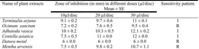
[image:4.595.56.546.441.720.2]Antibacterial activity of some plant extracts against isolated bacteria
Antibacterial activities of six different plants extracts were determined against the isolated bacterium. The extract of
Adhatoda vasica showed highest antibacterial activity with 12.1±0.2mm diameter of zone of inhibition at 30µl/disc concentration followed by 12±0.0mm diameter of zone of inhibition at 30µl/disc concentration by Centella asiatica plant extract. On the left hands, the extract of Datura metal showed lowest 6±0.0mm inhibition zone against the isolated bacteria at 10, 20, 30 µl/disc respectively (Fig. 4). The results are presented in Table 3.
DISCUSSION
Pumpkins are one of the important summer vegetable crops grown all around the world for a variety of reasons and mostly important to agricultural countries like Bangladesh. Pumpkin is affected by different types of viral, bacterial and fungal
diseases. Angular leaf spot disease is one of the most common bacterial diseases of pumpkin which is caused by
Pseudomonas syringae pv. lachrymans bacterium. Isolated bacterium was creamy in color on nutrient agar medium. Klement et al.,1990 and Narayanasamy, 1997 found that the
Pseudomonas syringae isolated from apicot trees were creamy color on nutrient agar medium. This results support our present finding. Bacterium isolated from angular leaf spot disease of pumpkin showed gram negative, in gram staining. According to Shila et al., 2013, Pseudomonas syringae associates with the cucurbits are gram negative. SIM test showed H2S, indole
[image:5.595.111.490.86.248.2]negative and non-motile on the medium. On contrast, Baron et al., 1990, reported a positive indole test and it gave red color on top of the agar medium within a second when Kovacs reagent was added. Isolated bacterium showed positive result to Simmon’s citrate agar medium and it gave blue color. Brown et al., 2015 found that some microbes grow on Simmons citrate agar medium. They are capable of using citrate as the sole carbon source, the ability to metabolize the ammonium salt in the medium and change the color into blue.
Table 2. Antibacterial activity of some antibiotic against the isolated bacteria
Name of antibiotic Symbol Disc potency
µl/disc
Zone of inhibition (in mm) Mean ± SE
Sensitivity pattern
Ampicillin AMP 10 6.6 ± 0 .2 R
Neomycin N 30 20.3 ± 0.5 S
Doxycycline DO 30 8.5 ± 0.4 R
Kanamycin K 30 15. 5 ± 0.5 I
Streptomycin S 10 13.6 ± 0.5 I
Erythromycin E 15 6.1 ± 0.2 R
Tetracycline TE 30 12.5 ± 0.5 I
Cefotaxime CTX 30 22 ± 0.0 S
Carbenicillin CB 100 20.6 ± 0.5 S
Azithromycin AZM 15 14.6 ± 0.5 I
Clarithromycin CLR 15 7 ± 0.0 R
Penicillin P 10 6.3 ± 0.2 R
Gentamicin GEN 10 20.6 ± 0.5 S
Amoxycillin AML 10 7.8 ± 0.2 R
Chloramphenicol C 30 19 ± 0.9 S
[image:5.595.90.513.276.386.2]Legend: R = Resistant (5-10 mm), I = Intermediate (11-15 mm), S = Susceptible (16 mm ≥) SE= Standard Error
Fig. 4. Showing antibacterial activity test of isolated bacteria, A) Adhatada vasica, B) Centella asiatica, C) Datura metel
Table 3. Antibacterial activity of some plant extracts against the isolated bacteria
Name of plant extracts Zone of inhibition (in mm) in different doses (µl/disc)
Mean ± SE
Sensitivity pattern
10µl/disc 20 µl/disc 30 µl/disc
Terminalia arjuna 9.1 ± 0.2 9.7 ± 0.6 11 ± 0.1 I
Ocimum sanctum 7.2 ± 0.2 7.6 ± 0.5 9.5 ± 0.4 R
Adhatoda vasica 10 ± 0.2 10.3 ± 0.5 12.1 ± 0.2 I
Centella asiatica 7.5 ± 0.5 11 ± 0.0 12 ± 0.0 I
Datura metal 6 ± 0.0 6 ± 0.0 6 ± 0.0 R
[image:5.595.128.477.439.526.2]This result confirmed our present finding. Catalase test was used to differentiate that bacterium that produces an enzyme catalase. Here, positive result was found when H2O2 was added
to the isolated bacterium and it produced air bubbles. Our work was confirmed by the work of Facklam and Elliott, 1995. The lactose fermenting capability of these strains was detected from the MacConkey agar test. Bacteria produce color around the colony so it was lactose fermenting. Isolated bacteria showed slants and butt, both were yellow in color, confirming glucose and lactose fermenting. TSI is most frequently used in the identification of the Enterobacteriaceae, although it is useful for other gram-negative bacterium. Isolated bacterium was glucose, lactose and or sucrose fermenting. KOH test was used to differentiate gram negative bacterium. Isolated bacteria from angular leaf spot infected pumpkin plant showed viscous appearance after adding KOH. According to Suslow et al., 1982, gram staining test was conducted with 3% (w/v) KOH. They found the similar result for Xanthomonas cynaraespp. as it appeared viscous after adding KOH. In urease test, the isolated bacterium showed negative result because no color change was found. Similar result was found by Bailey and Scott, 1974 and Christensen, 1946. The MR test was used to identify mixed acid fermenting bacteria that yield a stable acid as end product. So, the isolated bacterium was positive in methyl red test. Our result was confirmed the work of Crown
et al., 1998.
This result showed that isolated bacterium was highly sensitive against Cefotaxime with inhibition zone 22±0.0mm at 30µl/disc concentration. The isolated bacterium was highly susceptible against Cefotaxime antibiotic. Hasan and Sikdar, 2016 found the similar zone of inhibition by standard Kanamycin against Pseudomonas sp. Bharathi et al., 2014 reported a similar diameter of zone of inhibitions by Erythromycin against Pseudomonas aeruginosa. These results support our present findings. The use of medicinal plant as traditional medicine has been started several 1000 years ago (Chang et al., 2016). In this investigation, Adhatoda vasica and Centella asiatica exhibited broad spectrum activity against isolated bacterium with inhibition zone 12.1±0.2mm and 12±0.0mm at 30 µl/disc concentrations respectively. Bharathi
et al., 2010 Studied antimicrobial activity of Datura metel in 2010. In this investigation, Daura metel showed lowest antibacterial activity against isolated bacteria with inhibition zone 6±0.0mm at 10, 20, 30 µl/disc concentration respectively. Isolated bacterium showed 11±0.1mm zone of inhibition against Terminalia arjuna which are intermediate susceptible. Praba and Kumaresan, 2014worked on Allium sativum extract against Pseudomonas aeruginosa at 50% concentration. This findings also confirmed by Whitemore and Naidu, 2010 who found inhibitory action of garlic against gram positive and gram negative bacteria. Thus, this study confirms the efficacy of some antibiotic and plant extracts as natural antimicrobials and suggests the possibility of employing them in drugs for treatment of infectious diseases caused by Pseudomonas syringe pv. lachrymans.
Conclusion
Angular leaf spot is one of the most economically devastating bacterial disease of pumpkin caused by Pseudomonas syringe
pv. lachrymans bacterium. In the present investigation, we performed isolation, biochemical characterization and biological control measurement against the isolated bacteria. We found significant result in both antibiotic and antibacterial
sensitivity test. The plant extract had broad spectrum of antimicrobial activity against the isolated bacteria and this effect is increased by increasing the quantity of this compound, which can be used as an alternative for antibiotics.It would be helpful for future detection and control of this serious disease.
Acknowledgment
The authors wish to thank Ministry of Education (MoE) and Ministry of Science and Technology (MoST), People’s Republic of Bangladesh for providing financial support and other facilities during the whole research work.
Conflict of interest
The authors declare that there is no conflict of interests regarding the publication of this paper.
REFERENCES
Bailey, W. R. and Scott, E. G., 1974. Diagnostic microbiology: a textbook for the isolation and identification of pathogenic microorganisms. Mosby Incorporated;
Baron, E. and Finegold, S., 1990. Bailey and Scott’s diagnostic microbiology, 8th edition. The Mosby Company, St. Louis, MO.
Bharathi, B., Sharmiladevi, R., Swamidoss, D. G., 2010. Studies on Antibacterial Activity and Phytochemical Analysis of Datura metel L. against Bacterial Pathogens Associated with HIV. Advanced Biotechnology, 10(3): 21-25.
Bharathi, T., Kolanjinathan, K. and Saranraj, P., 2014. Antimicrobial activity of solvent extracts of Ocimum sanctum, Azadirachta indica and Phyllanthus amarus
against clinical Pathogens. Global Journal of Pharmacology, 22: 33-45.
Bhat, N. A., Bhat, K. A., Zargar, M. Y., Teli, M. A., Nazir, M., 2010. Current status of angular leaf spot of (Pseudomonas syringae pv. lachrymans) of cucumber: a review.
International Journal of Current Research. 8:1-11.
Bradbury, J. F., 1986. Guide to Plant Pathogenic Bacteria. CAB International Mycological Institute, Kew, UK. Brown, A. and Smith, K., 2015. Benson's Microbiological
Applications, Laboratory Manual in General Microbiology. 13th Ed. pp. 277.
Chang, J. D., Mantri, N., Sun, B., Jiang, L., Chen, P. and Jiang, B. et al., 2016. Effects of elevated CO2 and temperature on
Gynostemma pentaphyllum physiology and bioactive compounds. Journal of Plant Physiology, 19: 41–52. Christensen, W. B., 1946. Urea decomposition as a means of
differentiating Proteus and paracolon cultures from each other and from Salmonella and Shigella types. Journal of Bacteriology, 52: 461–466.
Crown, S.T., Gen, J., 1998. Micro method for the methyl red
test Microbiol, 9:101-109. doi:
10.5829/idosi.gjp.2014.8.3.83191.
Facklam and Elliott, J. A., 1995. Identification, classification, and clinical relevance of catalase-negative, gram-positive cocci, excluding the Streptococci and
Enterococci. Journal of Clinical Microbiology, 8(4): 479. Harley, J. P., 2005. Laboratory exercises in microbiology, 6th
ed. McGraw Hill, New York, NY.
Hasan, M. F., Sikdar, B., 2016. Screening of antimicrobial, cytotoxic and pesticidal activities of Coccinia grandis
Food Sciences, 5 (6): 584-588. DOI: 10.15414/jmbfs.2016.5.6.584-588.
Jacquelyn, G., 1993. Microbiology: Principles and Explorations. Black Prentice Hall, p. 65.
Klement, Z., Rudolph, K. and Sands, D. C., 1990. Methods in Phyto-bacteriology. Akademiai Kiado, Budapest, Hungary. MacConkey, A. T., 1905. Lactose fermenting bacteria in faeces. Journal of Hygiene, 5(3): 333-379. https://doi.org/ 10.1017/S002217240000259X.
MacFaddin, J. F., 2000. Biochemical tests for identification of medical bacteria, 3rd ed. Lippincott Williams & Wilkins, Philadelphia, PA.
Narayanasamy, P., 1997. Plant Pathogen Detection and Disease Diagnosis. Marcel Dekker., New York, NY, US. Praba, S. K. and Kumaresan, R., 2014. Efficacy of
antimicribial activity of aqueous garlic (Allium sativum) extract against different bacterial species. Journal of Chemical and Pharmaceutical Research, 6(10): 677-679. Provesi, J. G., Dias, C. O., Amante, E. R., 2011. Changes in
carotenoids during processing and storage of pumpkin puree. Food Chemistry. 128 (1): 195–202.
Riffaud, C. H., Glaux, C., Guilbaud, C., Prior, P., Morris, C. E., Dominguez, H., 2003. Epidemiological clues for developing methods of control of bacterial blight of cantaloupe caused by Pseudomonas syringae pv. aptata. In
Pseudomonas syringae and related pathogens (pp. 3-15). Springer, Dordrecht.
Shila, S. J., Islam, M. R., Ahmed, N. N., Dastogeer, K. M. G and Meah, M. B., 2013. Detection of Pseudomonas syringae pv. lachrymans associated with the seeds of cucurbits. Universal Journal of Agricultural Research, 1(1):1-8
Suslow, T. V., Schroth, M. N. and Isaka, M. H., 1982. Application of rapid method for gram differentiation of plant pathogenic and saprophytic bacteria without staining.
Phytopathology, 72: 917-91.
Vincent, J. M., Humphrey, B., 1970. Taxonomically significant group antigens in Rhizobium. Journal of
General Microbiology, 63: 379–382. DOI:
10.1099/00221287-63-3-379
Whitemore, B.B. and Naidu, A.S., 2000. Thiosulfinates. In: Naidu A.S. (Ed.), Natural food antimicrobial systems. Boca Raton, FL: CRC Press, p. 265-380.
Zitter, T. A., Hopkins, D. L., Thomas, C. E., 1996. Compendium of cucurbit diseases. Press, Saint Paul: APS. 1996.