Mahmood F. Mafee 1
Arvind Kumar
2Christina N. Tahmoressi1
Barry C. Levin
2Charles F. James1
Robert Kriz 1
Vlastimil Capek 1
This article appears in the March/ April 1988 issue of AJNR and the June 1988 issue of AJR.
Received February 20, 1986; accepted after r e-vision August 19, 1987.
Presented at the annual meeting of the American Society of Neuroradiology, San Diego, January 1986.
This work was supported in part by General Electric clinical application grant CT 2-5-37364.
, Department of Radiology, University of Illinois College of Medicine, and the University of Illinois Eye and Ear Infirmary, 1855 W. Taylor St., Chicago,
IL 60612. Address reprint requests to M. F. Mafee.
2 Department of Otolaryngology-Head and Neck
Surgery, University of Illinois College of Medicine, and the University of Illinois Eye and Ear Infirmary, Chicago, IL 60612
AJR 9:371-378, March/April 1988 0361-803X/88/0902-0371
© American Roentgen Ray Society
Direct Sagittal CT in the
Evaluation of Temporal Bo
n
e
Disease
371
The human temporal bone is an extremely complex structure. Direct axial and coronal CT sections are quite satisfactory for imaging the anatomy of the temporal bone; however, many relationships of the normal and pathologic anatomic detail of the temporal bone are better seen with direct sagittal CT sections. The sagittal projection is of interest to surgeons, as it has the advantage of following the plane of surgical approach. This article describes the advantages of using direct sagittal sections for studying various diseases of the temporal bone. The CT sections were obtained with the aid of a new headholder added to our GE CT 9800 scanner.
The direct sagittal projection was found to be extremely useful for evaluating diseases involving the vertical segment of the facial nerve canal, vestibular aqueduct, tegmen tympani, sigmoid sinus plate, sinodural angle, carotid canal, jugular fossa, external auditory canal, middle ear cavity, infra- and supra labyrinthine air cells, and temporo-mandibular joint.
CT has contributed greatly to an understanding of the complex
anatomy and
spatial relationship of the minute structures of the hearing and balance organs
,
which are packed into a small pyramid-shaped petrous temporal bone [1
, 2].
In the
past 6 years
,
high-resolution CT scanning has been rapidly replacing standard
tomography and has proved to be the diagnostic imaging method of choice for
studying the normal and pathologic details of the temporal bone
[3-14].
CT of the
temporal bone should always include at least two projections [5,
13, 15, 16]
. The
use of a single projection may lead to serious mistakes
, since structures parallel to
the plane of section are seen only partially or not at all
[13]
.
The basic projection
is of course the direct axial (horizontal) plane, since this is the most suitable and
practical as well as the easiest projection to obtain for the baseline
study of the
temporal bone [5, 13]. Direct coronal sections can be obtained with many scanners;
however, direct sagittal sections are hard or impossible to obtain because of the
limitations of the CT scanners. The purpose of our study was to review the potential
of direct sagittal CT (obtained with a new head-holder attached to our GE 9800
CT scanner)
[17]
in the diagnostic investigation of the diseases of
the temporal
bone and to demonstrate the important role of the sagittal projection in revealing
certain anatomic and pathologic details of the temporal bone.
Materials and Methods
372
MAFFE ET AL. AJNR:9, March/April 1988TABLE 1: Findings in Patients with Disease of the Ear or Temporomandibular Joint Who Were Studied with Direct Sagittal CT
Case Age Gender Clinical Diagnosis CT Diagnosis Final Diagnosis Comments No.
34
M Tender right mas- Aggressive infection vs Aggressive fibromatosis Erosion of FNC andsig-toid neoplasm moid plate best seen on
sagittal scans
2
70
M Hearing loss, mid- Mass in round window Cochlear schwan noma Location of tumor bestdie ear mass niche seen on sagittal scans
3
34
F Bulging red mass in Epidermoid cyst vs Cholesterol granuloma Extent and location of cyst right external ear glomus best seen on sagittalcanal; RIO glo- scans
mus
4
13
M SIP mastoidec- Brain hemiation and Brain herniation and scar Tegmental defect best tomy, AS: RIO RC in mastoid bowl tissue seen on sagittal scans RC5
56
F SIP mastoid sur- RC vs cholesterol Cholesterol granuloma, AD Lesion best seen on sagit-gery, AU: blue granuloma, AD tal scans
mass, AD
6
69
F Mastoid surgery, RC and encapsulated Cholesterol granuloma Bulging cyst best seen onAU cyst, AS sagittal scans
7
14
F Sensorineural hear- Large vestibular aque- Large vestibular aqueduct Sagittal view is view of ing loss, AD; RIO duct, AD choice for vestibularinner ear anom- aqueduct
aly
8
21
F Profound hearing Large vestibular aque- Large vestibular aqueduct Sagittal view is view ofloss AS; RIO in- duct, AS choice for vestibular
ner ear anomaly aqueduct
9
7
F Hearing loss, AU; Large vestibular aque- Large vestibular aqueduct Sagittal view is view ofRIO congenital duct, AS choice for vestibular
ear anomaly aqueduct
10
5
M Facial deformity; Deformity of ossicles, No surgery planned Large aqueduct best seen hearing loss, AU AU; large vestibular on sagittal scansaqueduct, AU
11
20
M Car accident; CHL, Ossicular dislocation; Fx, FNC; ossicular disloca- Fx of FNC best seen on facial paralysis, Fx, left temporal tion sagittal scans RIO Fx bone; Fx, FNC12
26
M Facial nerve paraly- Fx, dislocated ossicles, Fx, dislocation, AS Fx of tegmen and FNC sis, CHL, AS; Hx AS; Fx, FNC & teg- best seen on sagittalof trauma men scans
13
40
M Head trauma: CHL; Fx, dislocated ossicles Fx, dislocated ossicles Dislocation best seen onRIO, Fx, ossicu- axial and sagittal scans
lar, dislocation
14
45
F Tumor of EAC Tumor of EAC Squamous carcinoma of Extent of tumor best seen EAC on axial and sagittalscans
15
27
F Dislocation of right Dislocation, both TMJ Dislocation of both TMJ Sagittal view is view of and left TMJ disks disks confirmed at sur- choice for TMJdisks gery
16
2
9
F Dislocation of right Dislocation Dislocation confirmed at Sagittal view is view ofTMJ disk surgery choice for TMJ
17
11
M Vascular mass, AU High jugular bulb, AU Dehiscent bone of hypo- Sagittal view is important tympanic floor; protrud- view for jugular fossa ing jugular bulb, AU18
11
M COM, polyp, AS COM, AU; cholestea- COM, AU; cholesteatoma, Erosion of A TS best seentoma, AS AS on sagittal scans
19
42
M Cholesteatoma, Cholesteatoma, AS; Cholesteatoma, AS Normal FNC; facial paresisAS; facial paresis normal FNC resolved
2
0
10
F Cholesteatoma, AS Large cholesteatoma, Cholesteatoma, AS; teg- Tegmental defect best AS, with defect of mental defect seen on sagittal scans tegmen2
1
40
F COM, AU; maxi- Cholesteatoma, AU; Cholesteatoma, fisula, CT enabled definitive Ox; mum hearing fistula, LSC, AD; LSC, AD; surgery extent of lesion best loss, AU; RIO erosion of sigmoid planned for AS seen on sagittal scans tympanosclerosis plate, AD2
2
55
F CHL, AD; RIO tym- Attic cholesteatoma Intratympanic meningioma Ectopic intratympanic me-panosclerosis ningioma
[image:2.612.58.557.97.699.2]AJNR:9, March/April 1988
SAGITTAL CT OF TEMPORAL 80NE DISEASE
373
The group comprised 10 men and 12 women 5-70 years old. The
CT scans were obtained on a GE CT
IT
9800 unit by using 1 .5-mmcollimation with 0.25-mm2 pixels and extended gray scales to 4000
H (+3,000-1 ,000 H). The sections were obtained at 1.5-mm intervals. The technique and methodology of direct sagittal CT scanning has been described in another article [17]. Contrast infusion (meglumine
diatrizoate, 2 ml/kg body weight for children and a total of 150 ml for
adults) was used in those patients suspected of having tumor of the temporal bone or acute suppurative infection of the ear, including infected cholesteatoma. In these cases additional sections (10 mm) of the head were also obtained to rule out possible intracranial complications.
Results
The radiographic and clinical findings in our patients are
presented in Table 1. The sagittal CT scans were extremely
useful in demonstrating the destruction of the sigmoid plate
and facial nerve canal in a patient with aggressive fibromatosis
of the mastoid (Fig. 1) and in showing the extension of a
labyrinthine schwan noma into the round window niche in
Fig. 1.-Case 1: aggressive fibromatosis of right mastoid region.
A, Direct sagittal CT scan shows destructive enhancing mass (m) eroding sigmoid plate (ar-row) and nearly entire mastoid bone.
B, Direct sagittal CT scan shows proximal portion of mastoid segment of facial nerve canal (arrow) and complete destruction of its distal portion. At surgery, facial nerve was surrounded by lesion. Despite extensive facial nerve involve-ment, the patient did not have facial paralysis.
Fig. 2.-Case 2: cochlear schwan noma. A, Axial CT scan shows soft-tissue mass (ar-row) in middle ear, filling round window niche (arrowhead).
B, Direct sagittal CT scan shows soft-tissue mass (long black arrow) filling round window niche. Slight widening of round window mem-brane (large arrowhead) is a clue that tumor is coming from scala tympani of cochlea into round window niche of middle ear cavity. Note promon-tory (white arrow), cochlea (c), vestibule (v), common crus (short black arrows), and superior (s) and posterior (small arrowheads) semicircular canals. At surgery this histologically proved schwannoma was found to have arisen from the cochlea and to extend through round window membrane into round window niche.
A
another patient (Fig.
2)
.
In
one
patient
with cholesterol
gran-uloma of the middle ear
,
CT revealed
a
mass
in
the
e
x
ternal
canal
and middle ear
(Fig.
3). The
exact e
x
tent of the
mass
and erosion of the anterior
wall of the
middle
ear
was
dem-onstrated best on
sagittal scans (Fig
.
38)
.
Three patients (cases
4-6)
had previous mastoide
c
tomies
.
In case
4
a large tegmental defect with
associated brain
herniation was demonstrated on
sagittal
and
coronal CT
sections (Fig
.
4). The anatomic
relationships of the
mastoid
cavity
,
tegmental defect
,
brain herniation
,
and
facial nerve
canal were better appreciated on
sagittal scans (Fig
.
4).
In
cases 5 and 6, CT revealed a large bulging
mass
in
the
mastoid cavity
(Fig
.
5); the masses were interpreted
as
recur-rent cholesteatomas or cholesterol granulomas
.
At
surgery
both patients had cholesterol granulomas
.
The sagittal
scans
were most useful to the
surgeon
in demonstrating the rela
-tionship of these masses to the tegmen and
sigmoid
plate
(Fig
.
58)
.
Four patients were suspected of having
congenital ear
anomalies (cases 7-10)
.
Three of these
(cases
7-9) had
a
large vestibular aqueduct
,
which
was seen
best on
sagittal
[image:3.612.225.560.340.751.2]374
MAFFE ET AL.
AJNR:9, March/April 1988A
B
A
B
A
B
CT scans (Fig. 6).
One pat
i
ent (case 10)
had an anomaly of
the ossicles and
large vestibular aqueduct in both ears.
[image:4.613.58.528.74.679.2]In three
patients
with a history of
trauma
(cases 11-13),
CT revealed fractures of the temporal bone. Sagittal CT scans
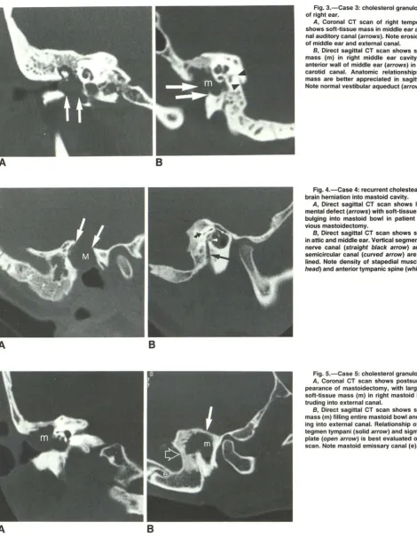
Fig. 3.-Case 3: cholesterol granuloma (cyst) of right ear.
A, Coronal CT scan of right temporal bone shows soft-tissue mass in middle ear and exter-nal auditory caexter-nal (arrows). Note erosion of floor of middle ear and external canal.
B, Direct sagittal CT scan shows soft-tissue mass (m) in right middle ear cavity, eroding anterior wall of middle ear (arrows) in region of carotid canal. Anatomic relationships of this mass are better appreciated in sagittal plane.
Note normal vestibular aqueduct (arrowheads).
Fig. 4.-Case 4: recurrent cholesteatoma and brain herniation into mastoid cavity.
A, Direct sagittal CT scan shows large teg-mental defect (arrows) with soft-tissue mass (M) bulging into mastoid bowl in patient with pre-vious mastoidectomy.
B, Direct sagittal CT scan shows soft tissue in attic and middle ear. Vertical segment of facial
nerve canal (straight black arrow) and lateral semicircular canal (curved arrow) are well out-lined. Note density of stapedial muscle (arrow-head) and anterior tympanic spine (white arrow).
Fig. 5.-Case 5: cholesterol granuloma cyst. A, Coronal CT scan shows postsurgical ap-pearance of mastoidectomy, with large bulging soft-tissue mass (m) in right mastoid bowl pro
-truding into external canal.
B, Direct sagittal CT scan shows soft-tissue mass (m) filling entire mastoid bowl and protrud
-ing into external canal. Relationship of mass to
tegmen tympani (solid arrow) and sigmoid sinus plate (open arrow) is best evaluated on sagittal
scan. Note mastoid emissary canal (e).
AJNR:9. March/April 1988
SAGITTAL CT OF TEMPORAL BONE DISEASE
375
[image:5.612.70.561.72.665.2]Fig. S.-Case 7: large vestibular aqueduct. Direct sagittal CT scan shows marked enlarge-ment of vestibular aqueduct (arrows). (Compare with normal vestibular aqueduct in Fig. 3B.)
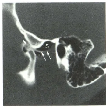
Fig. 7.-Case 11: comminuted fracture of tem-poral bone with involvement of facial nerve canal. Direct sagittal CT scan shows linear frac-ture of mastoid (solid black arrow), which ex-tends to and involves vertical segment of facial nerve canal (arrowhead). Bone fragment (open
arrow) compressing facial nerve was confirmed
at surgery. Note fracture of sigmoid plate (curved
arrow) and fractures of retrofacial air cells. In-creased soft tissue (S) in anterior tympanic cav-ity is from involvement of supratubal air cells and canal for tensor tympani. Clouding of supratubal and retrofacial air cells is from intraluminal and submucosal bleeding. Findings were confirmed at surgery.
Fig. B.-Case 12: fracture of temporal bone with ossicular dislocation.
A, Direct sagittal CT scan shows fracture of anterior wall of external canal (black arrow) and posterosuperior dislocation of incus (white ar-row).
B, Direct sagittal CT scan shows increased soft tissue in region of canal for tensor tympani with bone fragment (solid arrow). Note disconti-nuity of tegmen and fracture in region of genic-ulate ganglion (open arrow). Findings were con-firmed at surgery.
Fig. 9.-Case 13: incudomalleal dislocation. Direct sagittal CT scan shows separation of mal-leus (m) and incus (i). Note undisplaced fracture of anterior wall of middle ear cavity (straight
white arrow), pyramidal eminence (curved
ar-row), facial nerve canal (long black arrow), and lateral, superior, and posterior semicircular ca-nals (short black arrows).
Fig. 10.-Case 14: squamous cell carcinoma
of external auditory canal. Direct sagittal CT scan shows soft-tissue tumor (m) with erosion of in-ferior and anterior walls of external auditory canal. Isolated bone fragment (arrow).
6
A
9
informative in demonstrating
the tumor extent in a patient
with squamous
cell
carcinoma
of
the
external auditory canal
(Fig
.
10).
Dislocation of
the temporomandibular disk was
demonstrated best on
sagittal
CT
scans in cases 1 5 and 16
(Fig
.
11).
Protrusion of
the jugular veins into the middle ear
7
8
10
cavities from dehiscent jugular fossae and the relationship of
jugular veins to the round window membranes
were best
evaluated on sagittal CT scans in a patient
found to have
a
bluish mass behind both tympanic membranes (Fig
.
12)
.
376
MAFFE ET AL.
AJNR:9, March/April 1988A
B
A
B
attic cholesteatoma
after
CT showed
erosion of the
anterior
tympanic spine, which
was seen best on sagittal scans
.
Erosion of the facial
nerve canal
was clinically suspected
in a
patient
with a cholesteatoma
in
the
left ear
.
In
this patient
(case 19)
,
CT revealed a
large atticoantral cholesteatoma
eroding the
retrofacial
air cells and
thinning out
the
posterior
margin of
the
proximal
portion of the mastoid segment of
the
facial
nerve
canal.
The
anatomic
relationships of
the
choles-teatoma and facial nerve canal
were
delineated
best on
sag-ittal scans. At
surgery
,
the
CT
findings were
confirmed
,
with
the
c
holesteatoma
eroding
the
retrofacial
air
cells
but not
extending into
the
facial
nerve canal. One patient with a
cholesteatoma (case 20)
had erosion of the tegmen
tympani,
which was seen best on sagittal
CT
scans
.
Two patients were clinically diagnosed as having
tympa-nosclerosis (cases 21 and 22).
In
case 21, bilateral
cholestea-tomas were found with erosion of the
lateral
semicircular
cana
l
and
sigmoid sinus plate
.
The sigmoid sinus
plate defect
was depicted best on sagittal scans
.
In case 22 (the second
patient with the clinical
d
iagnosis
of tympanosclerosis),
CT
revealed
the
classic
find
ings
of an attic cholesteatoma
,
with
erosion
of the
lateral
attic wall, anterior tympanic spine,
and
Fig. 11.-Case 15: anterior dislocation of left temporomandibular joint meniscus without re-duction.
A, Direct sagittal CT air-positive contrast ar-throgram in closed-mouth position shows dislo-cated left meniscus (arrowhead). Air is in supe-rior joint space (long straight black arrow) and in inferior joint space (curved arrow), and air and contrast material are in anterior recess of supe-rior joint space (open arrow). Contrast material is also seen just superior and inferior to dis-placed meniscus (short straight black arrows).
B, Direct sagittal CT scan in open-mouth po-sition clearly shows unreduced meniscus
(ar-rowhead). In this position iodinated contrast ma-terial has replaced air in anterior recess of su-perior joint space (solid straight arrows). Note stretching of retrodiskal or bilaminar zone (open
arrow). Joint capsule is outlined by air and pos-itive contrast (curved arrow).
Fig. 12.-Case 17: high and dehiscent jugular fossa with protrusion of jugular bulb into tym-panic cavity, simulating vascular mass.
A, Direct sagittal CT scan shows jugular bulb (JB). Bone defect is along floor of hypotym-panum with jugular bulb bulging into middle ear up to level of round window membrane (arrow
-head). Note vestibule (V) and superior (black
arrow) and posterior (white arrow) semicircular
canals. _
B, Direct sagittal CT scan 4.5 mm medial to A
shows carotid canal (C), jugular fossa (J), basal turn of cochlea (solid arrow), and internal audi-tory canal (open arrow). Jugular fossa is quite high in position and reaches level of internal auditory canal.
ossicles. To our surprise, an intratympanic (ectopic)
meningi-oma was found at surgery
.
In
this
patient
,
postoperative
contrast-enhanced CT, including thin axial
,
coronal
,
and
sag-ittal sections
(superior
for jugular fossa and sublabyrinthine
air
cells)
of the posterior fossa and base of the skull and
temporal bone
,
did not show any mass in the posterior fossa
or base of the skull.
Discussion
The
radiographic anatomy
of the
complex
structures of the
human
temporal
bone has always been a challenge to
radi-ologists
.
The
introduction
of tomography
in the
late
1930s
as
a technique for
body-section imaging
was
an important step
in
the radiographic delineation
of
the
minute structures of the
ear
[18]
.
However
,
a practical application
for
studying the
temporal
bone
was
not available until 20 years later, when
multidirectional tomographic
units became commercially
avail-able [18, 19].
Before the
introduction of high-resolution
CT,
complex-motion
tomography
was the
diagnostic imaging
[image:6.615.63.490.81.467.2]AJNR:9, March/April 1988
SAGITTAL CT OF TEMPORAL BONE DISEASE
377
Fig. 13.-A, Normal facial nerve canal. Direct sagittal CT scan shows entire mastoid segment of facial nerve canal (open arrow). Immediately below lateral semicircular canal (L) is posterior portion of tympanic seg-ment of facial nerve canal. Note incus (i), malleus (m), anterior tympanic spine (white arrow), pyramidal eminence (white arrowhead), and low den-sity of stapedius canal (black arrowhead) in front of facial nerve canal. (Compare with S.)
S, Normal facial nerve. Sagittal partial-saturation T1-weighted MR scan (repetition time
=
800 msec, echo time=
25 msec) obtained with surface receiver coil shows superior semicircular canal (open arrow), posterioralteration of the bony structures of the temporal bone and
base of the skull [18
,
19]
.
In the past 6 years CT has rapidly been replacing
complex-motion tomography and has proved to be the diagnostic
imaging method of choice for assessing the temporal bone
[1-14]
.
Three prerequisites are necessary for the study of the
temporal bone: high definition
,
thin sections
,
and multiple
projections [13]
.
Direct axial and coronal CT sections are
quite satisfactory for demonstrating the anatomy of the
tem-poral bone; however, study of the axial or coronal sections
should be supplemented by direct sagittal sections
.
The
sag-ittal projection is of much interest to surgeons as it has the
advantage of following the plane of surgical approach [19].
Many relationships of the structure of the temporal bone are
better seen by studying direct sagittal CT sections of the
temporal bone
.
This projection is particularly satisfactory for
studying the normal and pathologic details of the mastoid
segment of the facial nerve canal (Figs. 7
,
13A
,
and 13B),
ossicles (Figs
.
8A, 9, and 13C), vestibular aqueduct (Figs
.
3
and 6)
,
tegmen tympani (Figs
.
4A and 8B), sigmoid plate
(Figs.
1 and 7)
,
external auditory canal (tympanic bone)
(Fig.
10),
mastoid (Fig
.
1), jugular fossa (Fig. 12A)
,
carotid canal
(Fig.
12B), supra-sublabyrinthine air cells (Figs. 8B and 9)
,
and
retrofacial air cells (Figs. 7 and 13A)
.
We recommend the
direct sagittal projection to evaluate the vestibular aqueduct
(Fig. 6); fractures of the temporal bone (Figs
.
7-9); patients
with infranuclear facial paralysis
;
and diseases involving the
round window niche (Fig
.
2)
,
tegmen tympani (Fig
.
4A)
,
sig-moid sinus plate (Fig
.
7)
,
carotid canal (Fig. 12B), jugular fossa
(Fig
.
12), external auditory canal (Fig
.
10)
,
and
temporoman-dibular joint (Fig
.
11)
.
In our institution the sagittal projection
has proved extremely valuable for assessing the extent of
portion of tympanic segment of facial nerve (curved arrow), mastoid (large arrowheads) and parotid (black arrows) segments of facial nerve. Note stapedius muscle (straight white arrows), which originates within pyramidal eminence in front of vertical (mastoid) portion of facial nerve. Small structure (small arrowhead) extending from facial nerve to stapedius mus-cle is believed to represent stapedius nerve.
C, Normal ossicles. Direct sagittal CT scan shows malleus (long arrow), incus (shorf arrow), inferior aspect of lateral wall of attic (arrowhead), and
temporomandibular joint.
Fig. 14.-Case 15: dislocated right temporoman-dibular joint meniscus. Direct sagittal CT air-positive contrast arthrogram in open-mouth position shows
dislocated disk (short arrow). Air is in superior (S) and inferior (i) joint spaces. Note stretching of
re-trodiskal zone (long arrows).
atticoantral cholesteatoma
and
its
relationship to the
s
igmoid
plate and
tegmen tympani
.
This projection is
es
s
ential for
evaluation of the mastoid
segment of the facial nerve canal
(Figs. 1
Band 7)
,
ves
t
ibular aqueduct
(Fig.
6)
,
and temporo
[image:7.612.58.560.85.266.2] [image:7.612.385.559.370.550.2]378
MAFFE ET AL.
AJNR:9, March/April 1988of
the
jugular fossa
to
the internal
auditory
canal (Fig
.
12B)
and
assess the relationship of
the
vertical segment of the
facial
nerve canal to the posterior wall of the external auditory
(tympanic
sulcus) canal
and sigmoid sinus plate (Fig
. 13A).
The
sagittal
plane in general
is complementary
to the
cor-onal
and axial
planes
, since
it
shows
the anterior to posterior
aspects
of
structures
seen laterally to medially on coronal
sections
(Fig. 3) and the inferior to superior aspects of
struc-tures seen anteriorly
to posteriorly on axial sections (Fig
.
2)
[15
, 16
,
18]. In our
series
,
the direct sagittal projection proved
to
be
extremely
valuable in several cases (Table 1)
.
Therefore
,
we believe this projection
should
be used to
complement the
ax
ial and coronal planes
,
depending on the disease and the
anatomic
details
to
be imaged.
Disadvantages of the direct sagittal technique are the
ad-ditional
radiation,
time
,
and effort
.
In many cases, the
infor-mation obtained with a
x
ial and
sagittal
planes may be
suffi-cient,
obviating coronal sections. In temporal bone fractures
,
lesions of
the
external auditory canal, and diseases involving
the
vestibular
aqueduct
,
round window niche
,
round window
membrane
,
tegmen tympani
,
and sigmoid sinus plate
,
axial
and sagittal
CT scans
are
recommended
.
Sagittal CT is essential for evaluating lesions of the
tem-poromandibular joint (Fig. 10)
.
The demarcation between disk
and retrodiskal
tissue (bilaminar zone)
can
be established
easily
on
sagittal
CT scans (Fig
.
14). CT arthrography is an
invasive method requiring technical skill and radiation
expo-sure
.
In our institution
,
CT arthrography has proved to be a
valuable
and complementary
diagnostic tool in the
examina-tion
and
treatment of patients with jaw dysfunction
.
The
application
of our
sagittal
head-holder is not limited to the
temporal
bone and
temporomandibular
joint. We have
found
it very useful for
the
evaluation of
the
other craniofacial
structures,
including sphenoethmoid complex
,
sella turcica
,
and the orbits (Fig. 1 A)
.
ACKNOWLEDGMENTS
We thank Ruben Stortzum for technical expertise and assistance
in manufacturing the sagittal head-holder and Mari Salazar for
sec-retarial assistance.
REFERENCES
1. Shaffer KA, Haughton
vw
, Wilson CR
. High resolution computed tomog-raphy of the temporal bone. Radiology 1980;134:409-4142. Valvassori GE, Mafee MF, Dobben GO. Computerized tomography of the temporal bone. In: Proceedings of the Sixth Shambaugh International
Workshop of Otomicrosurgery. Huntsville, AL: Strode, 1980:000-000
3. Valvassori GE, Mafee MF, Dobben GO. Computerized tomography of the temporal bone. Laryngoscope 1982;92:562-565
4. Lufkin R, Barni JJ, Glen W, Mancuso A, Canalis R, Hanafee W. Comparison of computed tomography and pleuridirectional tomography of the temporal bone. Radiology 1982;143:715-718
5. Mafee MF, Kumar A, Yannias D., Valvassori GE, Applebaum EL. Computed tomography of the middle ear in the evaluation of cholesteatomas and other soft tissue masses: comparison with pleuridirectional tomography. Radiology 1983;148:465-472
6. Mafee MF, Valvassori GE, Dobben GO. The role of radiology in surgery of the ear and skull base. Otolaryngol Clin North Am 1982;15:723-753 7. Mafee MF, Valvassori GE, Shu gar MA, Yannias DA. High resolution and
dynamic sequential computerized tomography in the evaluation of glomus complex tumors. Arch Otolaryngo/1983;109:691-696
8. Swartz JD, Goodman RS, Russell KB, Marlowe FI, Wolfson RJ. High-resolution computed tomography of the middle ear and mastoid. Part II:
Tubotympanic disease. Radiology 1983;148:455-459
9. Mafee MK, Aimi K, Valvassori GE. Computed tomography in the diagnosis
of primary tumors of the petrous bone. Laryngoscope 1984;94: 1423-1430 10. Mafee MF, Singleton EL, Valvassori GE, Espinosa GA, Kumar A, Aimi K.
Acute otomastoiditis and its complications: role of CT. Radiology
1985;155:391-397
11. Mafee MF, Henrickson GC, Deitch RL, et al. Use of CT in the stapedial otosclerosis. Radiology 1985;156:709-714
12. Mafee MF, Valvassori GE, Deitch RL, et al. Use of CT in the evaluation of cochlear otosclerosis. Radiology 1985;156: 703-708
13. Valvassori GE, Mafee MF. The temporal bone. In: Carter BL, ed. Computed
tomography of the head and neck. New York: Churchill Livingstone 1985: 171-205
14. Mafee MF, Aimi K, Kahen H, Capek V. Chronic otomastoiditis. A conceptual understanding of CT findings. Radiology 1986;160: 193-200
15. Zonneveld FW. The value of non-reconstructive multiplanar CT for the evaluation of the petrous bone. Neuroradiology 1983;25:1-10
16. Zonneveld FW, Van Waes PFG, Damsma P, Rabischong P, Vignaud J.
Direct multiplanar computed tomography of the petrous bone. Radiograph
-ies 1983;3: 41 0-449
17. Heffez L, Mafee MF, Langer B. Use of a new head holder for obtaining direct sagittal CT images of the TMJ. J Oral Maxillofac Surg 1987;45:822
-824
18. Valvassori GE, Buckingham RA. Tomography and cross-sections of the
ear. Stuttgart: George Thieme, 1975
19. Buckingham RA, Valvassori GE. Tomographic anatomy of the temporal