University of Warwick institutional repository: http://go.warwick.ac.uk/wrap This paper is made available online in accordance with
publisher policies. Please scroll down to view the document itself. Please refer to the repository record for this item and our policy information available from the repository home page for further information.
To see the final version of this paper please visit the publisher’s website. Access to the published version may require a subscription.
Author(s): Alison Atterbury and Mark J. Wall
Article Title: A population of immature cerebellar parallel fibre synapses are insensitive to adenosine but are inhibited by hypoxia Year of publication: 2011
Link to published article:
http://dx.doi.org/10.1016/j.neuropharm.2011.06.006
A population of immature cerebellar parallel fibre synapses are
insensitive to adenosine but are inhibited by hypoxia.
Alison Atterbury and Mark J. Wall* Neuroscience Group,
School of Life Sciences, University of Warwick, Coventry
CV4 7AL
*To whom correspondence should be addressed. Mark J. Wall
Neuroscience Group, School of Life Sciences, University of Warwick, Coventry
CV4 7AL
E-mail:[email protected]
Running title: heterogeneous adenosine signalling
Abstract
The purine adenosine plays an important role in a number of physiological and pathological processes and is neuroprotective during hypoxia and ischemia. The major effect of adenosine is to suppress network activity via the activation of A1 receptors. Here we report that in immature cerebellar slices, the activation of A1 receptors has variable effects on parallel fibre synaptic transmission, ranging from zero depression to an almost complete abolition of transmission. Concentration-response curves suggest that the heterogeneity of inhibition stems from differences in A1receptor properties which could include coupling to downstream effectors. There is less variation in the effects of adenosine at parallel fibre synapses in slices from older rats and thus adenosine signalling appears developmentally regulated.
1. Introduction
Adenosine plays important roles in a number of physiological and pathological processes in the brain (for review see Dunwiddie and Masino, 2001; Dale and Frenguelli, 2009). Adenosine accumulates during hypoxia and ischemia and has a neuroprotective role (Goldberg et al. 1988; Von Lubitz et al 1988; Cunha 2005) reducing the brains energy requirements during periods of metabolic stress. The actions of adenosine are well characterised, it acts via multiple cell surface G-protein coupled receptors: A1, A2A, A2Band A3(Fredholm et al. 2000). The major effect of adenosine is to dampen down network excitation via A1receptor activation.
Adenosine A1receptors inhibit Ca2+influx via voltage gated Ca2+channels leading to reduced transmitter release (Gundlfinger et al. 2007) and A1receptors activate
inwardly rectifying K+channels (GIRKs) leading to the hyperpolarisation of the membrane potential (Lüscher et al. 1997).
In many brain regions A1receptors are present on the terminals of
coupling to effectors rather than differences in adenosine metabolism or uptake.
These synapses are still depressed during hypoxia although the mechanism does
not appear to involve the activation of A1receptors by the increased extracellular
adenosine concentration.
2. Methods
2.1 Preparation of cerebellar slices
Transverse slices of cerebellar vermis (400 µm) were prepared from male Wistar rats, at postnatal days 9-28 (P9-28). As described previously (Wall et al. 2007) and in accordance with the U.K. Animals (Scientific Procedures) Act (1986), male rats were killed by cervical dislocation and then decapitated. The cerebellum was rapidly removed and slices were cut on a Microm HM 650V microslicer in cold (2-4ºC) high Mg2+, low Ca2+aCSF, composed of (mM): 127 NaCl, 1.9 KCl, 7-8 MgCl2, 0.5 CaCl2,
1.2 KH
2PO4, 26 NaHCO3, 10 D-glucose (pH 7.4 when bubbled with 95% O2 and 5% CO2, 300 mOSM). Slices were stored in normal aCSF (1.3 mM MgCl2, 2.4 mM
CaCl2) at room temperature for 1-6 hours before recording.
2.2 Extracellular recording
An individual slice was transferred to a recording chamber, submerged in normal aCSF and perfused at 6 ml/min (30-32C). The slice was placed upon a suspended
of parallel fibres, square voltage pulses (2- 5V, 200s duration) were delivered by an
isolated pulse stimulator (model 2100 AM systems Everett WA USA) via a concentric bipolar metal stimulating electrode (FHC, Bowdoin, ME, USA) placed on the surface of the molecular layer. The recording electrode (an aCSF filled microelectrode) was placed on the same track along which the parallel fibres travel (“on-beam”, Yuan and Atchison, 1999). A typical extracellular field potential consisted of an initial component which persisted in 5 mM kynurenate but was blocked by 1 M TTX
2.3 Drugs
All drugs were made up as 10-100 mM stock solutions, stored frozen and then thawed and diluted with aCSF on the day of use. Adenosine, baclofen, N-cyclopentyladenosine (CPA), tetrodotoxin (TTX) and 8-cyclopentyltheophylline (8CPT) were purchased from Sigma (Poole, UK). Kynurenic acid, CNQX (6-cyano-7-nitroquinoxaline-2,3-dione) and L-AP4 (L-(+)-2-Amino-4-phosphonobutyric acid) were purchased from Ascent Scientific (Bristol, UK).
2.4Statistics
All quoted data is mean ± SEM. For statistical analysis, paired and unpaired student’s
t tests were performed. Data was described with either sigmoid curves or linear fits using Origin 8.5 (Silverdale Scientific Ltd, Stoke Mandeville UK)
3. Results
3.1 Adenosine has heterogeneous effects on synaptic transmission at immature
parallel fibre synapses.
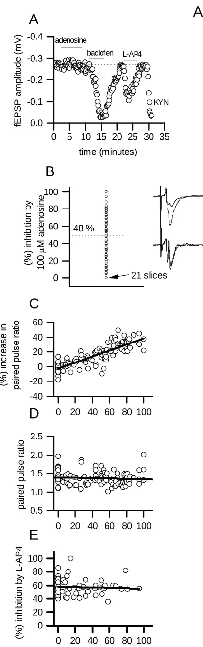
100M adenosine was 48 ± 2 %, which was accompanied by a significant (P<0.01)
increase in paired pulse facilitation (interval 50 ms) from 1.38 ± 0.02 to 1.67 ± 0.07. There was considerable variation (Fig. 1B) in the reduction of fEPSP amplitude produced by adenosine (co-efficient of variance 0.68) ranging from no effect in 21 slices (Fig. 1A, B), to an almost complete abolition of transmission in 3 slices (> 90 % reduction in fEPSP amplitude). The reduction in fEPSP amplitude strongly correlated with the increase in paired pulse ratio (r = 0.7, Fig. 1C) suggesting that the inhibition was presynaptic (Dunwiddie and Haas, 1985).
We investigated whether the degree of adenosine-mediated inhibition correlated with any synaptic properties. In all slices, synaptic transmission showed paired pulse facilitation before adenosine application (50 ms interval, paired pulse ratio 1.38 ± 0.02) and fEPSP amplitude could be greatly reduced (> 80 % reduction)
following block of glutamate receptors with either 5 mM kynurenic acid or 10M
CNQX. There was no correlation: between the reduction in fEPSP amplitude (100M
adenosine) and the initial (before adenosine application) fEPSP amplitude (r = 0.03, not illustrated), between the initial amount of paired pulse facilitation (before adenosine application) and reduction in fEPSP amplitude (r = 0.04, Fig. 1D) and between the magnitude of adenosine inhibition and the age of rat from which the cerebellar slices were made (P8-14, r = 0.04, not illustrated). Dunwiddie (1985)
the effects on the paired pulse ratio (Fig. 1C). Metabotropic glutamate receptors of the mGlu4 subtype are only expressed by parallel fibres in the cerebellum (Mateos et al. 1998) and thus inhibition of fEPSPs by their activation with the agonist L-AP4
confirms that parallel fibre synapses are activated. In all slices tested, L-AP4 (50M) significantly reduced fEPSP amplitude (mean inhibition 58 ± 2,n= 40 slices),
whether or not adenosine had any effect on fEPSP amplitude. There was no correlation between the reduction in fEPSP amplitude produced by L-AP4 and the
inhibition produced by 100M adenosine (Fig. 1A, E). Parallel fibres also express
GABABreceptors, which inhibit transmitter release (Batchelor and Garthwaite, 1992; Wall and Dale, 2007). In 9 slices, where adenosine (100M) produced little or no
inhibition (< 20 % reduction in fEPSP amplitude), activation of GABABreceptors
with 10-25M baclofen produced an almost complete abolition of synaptic
transmission (mean reduction in amplitude 97 ± 3 %, Fig. 1A). Thus synapses which were either weakly modulated or unaffected by adenosine were still strongly inhibited by either GABABreceptor or mGlu4 receptor activation.
3.2 There is less variation in adenosine inhibition in slices from older rats
To investigate if the adenosine-insensitivity of parallel fibre synapses persists
during cerebellar development, recordings were made from parallel fibre
synapses in slices from older rats (average age 23 ± 0.2 postnatal days). The
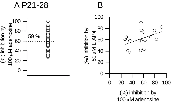
average inhibition produced by 100M adenosine was 59 ± 2 %, with the paired
pulse ratio increased from 1.5 ± 0.02 to 2 ± 0.08 (n=59 slices and see Wall et al.
2007). Although the degree of inhibition was variable, ranging from 27 to 100 %
adenosine (Fig. 2A). As with immature synapses there was no relationship
between the degree of inhibition with fEPSP amplitude (r = 0.002) or initial
paired pulse ratio (r = 0.04). In 14 slices, the inhibition produced by 100M
adenosine was compared to that produced by 50M L-AP4 (mean inhibition 62
± 4 %) and there was only a weak relationship (r = 0.14, Fig. 2B). Thus although
there is still variation in the effects of adenosine across synapses in older animals,
it appears less than at immature synapses.
3.3 Neither adenosine nor L-AP4 modulate Purkinje cell firing rate
It has been suggested that the field response evoked by parallel fibre stimulation consists of a combination of EPSPs and action potentials in Purkinje cells,
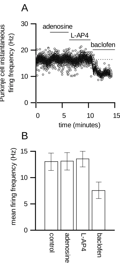
interneurones and Bergmann glia (Clark and Barbour, 1997). If the field response is contaminated with population spikes this could explain the variation in effects between baclofen, L-AP4 and adenosine. If baclofen and L-AP4 have some postsynaptic effect (moving the cell away from threshold) resulting in a loss of population spikes this could explain their greater efficacy and consistent depressant action on synaptic transmission. This seems unlikely as mGlu4 receptors only appear to be expressed on parallel fibres (and not on postsynaptic cells, Mateos et al. 1998) and the stimulation strengths are low and are unlikely to activate enough parallel fibres to fire a Purkinje cell. However to exclude this possibility, the firing rate of spontaneously active Purkinje cells was measured in control and following the
application of adenosine (100), L-AP4 (50M) and baclofen (25M). Neither
instantaneous firing rate, 13.0 ± 1.6 Hz, adenosine 13.1 ± 1.6, L-AP4 13.5 ± 1.6) although baclofen (7.6 ± 1.6 Hz) significantly (P<0.01) reduced the firing rate (Fig. 3). Thus the more consistent effect of L-AP4 on synaptic transmission compared to
adenosine does not appear to stem from a postsynaptic action.
3.4 The effects of adenosine are blocked by the A1receptor antagonist 8CPT
We have investigated whether the effects of adenosine are only through the activation of A1receptors. Recent evidence suggests that A2Areceptors (Brooks et al. 2008) may be expressed in the cerebellum and as they can facilitate synaptic transmission (Quarta et al. 2004) the variation in synaptic inhibition block could stem from the combined activation of inhibitory A1receptors and facilitatory A2Areceptors. However in 20 slices, we found that applying adenosine in the presence of the
selective A1receptor antagonist 8CPT (1-2) had no effect (data not shown), even
if adenosine had little effect on fEPSP amplitude in control. Thus the lack of effect on fEPSP amplitude cannot be explained by activation of multiple receptor subtypes with opposing effects.
3.5 Concentration responses at synapses sensitive and insensitive to adenosine
inhibition
Usually 100M adenosine would activate the majority of synaptic A1receptors and
Kimura et al. 2003). However, at synapses were 100M adenosine had little effect on
fEPSP amplitude, the concentration-response curve may be shifted to the right. Thus
the effects of applying higher concentrations (300M) of adenosine were investigated.
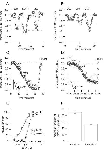
Slices could be divided into 2 groups: in the first group 300adenosine produced
little or no extra inhibition compared to 100M adenosine (n= 8, 54 ± 8 % Vs 50 ±
11 % inhibition, Fig. 4A) with effects comparable to L-AP4 (50M). In the second
group, 300M adenosine had a much larger effect on fEPSP amplitude than 100M
adenosine (12 ± 3.1 Vs 36 ± 9 %,n= 8, Fig. 4B). This large increase in inhibition
produced by 300M adenosine (~ 3 times that produced by 100M) suggests that at
these synapses 100M adenosine is not at the top of the concentration-response curve.
This could be the result of rapid extracellular adenosine breakdown so that the concentration of adenosine reaching the receptors is low or perhaps at some synapses there is expression of low potency receptors. To test for changes in receptor properties, concentration-response curves were constructed for the non-hydrolysable A1receptor
agonist CPA. Adenosine (100M) was first applied to identify synapses where there was either little or no inhibition (n= 6, mean reduction in fEPSP amplitude 15.8 ± 5 %) or synapses where adenosine produced at least a 50 % reduction in fEPSP amplitude (mean inhibition 68 ± 5 %,n= 7). The identity of all synapses was
confirmed by applying L-AP4 (50M) which markedly reduced fEPSP amplitude (<
50 % inhibition). Concentration response curves were then constructed by
sequentially adding increasing concentrations of CPA (0.001 µM to 100 µM). At synapses were adenosine produced a marked reduction in fEPSP amplitude, CPA almost completely inhibited synaptic transmission (90 ± 4.4 % block, Fig. 4C, E, F)
M). At synapses where adenosine had little or no effect on fEPSP amplitude, the
concentration response curve was shifted to the right (IC50of 250 nM, Fig 3D, E) with
a maximal inhibition (% inhibition of fEPSP amplitude with 100M CPA, 53 ±
0.6 %, Fig. 4F). The effects of CPA were readily reversed by 8CPT (2M)
suggesting that the affinity of the receptor for CPA was reduced. Thus synapses show variation in the potency of CPA suggesting that the variation stems from receptor properties (and possibly downstream signalling) rather than differences in adenosine uptake and metabolism.
3.6 Changes in the presynaptic volley can account for adenosine inhibition at
insensitive synapses
Adenosine has a small effect on the presynaptic volley, reducing it by ~ 10 %
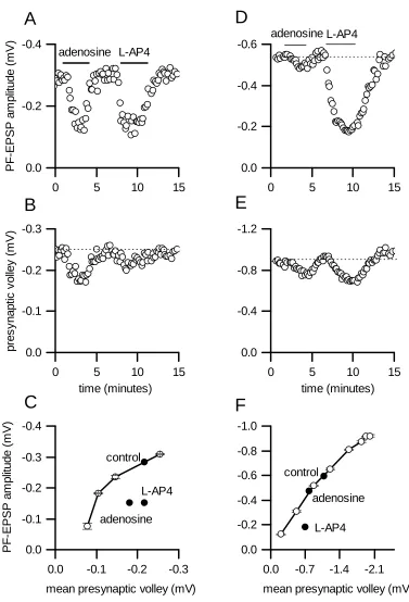
(Dunwiddie and Miller, 1993; Dittman and Regher, 1996; Wall et al. 2008). We have investigated whether this reduction in volley amplitude is sufficient to account for the reduction in fEPSP amplitude at synapses sensitive and relatively insensitive to adenosine inhibition. For each slice we varied the stimulus strength and then constructed a plot of volley amplitude against fEPSP amplitude. We then applied
adenosine (100M) and L-AP4 (50M) to inhibit transmission. If the reduction in volley amplitude (produced by adenosine and L-AP4) can account for the reduction in fEPSP amplitude then, when plotted, the point will fall upon the volley Vs fEPSP amplitude curve. If the reduction in volley amplitude is too small to account for the reduction in fEPSP amplitude, then the point will fall below the line. We confirmed the identity of the volley: it persists following application of the glutamate receptor
majority of slices (9 out of 12), the reduction in volley amplitude (produced by either adenosine or L-AP4) was much too small to account for the reduction in fEPSP amplitude and the points were well below the line (Fig. 5A, B, C also reported in hippocampus Dunwiddie, 1984). However, in 3 out of 12 slices where the effect of adenosine on fEPSP amplitude was small (~10-15 %) the reduction in fEPSP amplitude could be accounted for by the change in volley amplitude (with the point falling on the curve, Fig 5C, D, E). This suggests that the mechanisms of synaptic inhibition may differ between sensitive and insensitive synapses.
3.7 Effects of hypoxia on synapses sensitive and insensitive to adenosine
Starvation of O2(hypoxia) produces a marked increase in extracellular adenosine concentration in brain tissue, activating presynaptic A1receptors leading to the inhibition of transmitter release (Fowler, 1990; Winn et al. 1981; Katchman and Hershkowitz, 1993; Frenguelli et al. 2003). It has been reported that hypoxia-mediated adenosine release in the cerebellum depresses parallel fibre synaptic
transmission, an effect reversed by blocking A1receptors (Atterbury and Wall, 2009).
However these experiments were performed in slices where adenosine (100M) produced a large inhibition of transmission (< 50 % reduction in PF fEPSP amplitude). We have investigated if parallel fibre synapses, where adenosine has little effect on synaptic transmission, are either resistant or show a smaller response to hypoxia.
In confirmation of previous data (Atterbury and Wall, 2009), at synapses
where adenosine (100M) had a large effect on fEPSP amplitude (mean inhibition 68
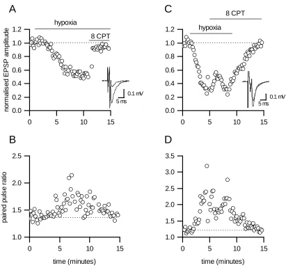
of transmission (steady state reduction in fEPSP amplitude 43 ± 3 %, delay to onset of inhibition 161 ± 37 s, time from start of depression to maximum inhibition 327 ± 34 s, Fig. 6A). This inhibition was accompanied by a significant (P<0.01) increase in paired pulse ratio (from 1.29 ± 0.05 to 1.7 ± 0.07, Fig. 6B) but there was no
significant change in the amplitude of the presynaptic volley (0.47 ± 0.07 Vs 0.47 ± 0.06 mV). Thus there is a reduction in transmitter release with no change in the number of active parallel fibres. The effects of hypoxia were almost completely
reversed (85 ± 3 %) by blocking A1receptors with 8-CPT (2M, Fig. 6A). Thus in
these slices, the major short-term effect of hypoxia is to activate presynaptic A1 receptors leading to reduced parallel fibre glutamate release.
In slices where 100M adenosine had a significantly (P<0.001) smaller effect
on fEPSP amplitude (mean inhibition 19 ± 3 % Vs ~68 %,n= 5), hypoxia still induced depression (mean inhibition of fEPSP amplitude 62 ± 5 %, Fig. 6C). This inhibition was significantly (P<0.01) larger than observed with synapses that were sensitive to adenosine and it was quicker to occur (delay 70.6 ± 6.2 vs 161 ± 37 s and time from start of depression to maximum inhibition 75 ± 44 Vs 327 ± 34 s, Fig. 6C). In 3 out of 5 slices the inhibition was accompanied by a large increase in the paired pulse ratio (Fig. 5D), in the other slices there was little change in the ratio (overall change in paired pulse ratio from 1.34 ± 0.06 to 1.78 ± 0.17). There was no significant (P = 0.4) change in the amplitude of the presynaptic volley during hypoxia (0.37 ± 0.1 Vs 0.44 ± 0.13 mV) and hence no change in the number of active parallel fibres. The inhibition produced by hypoxia was not reversed by 8 CPT (transient increase in fEPSP amplitude 21 ± 6 %, Fig. 6C) and thus most of the inhibition was not produced by extracellular adenosine acting via A1receptors.As activation of either mGlu4 or
GABABreceptor antagonists were applied (in the presence of 8CPT) to
investigate whether they could reverse the effects of hypoxia. Neither MSOP (100
M, mGlu4R antagonist,n= 3) or CGP5248 (20M, GABABreceptor
antagonist,n= 4) had any effect on fEPSP amplitude.The reduction in fEPSP
amplitude was almost completely removed by washing back in oxygenated aCSF.
4. Discussion
We have found considerable variation in the reduction of fEPSP amplitude produced by the application of adenosine to parallel fibre synapses in slices from immature rats (confirming previous results Atterbury and Wall, 2009). Differences in adenosine-sensitivity were observed in slices from the same rat and in recordings from different molecular layers in the same slice.Future experiments will investigate whether
these insensitive and sensitive synapses occur in specific cerebellar lobes.The
observation that activation of other presynaptic receptors present on parallel fibre terminals (mGluR4 and GABAB) produces consistent amounts of inhibition shows that the variation is specific to adenosine signalling. Furthermore, the consistent inhibitory effects of L-AP4 confirm that all the fEPSPs recorded result from parallel fibre stimulation (Mateos et al. 1998).
interneurones (Liu and Cull-Candy, 2000) and from Bergmann glia (Clark and Barbour, 1997; Bellamy and Ogden, 2005; Bellamy, 2007). One hypothesis for the variation in adenosine inhibition is that parallel fibres synapses onto different
postsynaptic cell types have different sensitivities to adenosine and that the variation in fEPSP inhibition stems from differences in the proportion of sensitive and
insensitive synapses recorded from. The evidence for such a hypothesis is relatively weak. Parallel fibre synapses onto Purkinje cells are modulated by adenosine (IC501.1 M, maximum block ~80 %, 100M adenosine close to the top of the concentration
4.1 What determines the degree of inhibition produced by adenosine?
Application of 100M adenosine produced different amounts of inhibition at
different parallel fibre synapses. This variation could be the result of different amounts of adenosine reaching the synaptic receptors due to differences in adenosine metabolism and uptake. The reported Kdvalue for adenosine at A1receptors is ~600-700 nM (hippocampus, Dunwiddie and Diao, 1994) and thus 100M adenosine
should saturate the receptors and produce a maximal effect. However adenosine is rapidly removed from the extracellular space (by uptake and by metabolism) and thus only a small fraction of exogenous adenosine actually reaches the receptors.
systems, enabling a number of transduction mechanisms to inhibit synaptic
transmission.Differences in the complement of presynaptic voltage-gated Ca2+ channels involved in transmitter release between individual parallel fibres could
underlie the variation in inhibition, if A1receptors only inhibit certain Ca2+
channel subtypes. It has been reported that there are multiple subtypes of Ca2+
channel involved in parallel fibre transmitter release (N, P and Q Doroshenko et
al 1997). At most synapses the actions of adenosine cannot be accounted for by an
action on the presynaptic volley (also reported by Dundwiddie 1984). However in synapses where the effects of adenosine on fEPSP amplitude were small, the inhibition could be explained by changes in volley amplitude. The volley is a compound action potential which scales with stimulus strength. Thus at these
synapses the reduction in transmitter release may simply stem from a reduction in the number of active fibres. Adenosine can activate GIRK K+channels which could cause action potential failures (by hyperpolarisation) or could reduce Ca2+influx via voltage gated Ca2+channels which may otherwise contribute to depolarisation.
4.2 Hypoxia
Hypoxia produces a rapid accumulation of extracellular adenosine which
activates presynaptic A1receptors leading to membrane potential
hyperpolarisation and the inhibition of excitatory synapses (reviewed in Krnjević
2008; Frenguelli and Dale, 2009). This helps to reduce the brains energy
requirements enabling it to maintain the minimal metabolism required for
survival and is thus neuroprotective in adult brain (Krnjević2008; Cunha 2005).
markedly depressed by hypoxia, as a result of the increased concentration of
extracellular adenosine activating A1receptors and reducing glutamate release
(also see Atterbury and Wall, 2009). It might be expected that synapses that
were not sensitive to inhibition by adenosine would be much more resistant to
hypoxia, equivalent to hypoxia induced in the presence of an A1receptor
antagonist where there is only slow and partial depression (for example see
Gribkoff et al 1990). Surprisingly, these synapses were not resistant to hypoxia
but instead appeared more sensitive (a quicker onset of depression and a larger
steady state reduction in fEPSP amplitude). This inhibition was not produced by
the activation of A1receptors, as it was not reversed by A1receptor antagonists
(which only had a small transient action). However the reduction in EPSP
amplitude could be reversed following reoxygenation.
What is the mechanism of hypoxia-mediated depression at these synapses?
The mean increase in paired-pulse ratio suggests that some of the effects are
presynaptic (reduction in glutamate release), although the effects of hypoxia on
paired-pulse ratio were not consistent, with no effect at some synapses. There
was no change in the presynaptic volley and thus the number of active parallel
fibres was not reduced. Receptors such as mGluR4 and GABABare present on
parallel fibre terminals and could reduce the release of glutamate if activated
(Mateos et al. 1998; Batchelor and Garthwaite, 1992). In the hippocampus,
following block of A1receptors, part of the remaining hypoxia-induced
depression can be relieved with a group III mGluR antagonist (MPPG, de
Mendonça and Ribeiro 1997b). There is also evidence that activation of mGluR
(Groups I and III) can attenuate the action of adenosine via A1 receptors (de
reduced by either mGlu4 or GABABreceptor antagonists and thus they do not
appear to be involved. There are a number of ion channels that can be acutely
modulated by hypoxia including Ca2+channels and K+channels which could
underlie the inhibition (see Krnjević 2008 for review).
Why are there multiple mechanisms of hypoxia-induced depression at
immature parallel fibre synapses? It has been suggested that, unlike adult brain,
the activation of A1receptors maybe ineffective or even detrimental to neuronal
survival following hypoxia in the immature brain (reviewed in Cunha 2005).
Thus the neurones less-sensitive to adenosine may be preferentially protected
following a hypoxic insult and thus may play an important role in maintaining
cerebellar function. Future work is required to determine the precise anatomical
location of these neurones in the cerebellar vermis providing a clue to their
function.
4.3 Conclusions
Acknowledgements
AA holds a BBSRC and Warwick Medical Research Fund studentship. We thank Prof Bruno Frenguelli for comments on earlier drafts.
References
Atterbury, A., Wall, M.J., 2009. Adenosine signalling at immature parallel fibre-Purkinje cell synapses in rat cerebellum. J. Physiol. 587, 4497-508. Batchelor, A.M., Garthwaite, J., 1992. GABAB Receptors in the Parallel Fibre
Pathway of Rat Cerebellum. Eur. J. Neurosci. 4, 1059-1064.
Bellamy, T.C., 2007. Presynaptic modulation of parallel fibre signalling to Bergmann glia. Neuropharm. 52, 368-75.
Bellamy, T.C., Ogden, D., 2005. Short-term plasticity of Bergmann glial cell
extrasynaptic currents during parallel fiber stimulation in rat cerebellum. Glia. 52, 325-35.
Brooks, D.J., Doder, M., Osman, S., Luthra, S.K
.
, Hirani, E., Hume, S., Kase, H., Kilborn, J., Martindill, S., Mori, A., 2008. Positron emission tomography analysis of [11C]KW-6002 binding to human and rat adenosine A2Areceptors in the brain. Synapse 62, 671-81.Clark, B.A., Barbour, B., 1997. Currents evoked in Bergmann glial cells by parallel fibre stimulation in rat cerebellar slices. J. Physiol. 502, 335-50.
Cunha, R.A., 2005. Neuroprotection by adenosine in the brain: From A(1) receptor activation to A (2A) receptor blockade. Purinergic Signal. 2005 1:111-34. Dale, N., Frenguelli, B.G., 2009. Release of Adenosine and ATP during ischemia and
epilepsy. Curr. Neuropharmacol. 7:160-179.
de Mendonça, A., Ribeiro, J.A., 1997a. Influence of metabotropic glutamate receptor agonists on the inhibitory effects of adenosine A1 receptor activation in the rat hippocampus. Br. J. Pharmacol. 121, 1541-8.
Dittman, J.S., Regehr, W.G., 1996. Contributions of dependent and calcium-independent mechanisms to presynaptic inhibition at a cerebellar synapse. J. Neurosci. 16, 1623-1633.
Doroshenko, P.A., Woppmann, A., Miljanich, G., Augustine, G.J. 1997 Pharmacologically distinct presynaptic calcium channels in cerebellar excitatory and inhibitory synapses. Neuropharm 36, 865-872.
Dunwiddie, T.V., 1984. Interactions between the effects of adenosine and calcium on synaptic responses in rat hippocampus in vitro. J. Physiol. 350:545-59.
Dunwiddie
,
T.
V.
, Haas,
H.
L.,
1985.
Adenosine increases synaptic facilitation in the in vitro rat hippocampus: evidence for a presynaptic site of action. J.
Physiol. 369:365-77.Dunwiddie, T.V., Diao, L., 1994. Extracellular adenosine concentrations in hippocampal brain slices and the tonic inhibitory modulation of evoked excitatory responses. J. Pharmacol. Exp. Ther. 268, 537-45.
Dunwiddie, T.V., Masino, S,A., 2001. The role and regulation of adenosine in the central nervous system. Annu Rev Neurosci. 24:31-55.
Dunwiddie, T.V., Miller, K.K., 1993. Effects of adenosine and cadmium on presynaptic fiber spikes in the CA1 region of rat hippocampus in vitro. Neuropharm 32:1061-1068.
Fowler, J.C., 1990. Adenosine antagonists alter the synaptic response to in vitro ischemia in the rat hippocampus. Brain. Res. 509, 331 334.
Fredholm, B.B., Arslan, G., Halldner, L., Kull, B., Schulte, G., Wasserman, W., 2000. Structure and function of adenosine receptors and their genes. Naunyn
Schmiedebergs Arch. Pharmacol. 362, 364-74.
Frenguelli, B.G., Llaudet, E., Dale, N., 2003. High-resolution real-time recording with microelectrode biosensors reveals novel aspects of adenosine release during hypoxia in rat hippocampal slices, J. Neurochem. 86, 1506–1515.
Goldberg, M., Monyer, H., Weiss J., Choi, D., 1988. Adenosine reduces cortical neuronal injury induced by oxygen or glucose deprivation in vitro. Neurosci. Lett. 89, 323–327.
Gribkoff, V.K., Bauman, L.A., VanderMaelen, C.P., 1990. The adenosine antagonist 8-cyclopentyltheophylline reduces the depression of hippocampal neuronal responses during hypoxia. Brain Research 512, 353-357.
Gundlfinger, A., Bischofberger, J., Johenning, F.W., Torvinen, M., Schmitz, D., Breustedt, J., 2007. Adenosine modulates transmission at the hippocampal mossy fibre synapse via direct inhibition of presynaptic calcium channels. J. Physiol. 582, 263-77.
Katchman, A.N., Hershkowitz, N., 1993. Adenosine antagonists prevent hypoxia-induced depression of excitatory but not inhibitory synaptic currents. Neurosci. Lett. 159, 123-126.
Kimura, M., Saitoh, N., Takahashi, T., 2003. Adenosine A(1) receptor-mediated presynaptic inhibition at the calyx of Held of immature rats. J Physiol. 553, 415-426.
Kocsis, J.D., Eng, D.L., Bhisitkul, R.B., 1984. Adenosine selectively blocks parallel-fiber-mediated synaptic potentials in rat cerebellar cortex. Proc. Natl. Acad. Sci. USA.81:6531-4.
KrnjevićK., 2008. Electrophysiology of cerebral ischemia. Neuropharmacology 55, 319-333.
Lüscher, C
.
, Jan, L.Y., Stoffel, M., Malenka, R.C.
, Nicoll, R,A., 1997. Gprotein-coupled inwardly rectifying K+channels (GIRKs) mediate postsynaptic but not presynaptic transmitter actions in hippocampal neurons. Neuron. 19, 687-9.
Mateos, J.M., Azkue, J., Sarría, R., Kuhn, R., Grandes, P., Knöpfel, T., 1998.
Localization of the mGlu4a metabotropic glutamate receptor in rat cerebellar cortex. Histochem. Cell. Biol. 109, 135-9.
Palmer, T.M., Stiles, G.L., 1997. Structure-function analysis of inhibitory adenosine receptor regulation. Neuropharm. 36, 1141-1147.
Quarta, D., Borycz, J., Solinas, M., Patkar, K., Hockemeyer, J., Ciruela, F., Lluis, C., Franco, R., Woods, A.S., Goldberg, S.R., Ferré, S., 2004. Adenosine receptor-mediated modulation of dopamine release in the nucleus accumbens depends on glutamate neurotransmission and N-methyl-D-aspartate receptor
stimulation. J. Neurochem. 91, 873-80.
Takahashi, M., Kovalchuk, Y., Attwell, D., 1995. Pre- and postsynaptic determinants of EPSC waveform at cerebellar climbing fiber and parallel fiber to Purkinje cell synapses. J. Neurosci. 15, 5693-702.
Von Lubitz, D., Dambrosia, J., Kempski, O., Redmond, D., 1988.
Cyclohexyladenosine protects against neuronal death following ischemia in the CA1 region of gerbil hippocampus, Stroke, 19, 1133-1139.
Wall, M.J., Atterbury, A., Dale, N., 2007. Control of basal extracellular adenosine concentration in rat cerebellum. J. Physiol. 582, 137-51.
Wall, M.J., Dale, N., 2007. Auto-inhibition of rat parallel fibre-Purkinje cell synapses by activity-dependent adenosine release. J. Physiol. 581, 553-65.
Winn, H.R., Rubio, R., Berne, R.M., 1981. Brain adenosine concentration during hypoxia in rats, Am. J. Physiol. 241, 235-242.
Legends
Fig. 1.Heterogeneous actions of adenosine at parallel fibre synapses in cerebellar
slices from P9-12 rats. A: Graph plotting the amplitude of individual parallel fibre
fEPSPs against time. Application of 100M adenosine had no effect on fEPSP
amplitude but baclofen (25M, GABABreceptor agonist) and L-AP4 (50M, mGluR4 agonist) greatly reduced fEPSP amplitude. At the end of the experiment, fEPSPs were blocked with 5 mM kynurenate. B: Graph illustrating the distribution of adenosine inhibition (% reduction in fEPSP amplitude) in 167 slices (dotted line is the mean inhibition, 47.7 %). Inset, superimposed average fEPSPs (15 fEPSPs) in control and in 100M adenosine recorded from 2 slices illustrating the range of inhibition
observed. C: Graph plotting the percentage increase in paired pulse ratio (produced by
100M adenosine) against percentage reduction in fEPSP amplitude produced by 100 M adenosine. The data can be described by a linear fit (slope 0.4, R = 0.7). D: Graph
plotting initial fEPSP paired-pulse ratio against percentage reduction in fEPSP
amplitude produce by adenosine (100M, the linear fit has an r value of 0.03). E:
Graph plotting the percentage reduction in fEPSP amplitude produced by 50M L-AP4 against the percentage reduction in fEPSP amplitude produced by adenosine
(100M). The linear fit has a slope of 0.35.
Fig. 2.Actions of adenosine at parallel fibre synapse in cerebellar slices from older
produced by 50M L-AP4 against the percentage reduction in fEPSP amplitude
produced by adenosine (100M). The linear fit has a slope of -0.03.
Fig. 3.Adenosine and L-AP4 do not change the firing rate of Purkinje cells recorded
in slices from P9-12 rats. A: Graph plotting the instantaneous firing rate (Hz) of a
Purkinje cell against time. Application of adenosine (100M) or L-AP4 (50M) had
no effect on the firing rate although addition of baclofen (25M) significantly
reduced the firing rate. The dotted line is the mean firing rate in control (16. 4 Hz). B: Graph summarising Purkinje cell firing rate data from 5 slices.
Fig. 4.Variation in adenosine receptors properties in slices from P9-12 rats. A: Graph
plotting the normalised amplitude of individual fEPSPs against time (fEPSPs were normalised by dividing by mean fEPSP amplitude in control). Application of 100 followed by 300M adenosine produced a similar percentage (~55 %) reduction of
fEPSP amplitude. Application of L-AP4 (50M) also reduced fEPSP amplitude
confirming that EPSPs were from parallel fibre synapses. B: Graph plotting the normalised amplitude of individual PF fEPSPs against time (fEPSPs were normalised by dividing by mean fEPSP amplitude in control). In this slice, 300M adenosine
produced a much larger reduction in fEPSP amplitude (~35 %) than 100M
adenosine (15 %). Application of L-AP4 (50M) also reduced fEPSP amplitude confirming that fEPSPs were from parallel fibre synapses. C: Graph plotting the amplitude of individual PF EPSPs against time for synapses sensitive to adenosine (100M adenosine abolished EPSPs). The amplitude of fEPSPs has been normalised
to the average fEPSP amplitude in control. Increasing concentrations of the A1
amplitude. Application of 10M 8 CPT (in the presence of 1M CPA) reversed the
inhibition. Inset, superimposed average fEPSPs in control (20 fEPSPs) and in the
presence of 100M adenosine for same slice as (C). D: Graph plotting the amplitude
of individual PF fEPSPs against time for synapses relatively insensitive to adenosine (100M adenosine reduced EPSP amplitude by ~30 %). The amplitude of fEPSPs
has been normalised to the average fEPSP amplitude in control. Increasing
concentrations of the A1receptor agonist CPA (from 0.01 to 10M) produced greater
reductions in fEPSP amplitude. Application of 2M 8 CPT (in the presence of 10M
CPA) reversed the inhibition Inset, superimposed average fEPSPs in control (15 fEPSPs) and in the presence of 100M adenosine. E: Log concentration-response
curves plotting percentage inhibition (relative to maxium inhibition) against log concentration of CPA (M). The open circles were from synapses where application
of adenosine (100M) produced at least 50 % inhibition (n= 7). The points are fitted
with a sigmoid curve which gives an EC50of 50 nM. The filled circles were from
slices (n= 5) where the inhibition from 100M adenosine was < 50 %. The points are fitted with a sigmoid curve which gives an EC50of 250 nM. F: Bar chart plotting
inhibition produced by supra-maximal concentrations of CPA (up to 100M) for
synapses which are sensitive and insensitive to adenosine inhibition.
Fig. 5.A reduction in volley amplitude can only account for the reduction in fEPSP
amplitude at a small minority of synapses where adenosine has little effect. A: Graph
plotting the amplitude of individual PF EPSPs against time. Both adenosine (100M)
and L-AP4 (50M) produced a marked reduction in EPSP amplitude. B: Graph
Both adenosine (100M) and L-AP4 (50M) produced a small reversible reduction
in volley amplitude. C: Graph plotting mean EPSP amplitude against mean presynaptic volley amplitude for the same slice in (A and B). To determine the relationship between presynaptic volley and EPSP amplitude, the stimulus strength was increased (from minimal stimulation strength) and the mean volley amplitude was plotted against mean EPSP amplitude for each stimulus. If the effects of adenosine and L-AP4 on fEPSP amplitude can be accounted for by the reduction in volley amplitude then fEPSP amplitude plotted against volley amplitude in adenosine and L-AP4 should fall on the line. The points fall well below the line (filled symbols) and thus the reduction in fEPSP amplitude cannot be accounted for by a fall in volley amplitude and by implication cannot be account for by a reduction in active parallel fibres. D: Graph plotting the amplitude of individual PF fEPSPs against time.
Adenosine (100M) had little effect but L-AP4 (50M) produced a marked
reduction in fEPSP amplitude. E: Graph plotting the amplitude of the presynaptic volley against time for the same slice as (D). Both adenosine (100M) and L-AP4
(50M) produced a small reversible reduction in volley amplitude. F: Graph plotting
mean fEPSP amplitude against mean presynaptic volley amplitude for the same slice in (D and E). The relationship between volley and EPSP amplitude was constructed by changing the stimulus strength (as above). The amplitude of fEPSPs plotted
Fig. 6.Hypoxia has variable effects on parallel fibre synaptic transmission in slices from P9-12 rats. A: Graph plotting the normalised amplitude of individual parallel fibre fEPSPs against time (fEPSPs were normalised by dividing by the mean control amplitude). Hypoxia (N2/CO2) induced a reduction in fEPSP amplitude (~ 47%)
which is almost completely reversed by blocking A1receptors with 8 CPT (2M).
Inset, superimposed averages of 15 fEPSPs in control and in 100M adenosine from
same experiment as (A, adenosine sensitivity measured before hypoxia was induced). Adenosine reduced fEPSP amplitude by ~ 80 %. B: Graph plotting the paired pulse ratio against time for fEPSPs in (A, paired pulse interval 50 ms). Hypoxia markedly increased the paired pulse ratio which was reversed by 8 CPT. C: Graph plotting the normalised amplitude of individual parallel fibre fEPSPs against time (fEPSPs were normalised by dividing by the mean control amplitude). Hypoxia induced a rapid reduction in EPSP amplitude (71 %) which was only partially and transiently reversed
by blocking A1receptors with 8 CPT (2M) but was fully reversed by washing in
oxygenated aCSF. Inset, superimposed averages of 15 fEPSPs in control and in adenosine (100M) from same experiment as (C, adenosine sensitivity measured
before hypoxia was induced). Adenosine reduced fEPSP amplitude by ~ 20 %. D: Graph plotting the paired pulse ratio against time for fEPSPs in (C, paired pulse ratio 50 ms). Hypoxia markedly increased the paired pulse ratio which was only partially
0 20 40 60 80 100 (% ) in h ib it io n b y 1 0 0 M a d e n o s in e 48 % 21 slices time (minutes)
0 20 40 60 80 100
0.5 1.0 1.5 2.0 2.5 (% ) in h ib it io n b y L -A P 4 p a ir e d p u ls e ra ti o
A
B
E
0 20 40 60 80 100
0 20 40 60 80 100
C
D
0 5 10 15 20 25 30 35
0.0 -0.1 -0.2 -0.3 -0.4 fE P S P a m p lit u d e (m V ) adenosine baclofen L-AP4 KYN
0 20 40 60 80 100
-40 -20 0 20 40 60
adenosine inhibition (%)
[image:32.595.95.304.89.753.2](% ) in c re a s e in p a ir e d p u ls e ra tio
Figure 1
0 20 40 60 80 100 (% ) in h ib it io n b y 1 0 0 M a d e n o s in e 59 %
A P21-28
0 20 40 60 80 100 0 20 40 60 80 100 (% ) in h ib it io n b y 5 0 M L -A P 4
(%) inhibition by 100M adenosine
[image:33.595.94.438.73.280.2]B
0 5 10 15 0 10 20 30 0 5 10 15 P u rk in je c e ll in s ta n ta n e o u s fi ri n g fr e q u e n c y (H z ) time (minutes) baclofen adenosine L-AP4
Figure 3 Atterbury and Wall
0.01 0.1 1 10 0 20 40 60 80 100 re la tiv e in h ib iti o n
CPA [M]
IC5050 nM
IC
50250 nM
A
B
D
0 20 40 60 80 100 m a x im u m in h ib iti o n o f E P S P a m p lit u d e (% ) sensitive insensitive0 10 20 30 40
0.0 0.2 0.4 0.6 0.8 1.0 1.2 n o rm a lis e d E P S P a m p lit u d e 0.03 n o rm a lis e d E P S P a m p lit u d e 0.01 0.1 0.31 + 8CPT time (minutes)
100 L-AP4 300
time (minutes)
5 ms 0.1 mV
[image:35.595.95.505.68.679.2]0 5 10 15 20 25 30 35 40 45 0.0 0.2 0.4 0.6 0.8 1.0 1.2 time (minutes) 0.03 0.01 0.1 0.3 1 + 8CPT 3 10
C
E
F
5 ms 0.1 mVFigure 4 Atterbury and Wall
10 20 30
0.0 0.2 0.4 0.6 0.8 1.0 1.2 n o rm a lis e d E P S P a m p lit u d e
0 10 20
0.0 0.2 0.4 0.6 0.8 1.0
1.2 100 300 L-AP4
0 5 10 15 0.0
-0.2 -0.4
0 5 10 15 0.0
-0.1 -0.2 -0.3
0 5 10 15 0.0
-0.2 -0.4 -0.6
0 5 10 15 0.0 -0.4 -0.8 -1.2 P F -E P S P a m p lit u d e (m V ) L-AP4 adenosine P F -E P S P a m p lit u d e (m V )
A
p re s y n a p ti c v o lle y (m V )B
0.0 -0.1 -0.2 -0.3 0.0
-0.1 -0.2 -0.3 -0.4
mean presynaptic volley (mV) L-AP4 adenosine control
C
L-AP4 adenosine time (minutes)D
E
0.0 -0.7 -1.4 -2.1 0.0 -0.2 -0.4 -0.6 -0.8 -1.0
mean presynaptic volley (mV) time (minutes)
L-AP4 adenosine control
[image:36.595.93.470.68.622.2]F
0 5 10 15 0.0 0.2 0.4 0.6 0.8 1.0 1.2
0 5 10 15
1.0 1.5 2.0 2.5
0 5 10 15
0.0 0.2 0.4 0.6 0.8 1.0 1.2
0 5 10 15
[image:37.595.93.504.70.455.2]1.0 1.5 2.0 2.5 3.0 3.5 n o rm a lis e d E P S P a m p lit u d e hypoxia 8 CPT p a ir e d p u ls e ra ti o time (minutes) 5 ms 0.1 mV 5 ms 0.1 mV hypoxia 8 CPT
Figure 6 Atterbury and Wall
time (minutes)