Review Article
Histone Acetylation Modifiers in the Pathogenesis
of Malignant Disease
Ulrich Mahlknecht and Dieter Hoelzer
Department of Hematology/Oncology, University of Frankfurt Medical Center,
Frankfurt, Germany
Address correspondence and reprint requests to: Ulrich Mahlknecht, MD, PhD, University of Frankfurt Medical Center, Department of Hematology/Oncology,
Theodor-Stern-Kai 7, D-60590 Frankfurt, Germany. Phone: +49-69-6301-5235. Fax: +49-69-6301-6131. E-mail: [email protected]
Abstract
Chromatin structure is gaining increasing attention as a potential target in the treatment of cancer. Relaxation of the chromatin fiber facilitates tran-scription and is regulated by two competing enzy-matic activities, histone acetyltransferases (HATs) and histone deacetylases (HDACs), which modify the acetylation state of histone proteins and other promoter-bound transcription factors. While HATs, which are frequently part of multisubunit coactivator complexes, lead to the relaxation of chromatin structure and transcriptional activation, HDACs tend to associate with multisubunit core-pressor complexes, which result in chromatin condensation and transcriptional repression of spe-cific target genes. HATs and HDACs are known to be involved both in the pathogenesis as well as in the suppression of cancer. Some of the genes encod-ing these enzymes have been shown to be rearranged in the context of chromosomal translocations in
human acute leukemias and solid tumors, where fu-sions of regulatory and coding regions of a variety of transcription factor genes result in completely new gene products that may interfere with regulatory cascades controlling cell growth and differentiation. On the other hand, some histone acetylation–modifying enzymes have been located within chromosomal regions that are particularly prone to chromosomal breaks. In these cases gains and losses of chromosomal material may affect the availability of functionally active HATs and HDACs, which in turn disturbs the tightly controlled equi-librium of histone acetylation. We review herein the recent achievements, which further help to eluci-date the biological role of histone acetylation modi-fying enzymes and their potential impact on our current understanding of the molecular changes involved in the development of solid tumors and leukemias.
rochromatin in the interphase nucleus, and of
mitotic chromosomes. This highly conserved
nu-cleoprotein complex occurs fundamentally every
200
40 bp throughout all eukaryotic genomes
(1). During mitosis, the tightly packed
meta-phase chromosomes need to be accurately
dis-tributed between two daughter cells, while the
DNA has to be accessible to various enzymatic
machineries during interphase, when DNA is
replicated, specific parts are transcribed, and
mutated DNA segments are repaired. Under
these circumstances, the nucleosomal
architec-ture represents a major structural obstacle that
DNA in chromatin is organized in arrays of
hete-limits the access of factors to
nucleosome-bound DNA (2). The interaction of DNA with
histone proteins is highly complex and may—
at least in part—be explained by electrostatic
interactions between negatively charged
phos-phate groups in the DNA backbone and
posi-tively charged amino acids in the histone
pro-teins (3–5). A number of post-translational
modifications of the histone components of
chromatin, including acetylation,
phosphory-lation, ubiquitination, methyphosphory-lation, and
ADP-ribosylation, which altogether affect
transcrip-tional regulation, have been described (6–8).
However, our focus in this review is on the role
of histone modification through acetylation in
the pathogenesis of cancer.
First observations linking transcriptional
activity with histone acetylation and
deacetyla-tion of the
-amino groups of conserved lysine
residues, which are present in the amino
termi-nal tails of all four core histones (H2A, H2B, H3,
and H4), were made more than three decades
ago (9). These observations have been reinforced
by studies that demonstrated transcriptionally
active euchromatin domains to be highly
acety-lated and/or hypomethyacety-lated (9–12), while
densely methylated inactive DNA has been
as-sociated with hypoacetylated histone proteins
(9,13,14). Notably, most DNA in mammals is
methylated at CpG dinucleotides, with the
exception of promoter elements, which contain
undermethylated CpG islands (15).
Methyl-CpG binding protein 2 (MeCP2) is a protein
that recognizes methylated DNA and interacts
with histone deacetylases, which are part of the
mSIN3A/histone deacetylases (HDAC)
multi-subunit repressor complex. This suggests that
MeCP2 mediates silencing of methylated DNA
through deacetylation (16–18) (Fig. 1).
It took more than three decades to test the
validity of the hypothesis that linked
transcrip-tional activity with the post-translatranscrip-tional
modi-fication of histone proteins, following the
identi-fication of the regulators of histone acetylation,
histone acetyltransferases, and histone
deacety-lases (19). These enzymes allow reversible
modification of histone proteins through the
addition or removal of acetyl groups, which
alter the strength of the bonding between
hi-stones and DNA, thereby modifying the
regu-lation of biological processes such as DNA
replication and repair, gene expression,
chro-matin assembly, condensation, and cell
divi-sion (see also 20,21 for reviews). In addition
to the effect of histone acetyltransferases (HATs)
and HDACs on the charge of the histone
oc-tamer, these enzymes may also directly alter
the activity of basal and sequence-specific
transcription factors as well as other cellular
regulators (cell-cycle regulators, signaling
cascades, etc.) (Fig. 2) (5,22,23).
Histone Modification and
Transcriptional Control
The work of many investigators during the last
few years has contributed to almost explosive
advances in our understanding of the
molecu-lar details of transcriptional regulation and
chromatin modification within the context of
the highly complex interplay of protein–DNA
binding factors and protein–protein
interac-tions. It is now becoming increasingly obvious
that most enzymes that regulate the acetylation
state of histone proteins and other
promoter-bound transcription factors (i.e., HATs and
HDACs) exert their enzymatic activities as
members of large multisubunit protein
com-plexes. A deregulation of the tightly controlled
equilibrium of acetylation and deacetylation
plays a causative role in the generation as well
as in the suppression of several types of cancer
(20,24–27). Depending on the specific target
promotors, hyperacetylation and deacetylation
may exert contradictive effects on gene
expres-sion (28) and suppress tumorigenesis in some
cases, while they facilitate cancer development
in others. This could be either (1) a consequence
of chromosomal translocations, where histone
acetylation modifiers may be fused to or
re-cruited by a newly generated transcription
fac-tor hybrid protein, which alters the expression
of specific target genes, or (2) an effect of
over-all changes in the concentrations of
function-ally available histone acetylation modifiers.
Histone Acetyltransferases
The first HAT gene to be cloned was
HAT1,
a
acetylation) may at least in part serve as an explanation for the highly complex mechanisms that are involved in chromatin com- paction. MeCP2, which recognizes methylated DNA, recruits HDACs, which are part of multisubunit repressor complexes (e.g., SIN3a or N-COR/SMR
T
-SIN3a) and mediates silencing of
methy-lated DNA through deacetylation (15–17). Under such conditions chromosomal DNA is inaccessible to DNA-binding factors, which are necessary for transcription, repair
, replication, etc. Conversely
,
demethylation and/or hyperacetylation are associated with transcrip- tional activation and the unfolding of chromatin, thereby allowing access to transcription factors and other regulators (231–233).
Fig. 1.
In a Giemsa-stained chromosome (left) the unstained
part of enhancer complexes and have predominantly been associated with transcriptional activation (19,41,151,235,236). In addition, HA
T
modifiers have been implicated in the regulation of diverse signaling cascades (122,237,238), in the alteration of protein conformation and protein activities (e.g., hormone receptors, transcription factors, DNA- associated regulators) (20,239–241), and in the regulation of cell- cycle events, whereas HDAC has been observed to result in a length- ening of G2 and M phases (20,242). Inhibition of HDAC arrested the cell cycle in G1 and G2 (20,212). HA
T
s, on the other hand, have been
connected mainly with cell-cycle progression (235,243–246).
Fig. 2.
Histone acetylation levels in cells result from a dynamic
equilibrium between the competing enzymatic activities of HA
T
s and HDACs.
Changes in histone acetylation levels have been
reported to affect transcriptional regulation, signal transduction cascades, cell survival, differentiation, and the activities of target pro- teins (e.g., transcript
io
n
f
a
ct
o
rs, cell-cycle regulators, etc.). While
HDACs, which may be recruited to specific promotors by trans- cription factor
-bound multisubunit
repressor complexes (e.g.,
N-COR/SMR
T
-SIN3A)
have mainly been associated with
transcrip-tional rep
ression (16,74,75,80–83,158,193,194,234), HA
T
s may
catalytic unit of both the yeast ADA
(Ada2-Ada3-Gcn5) and SAGA
(SPT-ADA-GCN5-acetyltransferase) coactivator complexes, which
exert HAT activity (32–34) and of the human
SAGA-homolog STAGA (SPT3-TAF
II31-GCN5
acetyltransferase) (35). Since then, a number of
enzymes with HAT activity have been identified
in humans, including CREB binding protein
(CBP)/p300 (36–38); p300/CBP associated factor
(p/CAF) (36,38,39); the p160 family of proteins
(NCOA1-3) (40–43); the MYST family, which
includes the human proteins monocytic
leuke-mia zinc finger (MOZ), monocytic leukeleuke-mia zinc
finger protein-related factor (MORF), Tat
inter-acting protein 60 (Tip60), and histone
acetyl-transferase binding to ORC (HBO1) (44–51);
hTF
IIIC90 (52,53); and TAF
II250 (54). For the
HAT proteins MOF (45), HAT-A4 (55), Esa1
(45), NuA3/NuA4 (56), and Elp3 (57), a human
homolog has not been identified to date.
So far, only one report for a
HAT
knock-out is
available, where
p300
nullizygous mice were
found to die early after gestation, exhibiting
de-fects in neurulation, cell proliferation, and heart
development and where heterozygous mice also
revealed considerable embryonic lethality. In the
same study, cells derived from
p300
-deficient
em-bryos displayed specific transcriptional defects
and proliferated poorly. Mice that were double
heterozygous for
p300
and
CBP
were consistently
associated with embryonic death (58). Taken
to-gether, HATs can be subdivided into two broad
categories, type A and type B, by virtue of their
subcellular localization. While type A HATs,
which are located in the nucleus, essentially are
believed to acetylate chromosomal histones,
thereby playing important roles in the
regula-tion of gene expression, type B HATs are found
in the cytoplasm, where they acetylate
cyto-plasmic histones prior to chromatin assembly
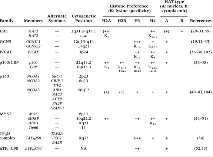
(for review see 20,21,27,59,60) (Table 1).
Histone Deacetylases
First links between the modification of histone
acetylation in conjunction with transcriptional
activity were observed in the early 1960s (9).
In the 1970s, an inhibition of histone
deacety-lase activity was shown to result in the
accu-mulation of acetylated histones in vivo (61).
Finally, the biochemical fractionation of yeast
extracts led to the discovery of two distinct yeast
histone deacetylating activities, HDA, the
cat-alytic subunit of a 350-kDa histone deacetylase
complex, which contains the histone
deacety-lase 1 (HDA1) protein and HDB, the catalytic
subunit of a 600-kDa histone deacetylase
com-plex, which contains reduced potassium
depen-dency 3 (RPD3). HDA1 and RPD3 share a
sig-nificant degree of sequence homology at the
protein level. Both proteins act mainly as
negative regulators of transcription. They may
however, counteract repression at telomeric
loci, where the general hyperacetylation of
histones is associated with gene activation.
Whether this observation, that HDAC
en-zymes repress genes in some parts of the
genome while they activate transcription in
other parts, reflects indirect mechanisms (e.g.,
reduction of the expression of genes, which
en-code other repressor proteins) or direct,
gene-specific effects, remains still to be elucidated
(28,62,63). Functionally, HDA1 and RPD3 have
nonoverlapping effects on the modulation of
lifespan: while the deletion of
RPD3
was
ob-served to extend life in yeast, this was not the
case for
HDA1,
unless it was combined with the
deletion of additional genes (e.g.,
SIR3
). The
si-multaneous deletion of both
HDA1
and
RPD3
has been shown to decrease lifespan and
be-cause the expression of both enzymes declines
with age, this could provide a possible
expla-nation for the increase in mortality during
senescence (64). While HDA activity in yeast
is strongly inhibited by Zn
2+, spermine, and
spermidine (65), RPD3 mutants are highly
sen-sitive to cycloheximide (63). Null mutants of
both
HDA1
and
RPD3
are viable and result in
a general increase of histone acetylation and
gene expression, except for genes located in
telomeric regions, where histone acetylation
has been associated with silencing (28).
repressor proteins, including the mammalian
heterodimeric repressors Mad/Max and Mxi/
Max, which are repressors in large part because
of their ability to recruit the RPD3/SIN3
complex to DNA-bound regulators of
tran-scription (66,72–79), while other repressors
(e.g., unliganded nuclear receptors) recruit the
RPD3/SIN3 complex via SMRT (silencing
me-diator of retinoic acid and thyroid hormone
re-ceptors) or nuclear corepressor (N-COR)
(78–82) and regulators like UME6 recognize
URS1 and bind SIN3, which in turn interacts
with RPD3 (75,83). RPD3, which contains the
catalytic deacetylase subunit of the SIN3/RPD3
complex, is clearly required for repression by
SIN3 (83). However, although it is likely that
the SIN3/RPD3 complex performs multiple
functions, some of which may play a more
prominent role in the repression of
transcrip-tion, it remains to be elucidated whether
his-tone deacetylation per se is the primary
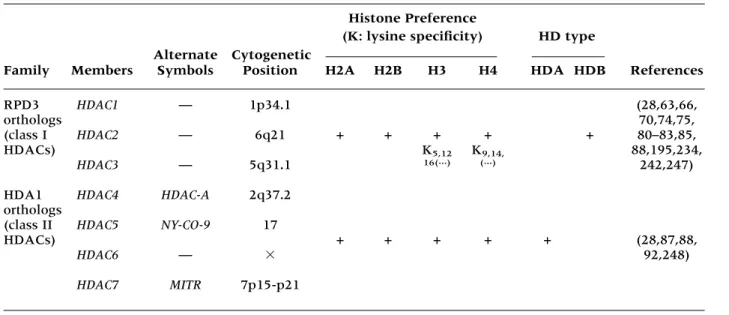
mech-anism of transcriptional repression (Table 2).
So far, seven human histone deacetylase
pro-teins, all of which share a highly conserved
cat-alytic domain, have been identified, of which
HDAC1, HDAC2, and HDAC3 are orthologs of
yeast RPD3 (66,67,84–86), while HDAC4,
HDAC5, HDAC6, and HDAC7 are yeast HDA1
orthologs (87,88). All human RPD3 orthologs
that have been reported so far repress
transcrip-tion when targeted to DNA via a DNA-binding
domain. They all bind transcription factor YY1,
which can act both as an activator and a
repres-sor of transcription (89). Accordingly, the
inhibi-tion of HDACs by trichostatin or trapoxin is
as-sociated with the activation or repression of
specific gene products (90). While mammalian
HDAC1 and HDAC2 have been shown to
inter-act with mSIN3 and the N-COR or SMRT
core-pressor complexes, which may associate
addi-tional proteins (e.g., SAP18, SAP30, RbAp48, or
RbAp46), HDAC3 does not appear to be part of
such multiprotein complexes (74,75,80–82,91).
Unlike the other deacetylases, HDAC4, which
Table 1. Human histone acetyltransferases
HAT type
Histone Preference (A: nuclear, B:
Alternate Cytogenetic
(K: lysine specificity) cytoplasmic)
Family Members Symbols Position H2A H2B H3 H4 A B References
HAT HAT1 — 2q31.2-q33.1 (++) ++ (+) + (29–31,55)
HAT2 — n.a. K5 K5,12
GCN5 GCN5L1 — 12q13-q14 +++ + + (19,32–35)
GCN5L2 — 17q21 K14 K8,16
P/CAF P/CAF — 3p24 ++ ++ + (36–38,102)
K14 K8
p300/CBP p300 — 22q13.2 ++ ++ ++ ++ + (36–38)
CBP — 16p13.3 K5 K5,12 K14 K5,8> 15,20 18,23 12,16
p160 NCOA1 SRC-1 2p23
NCOA2 GRIP-1 8q13
TIF2
NCOA3 AIB1 20q12 (+) (+) + + + (40–43,104)
RAC3 ACTR P/CIP TRAM-1
MYST MOZ — 8p11
MORF — 10q22.2 ++ ++ ++ + (44–51)
HBO1 — Xq21 K16
Tip60 — 11
TFIID TAF2A
complex TAFII250 CCG1 Xq13 +++ + + (54)
BA2R
belongs to the HDA1 family of HDACs, has been
reported to shuttle between the nucleus and the
cytoplasm in a process involving active nuclear
export (87,92). Unfortunately, human orthologs
of the yeast histone deacetylases HOS1 (28),
HOS2 (28,93) and HOS3 (28,94) have not been
reported so far.
Histone Acetylation, Solid Tumors,
and Leukemias: When HATs Are
the Key Players
More recently, an increasing number of
dis-ease processes have been observed to involve
abnormalities of the tightly regulated
inter-play of acetylating and deacetylating cellular
events, which are maintained by the enzymatic
activities of HATs and HDACs (95–100). A
de-crease in the amount of functionally available
HDACs (e.g., if
HDAC
loci are part of
chromo-somal deletions) or an increase of functionally
active HAT enzymatic activities (e.g., if
chro-mosomal segments, which encode HAT
pro-teins, are amplified) may shift the equilibrium
of histone acetylation toward acetylation,
which in turn has an effect on conformation
and activity of associated transcription factors
(e.g., GATA-1, TF
IIE
, TF
IIF, EKLF, and p53)
(23) and subsequently on gene expression,
in-teraction targets, and activities of downstream
signaling pathways (28,101–104). If, by contrast,
HATs are fused to a transcription factor in the
context of a chromosomal translocation (46–48,
105–110), which creates a novel chimeric
pro-tein, hyperacetylation may be confined to the
tar-get promoters of that specific transcription factor
and, because transcription factors have many
domains of protein–protein interaction, the
tran-scription factor/HAT-fusion may allow
acetyla-tion of associated regulators and result in the
transcriptional activation of a restricted number
of genes. As a consequence, chromosomal
re-gions that were silenced under normal
condi-tions may now be derepressed and change the
entire pattern of gene expression within affected
cells: genes that were previously silenced may
now be activated or even overexpressed,
whereas other genes, which were previously
ex-pressed, may now secondarily be repressed
(5,28,111). HAT enzymes have been found
am-plified, translocated, overexpressed, deleted, or
point mutated in several types of cancers:
■
An overexpression of HAT enzymatic
activi-ties has, for example, been described for the
steroid receptor coactivator-1 (SRC1)
ho-molog AIB1, which is involved in the
patho-genesis of both breast and ovarian cancers.
The associated
HAT
defect that has been
described for this particular type of cancer is
a more than 20-fold amplification of the
chro-mosomal region, which contains the AIB1
gene, and has been determined by
fluores-cence in situ hybridization (FISH) (104,112).
Table 2. Human histone deacetylases
Histone Preference
Alternate Cytogenetic
(K: lysine specificity) HD type
Family Members Symbols Position H2A H2B H3 H4 HDA HDB References
RPD3 HDAC1 — 1p34.1 (28,63,66,
orthologs 70,74,75,
(class I HDAC2 — 6q21 + + + + + 80–83,85,
HDACs) K5,12 K9,14, 88,195,234,
HDAC3 — 5q31.1 16(...) (...) 242,247)
HDA1 HDAC4 HDAC-A 2q37.2 orthologs
(class II HDAC5 NY-CO-9 17
HDACs) + + + + + (28,87,88,
HDAC6 — 92,248)
■
Changes in the availability of functionally
available HATs may also directly
affect-functions and activities of nonhistone
pro-teins, in view of the fact that some histone
acetyltransferases (p300, P/CAF, and
TAF-250) have been described to directly
acety-late general transcription factors (TF
IIF and
TF
IIE
) (113), sequence-specific
transcrip-tion factors (e.g., GATA-1 or EKLF) (114,
115), tumor suppressors (e.g., p53 or NF
B)
(116–118), architectural chromatin proteins
(e.g., HMGI(Y)) (119), and DNA repair
complexes, where it seems to increase
the DNA-binding capacity of the protein
(115,116).
■
Both p300 and CBP possess HAT activity,
which is partly intrinsic and partly the result
of association with other proteins (e.g.,
p/CAF) (36–38). The CBP/p300 HAT-protein
complex integrates many signaling
path-ways (e.g., the TGF-
signaling pathway)
(120–122) and is able to interact with a series
of transcription factors (e.g., CREB, c-Jun,
JunB, c-Fos, Myb, MyoD, YY1, nuclear
re-ceptors and basal components of the
tran-scriptional apparatus, etc.) (123) and to
par-ticipate in the direct or indirect stimulation
of transcription through scaffolding different
classes of transcriptional regulators onto
spe-cific chromatin domains (124,125). p300 and
CBP have been envisioned as negative
regu-lators of cell growth; mutations or
transloca-tions of the
p300
or of the
CBP
genes have
been found to be associated with several
solid tumors (e.g., point mutations of
p300,
which may be found in colorectal and gastric
carcinomas, are usually located within the
cysteine histidine-rich regions of the protein,
known to play important roles in the
biolog-ical activities of p300 (121,126,127); loss of
heterozygosity for
p300
in 80% of
glioblas-tomas (128) and acute leukemias (e.g., in the
M4/M5 subtype of acute myeloid leukemia
[AML] where
CBP
is found translocated and
fused to the putative acetyltransferase
MOZ
[t(8;16)(p11;p13)], which is a human
ho-molog of the yeast
SAS
genes [SAS: something
about silencing]) (48,109,129). Remarkably,
p300 mutations are located within the
cys-teine histidine-rich regions, which have
been observed to play an important role in
the biological activities of p300 (130). In
ad-dition, it has been reported that the
onco-genic viral proteins E1A and SV40 are able
to antagonize CBP-dependent transcription,
thereby promoting cellular proliferation
(38,131–133).
Leukemia-associated chromosome 8
inver-sions of the genotype inv(8)(p11;q13)
charac-teristically fuse
MOZ
to
TIF2/NCOA2/GRIP1
(tran-scriptional mediator/intermediary factor 2)
(134,135), a NR (nuclear hormone receptor)
coactivator that itself binds CBP/p300 (136,137).
The phenotype of the resulting MOZ-TIF2
fu-sion is therefore highly similar to the MOZ-CBP
fusion.
In other leukemias, particularly
therapy-related AML, myelodysplastic syndrome, and
chronic myelomonocytic leukemia,
CBP
may be
fused to
MLL
(mixed lineage leukemia), a gene
that has been associated with the
myelodysplas-tic syndrome [t(11;16)(q23;p13)](48,105–108).
In both translocations (
MLL–CBP
and
MOZ-CBP
)
the HAT domain of CBP remains intact within
the fusion protein and because both MOZ and
MLL have been implicated in the modification
of chromatin structure, it is likely that the
mole-cular mechanism through which the fusion
pro-teins perturb growth is by dysregulating gene
expression patterns (130).
In a separate subset of AMLs, which has
been reported to be associated with
therapy-induced leukemia, the
p300
gene was found
rearranged and fused in frame with the
MLL
gene [t(11;22)(q23;q13)] (106,138). This
sug-gests that alterations of CBP function may occur
in the later stages of leukemogenesis, possibly as
a way to eliminate cell-cycle checkpoints and
apoptotic responses (130). Notably, the MLL
fu-sion proteins described herein lack the carboxy
terminal SET (suppressor of variegation)
do-main, which is a hall-mark of many
chromatin-associated proteins (139,140). The MLL SET
domain interacts with the human SWI/SNF
(switch defective/sucrose nonfermenting)
chro-matin-remodeling complex, a powerful
tran-scriptional activator that belongs to a family of
DNA-stimulated ATPases that can either
dis-rupt the structure of nucleosome core particles
or influence the mobility and spacing of
nucle-osome arrays (141,142). Therefore, a fusion of
MLL and CBP results in the dysregulation of
transcription by failing to recruit SWI/SNF.
Conversely, the leukemia-associated
MLL–AF9
(143–146). Somatic mutations within the
SWI/
SNF
complex have been identified in several
ag-gressive pediatric malignant rhabdoid tumors
(147).
Humans lacking one functional allele of
the
CBP
gene, having point mutations or
mi-crodeletions within the 16p13.3 chromosomal
region that contains
CBP,
develop a condition,
which has been described as
“Rubinstein-Taybi
syndrome”
(autosomal
dominant).
Individuals exhibiting this condition have a
par-ticular propensity for malignancy, skeletal
ab-normalities, and growth retardation (128,148).
Concordantly,
CBP
-heterozygous mice reveal
skeletal abnormalities corresponding to the
changes that are seen in Rubinstein-Taybi
syn-drome (149). Interestingly, in spite of
appar-ently overlapping functions between CBP and
p300, patients with Rubinstein-Taybi
syn-drome have an intact
p300
allele, which is
po-tentially unable to sufficiently substitute for
CBP (130,150).
Unfortunately, it is not presently clear
ex-actly how p300 and CBP are involved in the
development of cancer. What is known,
how-ever, is that p300 and CBP, which have
well-documented activity as transcriptional
activa-tors, are important key players in cell-cycle
control, within apoptotic pathways, in the
pro-motion of differentiation, and in p53 signaling
and activation (116,124,151,152). In a
simpli-fied scenario the anti-oncogenic properties of
p300 and CBP seem to go hand in hand with
the anti-oncogenic activities that have been
proposed for p53; since p300 forms a complex
with p53 that exerts its anticancerogenic
activ-ity by negatively regulating cell growth. Several
promoter/enhancer elements, such as the AP1
and c-Fos elements, function in a
p300-depen-dent manner (153), which has been correlated
with a promotion of G1-S transition, resulting
thereby in cellular proliferation and potentially
transformation (154). Although the formation
of p53–p300 may in part be responsible for the
recruitment of p300 onto some promoters, it
may inhibit the transactivating effects of p300
on others (e.g., on promoters containing the
DNA binding sites for the transcription factor
AP1, where increased levels of p300 are able to
overcome p53-mediated inhibition of AP1).
Most interestingly, p53 binds to p300 in a
re-gion that is required for its intrinsic HAT
activ-ity (37). However, this region is distinct form
the domains that bind to c-Jun (155), P/CAF
(38), and TBP (156), all of which are important
modulators of transcription. It has therefore
been suggested that p53 might function through
direct protein–protein interactions via p300 and
possibly also through other p300-associated
fac-tors (124).
It has been observed that factors like the
nuclear hormone receptors (e.g., RAR/RXR)
mediate transcriptional repression by
recruit-ing HDAC complexes in their unliganded
form (80–82), while they exhibit
ligand-in-ducible transcriptional activator functions
through the recruitment of HAT-coactivator
complexes (p300/CAF) when hormone is
bound (Fig. 3) (4,5,40,150,157). Similarly,
E2F and Rb, for example, form a repressor
complex, which recruits HDAC1 and HDAC2
(158,159) and subsequently represses the
cy-clin E promoter. Frequently, phosphorylation
is a key event that induces a conformational
change within a transcription factor or other
regulatory elements (e.g., NF
B, the IFN
en-hancer complex or Rb), which then readily
recruits a HAT-coactivator complex and
stimu-lates transcription (160–162).
Histone Acetylation, Solid Tumors,
and Leukemias: When HDACs Are
the Key Players
Leukemias are generally associated with
char-acteristic chromosomal translocations, which
may result either in the generation of a chimeric
protein with novel functional properties or in
the aberrant expression of a regulatory element.
Usually, chromosomal translocations affect only
one allele of a gene. Therefore, to cause a
pheno-typic effect, the activity of the newly generated
fusion protein needs to be dominant over that of
the wild-type protein. Frequently, the
translo-cations found in leukemias target regulatory
transcription factors that control cellular
prolif-eration, survival, and differentiation and may
obviate the need for multistep mutation
path-ways because they are observed for
proto-oncogenes and tumor suppressor genes in solid
tumors (163,164). In accordance with the
dis-ease-linked
HAT
defects that have been
de-scribed above,
HDAC
defects may very similarly
of gene expression. Several transcriptional
re-pressors (e.g., Mad and members of the nuclear
receptor superfamily), trans-cription factors,
and cellular regulators have been described to
associate with HDAC activities (5).
■
The analysis of the transforming chimeric
proteins PML-RAR
[t(15;17)(q22;q21)]
(163,165–167) and PLZF-RAR
[t(11;17)
(q23;q21)] (167–170), which are found in
dif-ferent acute promyelocytic leukemias (APL),
has shown a clear connection between the
action of histone deacetylases and the
devel-opment of cancer. In these instances the
promyelocytic leukemia gene (
PML
) [t(15;17)
(q22;q21)] or the promyelocytic leukemia
zinc finger gene (
PLZF
) [t(11;17)(q23;q21)]
are fused to the retinoic acid receptor-alpha
(
RAR
) and are no longer responsive to
phys-iological levels of retinoic acid (167,171,172).
These chromosomal changes result in a
block of cellular differentiation (i.e., in the
clonal expansion of cells arrested in the
promyelocyte stage of development)
yield-ing the clinical picture of an acute leukemia
(78,171,172). While patients featuring the
PML-RAR
[t(15;17)(q22;q21)] translocation,
readily differentiate upon treatment with
all-trans-retinoic acid (ATRA) (78,171), patients
having the
PLZF
-
RAR
[t(11;17)(q23;q21)]
type of translocation do not respond
ade-quately to treatment with ATRA (78,171).
On the molecular level, RAR
represses
target genes by tethering corepressors such
as N-COR and SMRT to promoter DNA
(78,173). These corepressors are part of one
or more large complexes that also contain
mSIN3A and HDAC proteins (5). In cells
that express PML-RAR
, retinoic acids lead
to the dissociation of the
SMRT-mSIN3A-HDAC1 and N-COR-mSIN3A-SMRT-mSIN3A-HDAC1,2
complexes from RAR
(97,98,172). By
con-trast, cells that express PLZF-RAR
have
two N-COR binding sites, one in the RAR
region (which is responsive to retinoic
acids) and one in the PLZF amino terminal
region (which is nonresponsive to retinoic
acids) of the fusion protein (97). Because
PLZF binds N-COR and SMRT
indepen-dently from RAR
, the HDAC corepressor
complex is readily released from RAR
upon treatment with retinoic acids, but not
from PLZF (97). As a consequence,
tran-scriptional repression is preserved.
How-ever, the sensitivity of PLZF to ATRA may be
restored by the treatment with an HDAC
in-hibitor (e.g., trichostatin A, an antibiotic). The
PLZF-bound HDAC corepressor complex is
then readily released, allowing these leukemic
cells to differentiate (97,98,172,174, 175).
■
An additional example of a translocation
found in AML and that has been shown to
involve HDACs is the translocation t(8;21)
(q22;q22), which results in a fusion of
AML-1
and
ETO
(176–178) and accounts for
ap-proximately 10–12% of AMLs (164).
Simi-larly,
AML-1
may be fused to
MTG16
(myeloid
tumor gene 16) in the context of a
transloca-tion t(16;21)(q24;q22) (179–181), or to
EVI1,
a
transcriptional repressor, in association with
a translocation t(3;21)(q26;q22) (177,182,
183). In all these translocations of
AML-1,
the Runt homology domain, which is the
re-gion of AML-1 that interacts with both DNA
and the core binding factor CBF
, is
pre-served (178,184,185). Other translocations
that are frequently seen are an inversion of
chromosome 16 [inv(16)] in AML, where
the
CBF
gene, which forms a transcription
factor complex with
AML-1,
is fused to the
smooth muscle myosin heavy chain gene
MYH11
(164) and the translocation t(12;21)
(p12;q22), which is found in 15–35% of
pe-diatric B-lineage ALLs and where a
TEL-AML1
gene fusion yields a novel chimeric
protein (186,187). The ability of CBF
to
associate with AML-1, hereby increasing
the affinity of AML-1 for its DNA-binding
site, is retained even when chromosome 16
is inverted (184,185). Because all the
translo-cations mentioned in this paragraph [t(8;21)
(q22;q22), t(3;21) (q26;q22), t(12;21)(p12;
q22) and inv(16)] interfere with the
tran-scriptional regulation of AML-1 responsive
genes (177,178,188–191), evidence emerges
that transcriptional repression of AML-1
tar-get genes is critical in the pathogenesis of
AMLs. In the case of AML-1/ETO,
overex-pression of
AML-1
and anti
AML-1/ETO
anti-sense oligonucleotides can induce
differen-tiation in cells containing this fusion
protein (185,191). In analogy to the
obser-vations that have been made for
PML-RAR
and PLZF-RAR
, ETO has been
of ETO with N-COR both when
AML-1
is
fused to
ETO
[t(8;21)(q22;q22)] and when it is
fused to
MTG16
[t(16;21)(q24;q22)] are
highly conserved, suggesting that t(16; 21)
(q24;q22) equally represses transcription
through the recruitment of HDACs. Besides
CBF
, AML-1 associates with other
tran-scription factors and regulators and
acti-vates transcription if bound to HATs (e.g.,
CBP and p300) (191) or represses
transcrip-tion when it interacts with mSIN3 (192)
(Fig. 3).
■
Other than the indirect recruitment of
HDAC with the assistance of protein
com-plexes, which include mSIN3A, N-COR,
and SMRT or Rb, several transcription factors
(e.g., YY1) and regulators are able to recruit
HDAC enzymes directly (reviewed in 21),
thereby interfering with the generation of a
functional initiation complex. These
HDAC-associated effects on transcriptional
regula-tion may be abrogated by HDAC-inhibitors
(trapoxin, trichostatin A, etc.). The
retino-blastoma protein (RB), which is important
in the induction of cell-cycle arrest under
unfavorable growth conditions, mediates
E2F-bound promoter repression through its
in-teraction with HDAC1. This binding of
HDAC to RB has been highlighted by the
observations that tumor-specific mutations
found in RB disrupt its association with
HDAC (159,193,194) and that viral
oncopro-teins (e.g., HPV16 E7 or the SV40 T-antigen)
are able to displace HDAC from RB (193,194).
These findings suggest a fundamental role for
histone modification in the suppression of
cancer.
■
So far, the chromosomal localizations of the
human
RPD3
orthologs
HDAC1-3
(102,195)
and the
HDA1
orthologs
HDAC4-7
have
been identified more or less precisely (87)
(Table 2) and, interestingly,
HDAC1-3
and
HDAC7
localize to chromosomal sites,
which are particularly fragile and frequently
altered through mutations, translocations,
and deletions, particularly in
myeloprolifera-tive disorders and solid tumors (101–103,
196). This may potentially result in a
shift of the acetylation equilibrium toward
acetylation.
It had long been epidemiologically postulated
that a diet high in fiber was associated with a
low incidence of colon cancer (197–200) until
the Nurses’ Health Study at Harvard, which
was conducted on 88,757 middle-aged women,
proved that the “protective effect” of dietary
fiber against colorectal cancer or adenoma was
not significant (198). Even though there is
con-siderable and to some extent inconclusive
liter-ature on dietary fiber in connection with colon
cancer, it appears that butyrate, a fiber
fermen-tation product, could in fact have a protective
effect against colon cancer (201–204). More
re-cently, several investigators demonstrated that
butyrate administration effectively reduced
in-cidence and size of colonic tumors (205,206),
their likelihood to metastasize (207), and that it
shifted their histological phenotype to one that
appeared less aggressive (206). Even though
the molecular mechanisms by which butyrate
mediates its protective effects are still very
unclear, butyrate has been shown to induce
both histone and nonhistone
hyperacetyla-tion through a noncompetitive and
nonspe-cific inhibition of HDACs via a
serine-threo-nine protein phosphatase of the PP1 type
(208–210). In addition to its capability to
in-duce differentiation, butyrate has been found
to cause a G1 cell-cycle arrest (211,212), which
is mediated through induction of the G1
cell-cycle inhibitor
p21
gene (213), thereby
requir-ing an inhibition of HDAC1. The fact that
p21
is deleted in the human colon carcinoma cell
line HCT1116 further supports that p21 is
es-sentially involved in butyrate-mediated
cellu-lar growth arrest (214). Because butyrate is
rapidly metabolized and it has not been
possi-ble to maintain adequate butyrate
concentra-tions in patients, butyrate homologs and
alter-native substrates [e.g., trichostatins and trapoxins
(215,216), depudecin (217), oxamflatin (218),
benzamide derivatives (219)], which appear
more promising than butyrate itself (e.g.,
phenyl-butyrate and tributyrin) are currently under
study (220–224).
Fig. 3.
Some factors that associate with histone modifying-enzymes (nuclear hormone receptors, transcription factors, and other regulat
ors) have the capacity
to mediate both transcriptional activation and repression, depending on whether they associate with HA
T
accepted concepts that reversible modification
of chromatin influences its transcriptional
com-petence and that promoters may be targeted
specifically by either activating or repressing
complexes. Histone acetylation modifiers have
been found to be engaged and mutated in
sev-eral types of cancer. Although genes encoding
HAT
enzymes have been found to be
preferen-tially translocated, amplified, overexpressed,
or point mutated,
HDACs
have repeatedly been
identified to mediate the function of oncogenic
translocation products, thereby accounting for
at least 30% of AMLs, 25% of childhood
B-ALLs, and more than 99% of APLs (26). They
have also been found to associate with tumor
suppressor proteins, which themselves are
fre-quently mutated (e.g., RB). Many questions
persist regarding the molecular mechanisms
that involve histone-modifying enzymes. The
further characterization of HATs and HDACs
will therefore not only continue to unravel the
role that these enzymes play in transcription, it
will also help to identify the molecular
mecha-nisms that promote leukemogenesis.
Addition-ally, it looks like acetylation is not just limited
to histones. It could therefore, in analogy to
phosphorylation, be a process that influences
the function of many proteins and cellular
processes. The identification of proteins that
interact with histone-modifying enzymes and
target genes, which are misregulated as a
conse-quence of mistargeted, or defective
histone-modifying enzymes may help to hasten the
development of less toxic, more refined and
specific forms of pharmacological interventions
for some forms of cancer and leukemias. First
ex-periences with histone deacetylase inhibitors
as “differentiation therapy” reagents have
shown promising results for several types of
leukemias (96,172,221–224) and solid tumors
(220,225–227) with few, if any, significant side
effects, indicating that such treatment could
have great therapeutic advantages when
com-pared to conventional chemotherapeutic agents.
Even though almost explosive advances in the
understanding of the molecular details of
tran-scriptional regulation and chromatin
modifica-tion through acetylamodifica-tion have been reported in
the last few years, many questions remain. The
identification of novel HAT/HDAC interaction
targets, the analysis of HAT/HDAC levels in
primary cells, and the response and tolerability
of histone deacetylase inhibitors in patients
may help to answer and generate new
ques-tions, such as how far a therapeutic modulation
of intracellular acetylation levels could
com-plement or replace existing chemotherapeutic
strategies in the treatment of cancer.
Acknowledgments
We wish to apologize to those investigators
whose relevant work was not discussed or cited
directly in this manuscript due to space
limita-tions. This work was partly supported by the
German National Science Foundation (Deutsche
Forschungsgemeinschaft, MA 2057
1-1) and
institutional funds from the University of
Fran-kfurt Medical Center.
References
1. Luger K, Mader AW, Richmond RK, et al. (1997) Crystal structure of the nucleosome core particle at 2.8 A resolution. Nature389:251–260. 2. Loidl P. (1994) Histone acetylation: facts and
questions. Chromosoma103:441–449.
3. Jeppesen P. (1997) Histone acetylation: a possi-ble mechanism for the inheritance of cell mem-ory at mitosis. Bioessays19:67–74.
4. Wade PA, Pruss D, Wolffe AP. (1997) Histone acetylation: chromatin in action. Trends Biochem. Sci.22:128–132.
5. Struhl K. (1998) Histone acetylation and tran-scriptional regulatory mechanisms. Genes Dev.
12:599–606.
6. Spencer VA, Davie JR. (1999) Role of covalent modifications of histones in regulating gene ex-pression. Gene240:1–12.
7. Bradbury EM. (1992) Reversible histone modi-fications and the chromosome cell cycle. Bioessays
14:9–16.
8. Barratt MJ, Hazzalin CA, Cano E, et al. (1994) Mitogen-stimulated phosphorylation of histone H3 is targeted to a small hyperacetylation-sensi-tive fraction. Proc. Natl. Acad. Sci. U.S.A.91:4781– 4785.
9. Allfrey VG, Faulkner R, Mirsky AE. (1964) Acetylation and methylation of histones and their possible role in the regulation of RNA syn-thesis. Proc. Natl. Acad. Sci. U.S.A.51:786–794. 10. Cameron EE, Bachman KE, Myohanen S, et al.
(1999) Synergy of demethylation and histone deacetylase inhibition in the re-expression of genes silenced in cancer. Nat. Genet.21:103–107. 11. Hebbes TR, Thorne AW, Crane-Robinson C. (1988) A direct link between core histone acety-lation and transcriptionally active chromatin.
Embo J.7:1395–1402.
12. Grunstein M. (1997) Histone acetylation in chro-matin structure and transcription. Nature 389:
13. Braunstein M, Rose AB, Holmes SG, et al. (1993) Transcriptional silencing in yeast is associated with reduced nucleosome acetylation. Genes Dev.7:
592–604.
14. Johnson CA, O’Neill LP, Mitchell A, et al. (1998) Distinctive patterns of histone H4 acetylation are associated with defined sequence elements within both heterochromatic and euchromatic regions of the human genome. Nucleic Acids Res.
26:994–1001.
15. Ng HH, Bird A. (1999) DNA methylation and chromatin modification. Curr. Opin. Genet. Dev.9:
158–163.
16. Nan X, Ng HH, Johnson CA, et al. (1998) Transcriptional repression by the methyl-CpG-binding protein MeCP2 involves a histone deacetylase complex. Nature393:386–389. 17. Jones PL, Veenstra GJ, Wade PA, et al. (1998)
Methylated DNA and MeCP2 recruit histone deacetylase to repress transcription. Nat. Genet.
19:187–191.
18. Wong CW, Privalsky ML. (1998) Transcriptional repression by the SMRT-mSin3 corepressor: multiple interactions, multiple mechanisms, and a potential role for TFIIB. Mol. Cell. Biol.18:
5500–5510.
19. Brownell JE, Zhou J, Ranalli T, et al. (1996) Tetrahymena histone acetyltransferase A: a ho-molog to yeast Gcn5p linking histone acetyla-tion to gene activaacetyla-tion. Cell84:843–851. 20. Kouzarides T. (1999) Histone acetylases and
deacetylases in cell proliferation. Curr. Opin. Genet. Dev.9:40–48.
21. Davie JR, Chadee DN. (1998) Regulation and regulatory parameters of histone modifications.
J. Cell. Biochem. Suppl.31:203–213.
22. Muscat GE, Burke LJ, Downes M. (1998) The corepressor N-CoR and its variants RIP13a and RIP13Delta1 directly interact with the basal transcription factors TFIIB, TAFII32 and TAFII70. Nucleic Acids Res.26:2899–2907. 23. Boyes J, Byfield P, Nakatani Y, et al. (1998)
Reg-ulation of activity of the transcription factor GATA-1 by acetylation. Nature396:594–598. 24. Minucci S, Pelicci PG. (1999) Retinoid receptors
in health and disease: co-regulators and the chro-matin connection. Semin. Cell Dev. Biol.10:215–225. 25. Behre G, Zhang P, Zhang DE, et al. (1999) Analy-sis of the modulation of transcriptional activity in myelopoiesis and leukemogenesis. Methods 17:
231–237.
26. Fenrick R, Hiebert SW. (1998) Role of histone deacetylases in acute leukemia. J. Cell. Biochem. Suppl.31:194–202.
27. Magnaghi-Jaulin L, Ait-Si-Ali S, Harel-Bellan A. (1999) Histone acetylation in signal transduc-tion by growth regulatory signals. Semin. Cell Dev. Biol.10:197–203.
28. Rundlett SE, Carmen AA, Kobayashi R, et al. (1996) HDA1 and RPD3 are members of distinct
yeast histone deacetylase complexes that regu-late silencing and transcription. Proc. Natl. Acad. Sci. U.S.A.93:14503–14508.
29. Kleff S, Andrulis ED, Anderson CW, et al. (1995) Identification of a gene encoding a yeast histone H4 acetyltransferase. J. Biol. Chem. 270:
24674–24677.
30. Parthun MR, Widom J, Gottschling DE. (1996) The major cytoplasmic histone acetyltransferase in yeast: links to chromatin replication and his-tone metabolism. Cell87:85–94.
31. Chang L, Loranger SS, Mizzen C, et al. (1997) Histones in transit: cytosolic histone com-plexes and diacetylation of H4 during nucleo-some assembly in human cells. Biochemistry36:
469–480.
32. Kuo MH, Brownell JE, Sobel RE, et al. (1996) Transcription-linked acetylation by Gcn5p of hi-stones H3 and H4 at specific lysines. Nature383:
269–272.
33. Grant PA, Duggan L, Cote J, et al. (1997) Yeast Gcn5 functions in two multisubunit complexes to acetylate nucleosomal histones: characterization of an Ada complex and the SAGA (Spt/Ada) com-plex. Genes Dev.11:1640–1650.
34. Eberharter A, Sterner DE, Schieltz D, et al. (1999) The ADA complex is a distinct histone acetyl-transferase complex in Saccharomyces cerevisiae. Mol. Cell. Biol.19:6621–6631.
35. Martinez E, Kundu TK, Fu J, et al. (1998) A hu-man SPT3-TAFII31-GCN5-L acetylase complex distinct from transcription factor IID. J. Biol. Chem.
273:23781–23785.
36. Bannister AJ, Kouzarides T. (1996) The CBP co-activator is a histone acetyltransferase. Nature384:
641–643.
37. Ogryzko VV, Schiltz RL, Russanova V, et al. (1996) The transcriptional coactivators p300 and CBP are histone acetyltransferases. Cell 87:
953–959.
38. Yang XJ, Ogryzko VV, Nishikawa J, et al. (1996) A p300/CBP-associated factor that competes with the adenoviral oncoprotein E1A. Nature
382:319–324.
39. Blanco JC, Minucci S, Lu J, et al. (1998) The hi-stone acetylase PCAF is a nuclear receptor coac-tivator. Genes Dev.12:1638–1651.
40. Glass CK, Rose DW, Rosenfeld MG. (1997) Nuclear receptor coactivators. Curr. Opin. Cell Biol.
9:222–232.
41. Chen H, Lin RJ, Schiltz RL, et al. (1997) Nuclear receptor coactivator ACTR is a novel histone acetyltransferase and forms a multimeric activa-tion complex with P/CAF and CBP/p300. Cell
90:569–580.
42. Spencer TE, Jenster G, Burcin MM, et al. (1997) Steroid receptor coactivator-1 is a histone acetyl-transferase. Nature389:194–198.
the steroid hormone receptor superfamily. Science
270:1354–1357.
44. Champagne N, Bertos NR, Pelletier N, et al. (1999) Identification of a human histone acetyl-transferase related to monocytic leukemia zinc finger protein. J. Biol. Chem.274:28528–28536. 45. Smith ER, Eisen A, Gu W, et al. (1998) ESA1 is
a histone acetyltransferase that is essential for growth in yeast. Proc. Natl. Acad. Sci. U.S.A. 95:
3561–3565.
46. Kamine J, Elangovan B, Subramanian T, et al. (1996) Identification of a cellular protein that specifically interacts with the essential cysteine region of the HIV-1 Tat transactivator. Virology
216:357–366.
47. Reifsnyder C, Lowell J, Clarke A, et al. (1996) Yeast SAS silencing genes and human genes as-sociated with AML and HIV-1 Tat interactions are homologous with acetyltransferases. Nat. Genet.14:42–49.
48. Borrow J, Stanton VP, Jr., Andresen JM, et al. (1996) The translocation t(8;16)(p11;p13) of acute myeloid leukaemia fuses a putative acetyl-transferase to the CREB-binding protein. Nat. Genet.14:33–41.
49. Hilfiker A, Hilfiker-Kleiner D, Pannuti A, et al. (1997) mof, a putative acetyl transferase gene re-lated to the Tip60 and Moz human genes and to the Sas genes of yeast, is required for dosage com-pensation in Drosophila. Embo J.16:2054–2060. 50. Iizuka M, Stillman B. (1999) Histone
acetyl-transferase HBO1 interacts with the ORC1 sub-unit of the human initiator protein. J. Biol. Chem.
274:23027–23034.
51. Yamamoto T, Horikoshi M. (1997) Novel sub-strate specificity of the histone acetyltransferase activity of HIV-1-Tat interactive protein Tip60.
J. Biol. Chem.272:30595–30598.
52. Hsieh YJ, Kundu TK, Wang Z, et al. (1999) The TFIIIC90 subunit of TFIIIC interacts with multi-ple components of the RNA polymerase III ma-chinery and contains a histone-specific acetyl-transferase activity. Mol. Cell. Biol.19:7697–7704. 53. Kundu TK, Wang Z, Roeder RG. (1999) Human
TFIIIC relieves chromatin-mediated repression of RNA polymerase III transcription and con-tains an intrinsic histone acetyltransferase activ-ity. Mol. Cell. Biol.19:1605–1615.
54. Mizzen CA, Yang XJ, Kokubo T, et al. (1996) The TAF(II)250 subunit of TFIID has histone acetyltransferase activity. Cell87:1261–1270. 55. Ruiz-Garcia AB, Sendra R, Galiana M, et al.
(1998) HAT1 and HAT2 proteins are compo-nents of a yeast nuclear histone acetyltransferase enzyme specific for free histone H4. J. Biol. Chem.
273:12599–12605.
56. Ohba R, Steger DJ, Brownell JE, et al. (1999) A novel H2A/H4 nucleosomal histone acetyltrans-ferase in Tetrahymena thermophila. Mol. Cell. Biol.19:2061–2068.
57. Wittschieben BO, Otero G, de Bizemont T, et al. (1999) A novel histone acetyltransferase is an integral subunit of elongating RNA polymerase II holoenzyme. Mol. Cell4:123–128.
58. Yao TP, Oh SP, Fuchs M, et al. (1998) Gene dosage-dependent embryonic development and proliferation defects in mice lacking the tran-scriptional integrator p300. Cell93:361–372. 59. Workman JL, Kingston RE. (1998) Alteration of
nucleosome structure as a mechanism of transcrip-tional regulation. Annu. Rev. Biochem.67:545–579. 60. Grant PA, Berger SL. (1999) Histone
acetyltrans-ferase complexes. Semin. Cell Dev. Biol.10:169–177. 61. Boffa LC, Vidali G, Mann RS, et al. (1978) Sup-pression of histone deacetylation in vivo and in vitro by sodium butyrate. J. Biol. Chem. 253:
3364–3366.
62. Carmen AA, Rundlett SE, Grunstein M. (1996) HDA1 and HDA3 are components of a yeast hi-stone deacetylase (HDA) complex. J. Biol. Chem.
271:15837–15844.
63. Vidal M, Gaber RF. (1991) RPD3 encodes a sec-ond factor required to achieve maximum positive and negative transcriptional states in Saccha-romyces cerevisiae. Mol. Cell. Biol.11:6317–6327. 64. Kim S, Benguria A, Lai CY, et al. (1999)
Modu-lation of life-span by histone deacetylase genes in saccharomyces cerevisiae. Mol. Biol. Cell 10:
3125–3136.
65. Vu QA, Zhang DE, Chroneos ZC, et al. (1987) Polyamines inhibit the yeast histone deacety-lase. FEBS Lett.220:79–83.
66. Taunton J, Hassig CA, Schreiber SL. (1996) A mammalian histone deacetylase related to the yeast transcriptional regulator Rpd3p. Science
272:408–411.
67. Yang WM, Inouye C, Zeng Y, et al. (1996) Tran-scriptional repression by YY1 is mediated by in-teraction with a mammalian homolog of the yeast global regulator RPD3. Proc. Natl. Acad. Sci. U.S.A.93:12845–12850.
68. McKenzie EA, Kent NA, Dowell SJ, et al. (1993) The centromere and promoter factor, 1, CPF1, of Saccharomyces cerevisiae modulates gene activ-ity through a family of factors including SPT21, RPD1 (SIN3), RPD3 and CCR4. Mol. Gen. Genet.
240:374–386.
69. Vidal M, Strich R, Esposito RE, et al. (1991) RPD1 (SIN3/UME4) is required for maximal ac-tivation and repression of diverse yeast genes.
Mol. Cell. Biol.11:6306–6316.
70. Pazin MJ, Kadonaga JT. (1997) What’s up and down with histone deacetylation and transcrip-tion? Cell89:325–328.
71. Wolffe AP. (1997) Transcriptional control. Sinful repression. Nature387:16–17.
73. Schreiber-Agus N, Chin L, Chen K, et al. (1995) An amino-terminal domain of Mxi1 mediates anti-Myc oncogenic activity and interacts with a homolog of the yeast transcriptional repressor SIN3. Cell80:777–786.
74. Hassig CA, Fleischer TC, Billin AN, et al. (1997) Histone deacetylase activity is required for full transcriptional repression by mSin3A. Cell 89:
341–347.
75. Laherty CD, Yang WM, Sun JM, et al. (1997) Hi-stone deacetylases associated with the mSin3 corepressor mediate mad transcriptional repres-sion. Cell89:349–356.
76. Amati B, Dalton S, Brooks MW, et al. (1992) Transcriptional activation by the human c-Myc oncoprotein in yeast requires interaction with Max. Nature359:423–426.
77. Kretzner L, Blackwood EM, Eisenman RN. (1992) Myc and Max proteins possess distinct transcriptional activities. Nature359:426–429. 78. Chen JD, Evans RM. (1995) A transcriptional
co-repressor that interacts with nuclear hor-mone receptors. Nature377:454–457.
79. Horlein AJ, Naar AM, Heinzel T, et al. (1995) Ligand-independent repression by the thyroid hormone receptor mediated by a nuclear recep-tor co-repressor. Nature377:397–404.
80. Alland L, Muhle R, Hou H, Jr., et al. (1997) Role for N-CoR and histone deacetylase in Sin3-medi-ated transcriptional repression. Nature387:49–55. 81. Heinzel T, Lavinsky RM, Mullen TM, et al. (1997) A complex containing N-CoR, mSin3 and histone deacetylase mediates transcriptional repression. Nature387:43–48.
82. Nagy L, Kao HY, Chakravarti D, et al. (1997) Nu-clear receptor repression mediated by a complex containing SMRT, mSin3A, and histone deacety-lase. Cell89:373–380.
83. Kadosh D, Struhl K. (1997) Repression by Ume6 involves recruitment of a complex con-taining Sin3 corepressor and Rpd3 histone deacetylase to target promoters. Cell 89:
365–371.
84. Yang WM, Yao YL, Sun JM, et al. (1997) Isola-tion and characterizaIsola-tion of cDNAs correspond-ing to an additional member of the human his-tone deacetylase gene family. J. Biol. Chem.272:
28001–28007.
85. Emiliani S, Fischle W, Van Lint C, et al. (1998) Characterization of a human RPD3 ortholog, HDAC3. Proc. Natl. Acad. Sci. U.S.A.95:2795–2800. 86. Dangond F, Hafler DA, Tong JK, et al. (1998) Differential display cloning of a novel human histone deacetylase (HDAC3) cDNA from PHA-activated immune cells. Biochem. Biophys. Res. Commun.242:648–652.
87. Wang AH, Bertos NR, Vezmar M, et al. (1999) HDAC4, a human histone deacetylase related to yeast HDA1, is a transcriptional corepressor.
Mol. Cell. Biol.19:7816–7827.
188. Grozinger CM, Hassig CA, Schreiber SL. (1999) Three proteins define a class of human histone deacetylases related to yeast Hda1p.
Proc. Natl. Acad. Sci. U.S.A.96:4868–4873. 189. Shi Y, Seto E, Chang LS, et al. (1991)
Transcrip-tional repression by YY1, a human GLI-Kruppel-related protein, and relief of repression by adenovirus E1A protein. Cell67:377–388. 190. Van Lint C, Emiliani S, Ott M, et al. (1996)
Transcriptional activation and chromatin re-modeling of the HIV-1 promoter in response to histone acetylation. Embo J.15:1112–1120. 191. Zhang Y, Iratni R, Erdjument-Bromage H, et al.
(1997) Histone deacetylases and SAP18, a novel polypeptide, are components of a human Sin3 complex. Cell89:357–364.
192. Miska EA, Karlsson C, Langley E, et al. (1999) HDAC4 deacetylase associates with and re-presses the MEF2 transcription factor. Embo J.
18:5099– 5107.
193. Aiba H, Kawaura R, Yamamoto E, et al. (1998) Isolation and characterization of high-osmolarity-sensitive mutants of fission yeast. J. Bacteriol.180:5038–5043.
194. Carmen AA, Griffin PR, Calaycay JR, et al. (1999) Yeast HOS3 forms a novel trichostatin A-insensitive homodimer with intrinsic his-tone deacetylase activity. Proc. Natl. Acad. Sci. U.S.A.96:12356–12361.
195. Wang J, Hoshino T, Redner RL, et al. (1998) ETO, fusion partner in t(8;21) acute myeloid leukemia, represses transcription by interaction with the human N-CoR/mSin3/HDAC1 com-plex. Proc. Natl. Acad. Sci. U.S.A.95:10860– 10865. 196. Wang J, Saunthararajah Y, Redner RL, et al. (1999) Inhibitors of histone deacetylase relieve ETO-mediated repression and induce differen-tiation of AML1-ETO leukemia cells. Cancer Res.59:2766–2769.
197. Grignani F, De Matteis S, Nervi C, et al. (1998) Fusion proteins of the retinoic acid receptor-alpha recruit histone deacetylase in promyelo-cytic leukaemia. Nature391:815–818.
198. Lin RJ, Nagy L, Inoue S, et al. (1998) Role of the histone deacetylase complex in acute promyelocytic leukaemia. Nature391:811–814. 199. Kosugi H, Towatari M, Hatano S, et al. (1999) Hi-stone deacetylase inhibitors are the potent in-ducer/enhancer of differentiation in acute myeloid leukemia: a new approach to anti-leukemia therapy. Leukemia13:1316–1324. 100. Horiuchi K, Fujimoto D, Fukushima M, et al.
(1981) Increased histone acetylation and deacety-lation in rat ascites hepatoma cells. Cancer Res.41:
1488–1491.
101. Le Beau MM, Espinosa Rd, Neuman WL, et al. (1993) Cytogenetic and molecular delineation of the smallest commonly deleted region of chromosome 5 in malignant myeloid diseases.
102. Randhawa GS, Bell DW, Testa JR, et al. (1998) Identification and mapping of human histone acetylation modifier gene homologues. Ge-nomics51:262–269.
103. Mahlknecht U, Bucala R, Hoelzer D, et al. (1999) High resolution physical mapping of human HDAC3, a potential tumor suppressor gene in the 5q31 region. Cytogenet. Cell Genet.86:237–239. 104. Anzick SL, Kononen J, Walker RL, et al. (1997) AIB1, a steroid receptor coactivator amplified in breast and ovarian cancer. Science277:965–968. 105. Rowley JD, Reshmi S, Sobulo O, et al. (1997)
All patients with the T(11;16) (q23;p13.3) that involves MLL and CBP have treatment-related hematologic disorders. Blood 90: 535– 541.
106. Taki T, Sako M, Tsuchida M, et al. (1997) The t(11;16)(q23;p13) translocation in myelodys-plastic syndrome fuses the MLL gene to the CBP gene. Blood89:3945–3950.
107. Sobulo OM, Borrow J, Tomek R, et al. (1997) MLL is fused to CBP, a histone acetyltransferase, in therapy-related acute myeloid leukemia with a t(11;16)(q23;p13.3). Proc. Natl. Acad. Sci. U.S.A.
94:8732–8737.
108. Satake N, Ishida Y, Otoh Y, et al. (1997) Novel MLL-CBP fusion transcript in therapy-related chronic myelomonocytic leukemia with a t(11;16)(q23;p13) chromosome translocation.
Genes Chromosomes Cancer20:60–63.
109. Quesnel B, Kantarjian H, Bjergaard JP, et al. (1993) Therapy-related acute myeloid leukemia with t(8;21), inv(16), and t(8;16): a report on 25 cases and review of the literature.
J. Clin. Oncol.11:2370–2379.
110. Dutnall RN, Tafrov ST, Sternglanz R, et al. (1998) Structure of the histone acetyltransferase Hat1: a paradigm for the GCN5-related N-acetyltransferase superfamily. Cell94:427–438. 111. Mahlknecht U, Ottmann OG, Hoelzer D. (2000)
When the band begins to play: Histone acety-lation caught in the crossfire of gene control.
Mol. Carcinog.(in press).
112. Lee SK, Anzick SL, Choi JE, et al. (1999) A nu-clear factor, ASC-2, as a cancer-amplified tran-scriptional coactivator essential for ligand-de-pendent transactivation by nuclear receptors in vivo. J. Biol. Chem.274:34283–34293.
113. Imhof A, Yang XJ, Ogryzko VV, et al. (1997) Acetylation of general transcription factors by histone acetyltransferases. Curr. Biol. 7: 689– 692.
114. Boyes J, Byfield P, Nakatani Y, et al. (1998) Regulation of activity of the transcription factor GATA-1 by acetylation. Nature396:594–598. 115. Zhang W, Bieker JJ. (1998) Acetylation and
modulation of erythroid Kruppel-like factor (EKLF) activity by interaction with histone acetyltransferases. Proc. Natl. Acad. Sci. U.S.A.95:
9855–9860.
116. Gu W, Roeder RG. (1997) Activation of p53 se-quence-specific DNA binding by acetylation of the p53 C-terminal domain. Cell90:595–606. 117. Sakaguchi K, Herrera JE, Saito S, et al. (1998)
DNA damage activates p53 through a phospho-rylation-acetylation cascade. Genes Dev.12:2831– 2841.
118. Perkins ND, Felzien LK, Betts JC, et al. (1997) Regulation of NF-kappaB by cyclin-dependent kinases associated with the p300 coactivator.
Science275:523–527.
119. Munshi N, Merika M, Yie J, et al. (1998) Acetylation of HMG I(Y) by CBP turns off IFN beta expression by disrupting the enhanceo-some. Mol. Cell2:457–467.
120. Feng XH, Zhang Y, Wu RY, et al. (1998) The tu-mor suppressor Smad4/DPC4 and transcrip-tional adaptor CBP/p300 are coactivators for smad3 in TGF-beta-induced transcriptional ac-tivation. Genes Dev.12:2153–2163.
121. Janknecht R, Wells NJ, Hunter T. (1998) TGF-beta-stimulated cooperation of smad proteins with the coactivators CBP/p300. Genes Dev.12:
2114–2119.
122. Luo K, Stroschein SL, Wang W, et al. (1999) The Ski oncoprotein interacts with the Smad proteins to repress TGFbeta signaling. Genes Dev.13:2196–2206.
123. Janknecht R, Hunter T. (1996) Transcription. A growing coactivator network. Nature383:22–23. 124. Avantaggiati ML, Ogryzko V, Gardner K, et al. (1997) Recruitment of p300/CBP in p53-de-pendent signal pathways. Cell89:1175–1184. 125. Martinez-Balbas MA, Bannister AJ, Martin K,
et al. (1998) The acetyltransferase activity of CBP stimulates transcription. Embo J.17:2886– 2893.
126. Zhou S, Buckhaults P, Zawel L, et al. (1998) Targeted deletion of Smad4 shows it is re-quired for transforming growth factor beta and activin signaling in colorectal cancer cells. Proc. Natl. Acad. Sci. U.S.A.95:2412–2416.
127. Muraoka M, Konishi M, Kikuchi-Yanoshita R, et al. (1996) p300 gene alterations in colorectal and gastric carcinomas. Oncogene 12: 1565– 1569.
128. Giles RH, Peters DJ, Breuning MH. (1998) Conjunction dysfunction: CBP/p300 in human disease. Trends Genet.14:178–183.
129. Aguiar RC, Chase A, Coulthard S, et al. (1997) Abnormalities of chromosome band 8p11 in leukemia: two clinical syndromes can be distinguished on the basis of MOZ involve-ment. Blood90:3130–3135.
130. Giordano A, Avantaggiati ML. (1999) p300 and CBP: partners for life and death. J. Cell Physiol.
181:218–230.
132. Lundblad JR, Kwok RP, Laurance ME, et al. (1995) Adenoviral E1A-associated protein p300 as a functional homologue of the tran-scriptional co-activator CBP. Nature 374: 85– 88.
133. Eckner R, Ludlow JW, Lill NL, et al. (1996) As-sociation of p300 and CBP with simian virus 40 large T antigen. Mol. Cell. Biol.16:3454–3464. 134. Carapeti M, Aguiar RC, Goldman JM, et al.
(1998) A novel fusion between MOZ and the nuclear receptor coactivator TIF2 in acute myeloid leukemia. Blood91:3127–3133. 135. Liang J, Prouty L, Williams BJ, et al. (1998)
Acute mixed lineage leukemia with an inv(8)(p11q13) resulting in fusion of the genes for MOZ and TIF2. Blood92:2118–2122. 136. Voegel JJ, Heine MJ, Zechel C, et al. (1996)
TIF2, a 160 kDa transcriptional mediator for the ligand-dependent activation function AF-2 of nuclear receptors. Embo J.15:3667–3675. 137. Voegel JJ, Heine MJ, Tini M, et al. (1998) The
coactivator TIF2 contains three nuclear recep-tor-binding motifs and mediates transactiva-tion through CBP binding-dependent and -in-dependent pathways. Embo J.17:507–519. 138. Ida K, Kitabayashi I, Taki T, et al. (1997)
Adenoviral E1A-associated protein p300 is in-volved in acute myeloid leukemia with t(11;22) (q23;q13). Blood90:4699–4704. 139. Stassen MJ, Bailey D, Nelson S, et al. (1995)
The Drosophila trithorax proteins contain a novel variant of the nuclear receptor type DNA binding domain and an ancient conserved mo-tif found in other chromosomal proteins. Mech. Dev.52:209–223.
140. Nislow C, Ray E, Pillus L. (1997) SET1, A Yeast Member of the Trithorax Family, Func-tions in Transcriptional Silencing and Diverse Cellular Processes. Mol. Biol. Cell8:2421–2436. 141. Rozenblatt-Rosen O, Rozovskaia T, Burakov D, et al. (1998) The C-terminal SET domains of ALL-1 and TRITHORAX interact with the INI1 and SNR1 proteins, components of the SWI/SNF complex. Proc. Natl. Acad. Sci. U.S.A.95:4152–4157. 142. Pollard KJ, Peterson CL. (1998) Chromatin re-modeling: a marriage between two families?
Bioessays20:771–780.
143. Welch MD, Drubin DG. (1994) A nuclear pro-tein with sequence similarity to propro-teins im-plicated in human acute leukemias is impor-tant for cellular morphogenesis and actin cytoskeletal function in Saccharomyces cere-visiae. Mol. Biol. Cell5:617–632.
144. Jacobson S, Pillus L. (1999) Modifying chro-matin and concepts of cancer. Curr. Opin. Genet. Dev.9:175–184.
145. Rubnitz JE, Morrissey J, Savage PA, et al. (1994) ENL, the gene fused with HRX in t(11;19) leukemias, encodes a nuclear protein with transcriptional activation potential in
ly