Primary spontaneous pneumothorax presenting
to a chiropractic clinic as undifferentiated thoracic
spine pain: a case report
Ryan Larson, BSc, DC
11 Private Practice, Elmira ON
Corresponding author: Ryan Larson
Private Practice, 55 Roberta St. Elmira ON, CANADA N3B 0A2 Tel: 226-338-5550
Email: [email protected]
Written consent to participate in this case study was provided. © JCCA 2015
Objective: To present a case of primary spontaneous
pneumothorax presenting to a chiropractic clinic as undifferentiated thoracic spine pain.
Clinical Features: A tall thin 25-year-old male anxiously presented to a chiropractic clinic with six days of sudden unexplained left thorax pain. His breathing was laboured and his dry cough aggravating. After assessment a high clinical suspicion of primary spontaneous pneumothorax prevailed.
Intervention and Outcome: The patient was referred to hospital for further investigation and primary spontaneous pneumothorax was confirmed on chest radiograph. He underwent immediate tube thoracostomy to drain the air from his pleural space and to re-inflate his lung. After three days the tube was removed. By two weeks the lung had returned to full size. No recurrences have occurred to date.
Conclusions: Primary spontaneous pneumothorax is a medical emergency in the presence of shortness of breath. The focus of treatment is to drain air from the pleural linings and to prevent recurrences. In less severe cases, patients may believe they have thoracic spine pain and seek manual therapy care. This case highlights the
Objectif : Présenter un cas de pneumothorax spontané
primaire présenté à une clinique chiropratique comme douleur de la colonne thoracique indifférenciée.
Caractéristiques cliniques : Un homme grand et mince de 25 ans s’est présenté anxieusement à une clinique de chiropratique en se plaignant d’une douleur soudaine inexpliquée dans le thorax gauche pendant six jours. Sa respiration était laborieuse et sa toux sèche s’aggravait. Après l’examen, une forte suspicion clinique de
pneumothorax spontané primaire s’est imposée.
Intervention et résultats : Le patient a été orienté
à l’hôpital pour des examens supplémentaires, et le pneumothorax spontané primaire a été confirmé à la suite de la radiographie thoracique. Il a immédiatement subi une insertion du drain thoracique afin de drainer l’air à partir de sa cavité pleurale et de regonfler son poumon. Au bout de trois jours, le drain a été retiré. En deux semaines, le poumon a retrouvé sa taille normale. Pas de récidive à ce jour.
Conclusions : Le pneumothorax spontané primaire est
important role chiropractors have as primary contact health care providers.
(JCCA. 2016;60(1):66-72)
k e y w o r d s: chiropractic, pneumothorax, chest
les patients peuvent croire qu’ils ont une douleur à la colonne thoracique et chercher des soins de thérapie manuelle. Ce cas met en évidence le rôle important des chiropraticiens en tant que fournisseurs de soins de santé primaires.
(JCCA. 2016;60(1):66-72)
m o t s c l é s : chiropratique, pneumothorax, poitrine
Introduction
Primary spontaneous pneumothorax remains a significant health problem.1,2 It ranks high on the list of common medical conditions, especially in the emergency depart-ment. A pneumothorax is defined as the presence of air in the pleural cavity which leads to a collapsed lung.
There are several types of pneumothoraces. Over half pneumothoraces are traumatic, either accidental or iatro-genic; the remaining occur without any preceding trauma and are labelled spontaneous pneumothorax (SP). SP can be divided into two types – primary and secondary. Pri-mary Spontaneous Pneumothorax (PSP) is the idiopathic variety which occurs in the otherwise healthy person. In Secondary Spontaneous Pneumothorax (SSP) an under-lying disease state responsible for the pneumothorax can be identified. SSP is associated with underlying lung dis-eases such as cystic fibrosis, Chronic Obstructive Pul-monary Disease, Acquired Immune Deficiency Syndrome and tuberculosis, with peak incidence with those aged >55 yrs.1 The consequences of pneumothorax in patients with pre-existing lung disease are significantly greater and management is potentially more difficult. The develop-ment of a one-way air valve leads to tension pneumothor-ax and can occur in either traumatic or spontaneous cases. Unless reversed by effective treatment, this situation can progress and cause death.3
The most likely pneumothorax presenting to chiroprac-tic clinic is SP. SP remains a significant health problem, with an annual incidence of 18-28 per 100,000 population in males and 1.2-6.0 per 100,000 population in females.2 Mortality rates of 1.26/million for men and 0.62/million for women per annum have been reported.1 The mortality of SP can be high, especially in older subjects and those
with SSP.4 The course of SP remains unpredictable, with a recurrence rate ranging from 25-54%.5 Smoking is an important risk factor for PSP. The lifetime risk of devel-oping pneumothorax in smoking males is 12%, compared with 0.1% in non-smoking males.6,7 Patients with PSP tend to be taller than control patients.5,8 The gradient of negative pleural pressure increases from the lung base to the apex, so that alveoli at the lung apex in tall individ-uals are subject to significantly greater distending pres-sure than those at the base of the lung, and the vectors in theory, predispose to the development of apical subpleur-al blebs.9 Although it is to some extent counterintuitive, there is no evidence that a relationship exists between the onset of pneumothorax and physical activity, the onset being as likely to occur during sedentary activity.10 Clin-ical signs and symptoms of SP include sudden sharp chest pain worse with breathing and coughing, chest tightness, dyspnea, easily fatigued, nasal flaring, anxiety, reduced lung expansion, hyper-resonance and diminished breath sounds on the side of the pneumothorax, cyanosis, sweat-ing, sever tachypnea and hypotension.3,11
dem-onstrates the importance of thorough history taking and assessment.
Case Presentation
Believing that he had spine pain and that chiropractic ma-nipulation would be helpful, a 25-year-old active male cabinetmaker, presented with sudden sharp progressing chest and rib pain of 6 days duration. This gentleman was very tall and thin (6’9”/205.7 cm and 180lbs/81.6 kg). Onset was insidious and his symptoms were localized to the left chest, rib cage, thoracolumbar spine and posterior shoulder. His breathing was laboured, painful and short. A dry cough was present and aggravated his symptoms. Over-the-counter ibuprofen was not helping. He reported smoking one pack per week. Due to his symptoms, he was unusually anxious. His sleep was significantly dis-turbed and limited. Auscultation did not reveal obvious abnormality; the clinician admits to limited experience with lung auscultation and therefore considered this as
a possible false negative. Global chest compression and thoracic joint palpation were aggravating. Global active/ passive/resisted thoracic ranges of motion were severe-ly limited and painful. Based on the history and physical findings a high clinical suspicion of primary spontaneous pneumothorax prevailed and the patient was referred to hospital for chest radiography and further investigation. At the hospital, chest radiographs were performed and a 40% spontaneous pneumothorax was confirmed in the left lung (Figure 1A). Immediate tube thoracostomy was performed to drain the trapped air in the pleural linings and to allow for the lung to re-inflate. Shortly after tube insertion, additional radiographs were taken demonstrat-ing an immediate decrease in the size of the pneumothor-ax to 15% (Figure 1B). The patient remained in hospital one day under supervision and then was released with chest tube still inserted. The tube was removed on the third day and follow up radiographs were taken, which still demonstrated the presence of a small pneumothorax.
Figure 1.
Expiration chest radiographs taken at hospital day of presentation. 1A. Demonstrating 40% left lung pneumothorax (arrows). 1B. Same day, chest tube inserted and pneumothorax decreased to 15% (arrows).
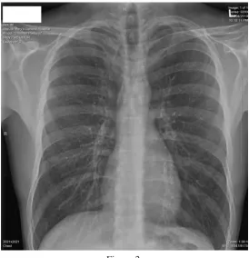
Figure 2.
Two weeks after tube inserted there is a complete resolution of pneumothorax.
At two weeks follow up radiographs revealed complete resolution of the pneumothorax (Figure 2). The patient has had no recurrences.
Discussion
The pathophysiology of pneumothorax remains unknown. Subpleural blebs and bullae are found at the lung apices at thoracoscopy and on CT scan in up to 90% of cases of PSP, and are thought to play a role.18,19 Pulmonary blebs are small subpleural thin walled air containing spaces, not larger than 1-2cm in diameter; their walls being less than 1 mm think. It’s thought if a bleb ruptures it can allow air to escape into the pleural space. Blebs are also observed in cell apoptosis20 which is a cells self-execution plan to guided rupture. It is theorized that the pleural lining cells are committing apoptosis leading to the creation of spon-taneous pneumothorax. Pulmonary bullae are focal re-gions of emphysema measuring more than 1cm in diam-eter with very thin cell walls.21 The location of the unique
or diffuse sites of air leakage leading to PSP is usually unknown. Distal airway inflammation due to cigarette smoking seems to play a key role. No clinician should miss the opportunity, especially in young people, to en-courage smoking cessation. Most young patients continue to smoke after their first episode of PSP, showing that clinical strategies need to be improved in order to better address the needs of this particular age group.22
increase in the intrapleural pressure such that it exceeds atmospheric pressure for much of the respiratory cycle. As a result, impaired venous return and reduced cardiac output results in the typical features of hypoxaema and haemodynamic compromise.23,24
Any clinical suspicion of pneumothorax must be as-sessed with a stethoscope specifically listening for de-creased or absent breathing sounds on the affected side. A diagnosis of PSP is usually made by expiration chest radi-ography. Standing erect PA and lateral chest radiographs are the mainstay. CT scan is the gold standard in the de-tection of small pneumothoraces and in size estimation.25 It is important to always check for associated rib fracture in all cases of pneumothorax. It is not unusual for patients with PSP to present several days after onset of symptoms because of the relatively minor severity.26
There are two main aims when treating pneumothor-ax; first, insure the trapped air is evacuated and that the lung is re-expanded; secondly, prevent recurrence. In first episodes of PSP observation and simple needle aspiration are established first-line therapies.27 If the PSP is small without significant breathlessness, observation is the treatment of choice. Adequate analgesia and high concentration oxygen therapy without any interventional therapy are usually enough to relieve the patients’ symp-toms.11 Observation is the guideline recommendation from the British Thoracic Society for first line treatment of patients with small closed PSP (<15% of lung size) with minimal symptoms. A 70-80% resolution rate can be achieved in these patients in about 7 weeks.29 Obser-vation alone is inappropriate for breathless patients who require active intervention, needle aspiration or chest tube drainage. If observation is unwarranted, needle aspiration is a more conservative second choice. How-ever, in the case of failed aspiration, a chest tube should be inserted. Within 24hrs of admission patients should be referred to a respiratory specialist since intercostal tube placement can lead to serious complications, even death.30 Patients should be hospitalized while a tube is in chest, at least until stable. Video-assisted Thoracosop-ic Surgery (VATS) is a newer alternative to chest tube drainage and has been shown to be more cost-effective31 with similar result.
Recurrences are common and prevention is thera-peutically challenging.32 The risk of recurrence of PSP is as high as 54% within the first four years, with
iso-lated risk factors including smoking, height and age >60 years.5,9,33 After a first recurrence, the likelihood of subse-quent recurrences increases progressively, up to 62% for a second recurrence and 83% for a third.34 Patients should avoid air travel until full resolution of pneumothorax has been confirmed by chest radiograph. The consequence of a recurrence during air travel may be serious.35 After a pneumothorax scuba diving should be permanently avoided.36 There is no evidence to link recurrence with physical exertion.10 The patient can be advised to return to work and resume normal physical activities once all symptoms have resolved, although it is reasonable to ad-vise that sports that involve extreme exertion and physical contact should be avoided until full resolution.11
To prevent recurrences of SP pleurodesis should be con-sidered.18 Pleurodesis is a medical procedure performed surgically or chemically in which the space between the parietal and visceral pleural is artificially obliterated. It involves the adhesion of the two pleurae producing a dif-fuse pleural symphysis.
If the PSP is small without significant breathlessness, the patients’ chief complaint can be misrepresented as unspecific Thoracic Spine Pain (TSP). This presentation can lead patients to seek chiropractic care for non-specif-ic mechannon-specif-ical TSP as this is a common presentation in clinical chiropractic practice. It is necessary to be clin-ically aware of differentials when an undifferentiated thoracic spine condition is present, as serious cases, such as pneumothorax, cancer and heart and lung disease may require emergency medical care.
In summary, a young tall and thin male who was a smoker, presented to a chiropractor with insidious left sided thorax pain thinking he required manual therapy for a thoracic spine complaint. He was experiencing Primary Spontaneous Pneumothorax. It is imperative that when pneumothorax is suspected that immediate referral be made to the hospital for further investigation and possibly acute emergency management.
chiro-practic researchers focusing on less traditional chiroprac-tic research pursuits, including innovative collaborative research efforts in undifferentiated chest pain.37 There-fore, it appears that an initiative of this nature would be in align with the efforts to move beyond simply the spine and into other primary contact musculoskeletal areas that benefit both the patient and the profession. Successful management of emergency care cases can only emphasize the role the chiropractic profession has in the health care system.
References:
1. Gupta DJ, et al. Epidemiology of pneumothorax in England. Thorax. 2000; 55:666-671.
2. Melton LJ, Hepper NCG, Offord KP. Incidence of spontaneous pneumothorax in Olmsted County, Minnesota: 1950-1974. Am Rev Respir Dis. 1987; 29: 1379-1382.
3. Zarogoulidis P, et al. Pneumothorax from definition to diagnosis and treatment. J Thorac Dis 2014; 69(S4): S372-376.
4. Wait MA, Estrera A. Changing clinical spectrum of spontaneous pneumothorax. Am J Surg. 1992; 164(5): 528-531.
5. Sadikot RT et al. Recurrence of primary spontaneous pneumothorax. Thorax. 1997; 52(9): 805-809. 6. Bense L et al. Smoking and the increased risk of
contracting spontaneous pneumothorax. Chest. 1987;92(6):1009-1112.
7. Jansveld CA, Dijkman JH. Primary spontaneous pneumothorax and smoking. BMJ. 1975; 4:559-560. 8. Withers JN et al. Spontaneous pneumothorax. Suggested
etiology and comparison of treatment methods. Am J Surg.1964; 108:772-776.
9. West JB. Distribution of mechanical stress in the lung, a possible factor in localisation of pulmonary disease. Lancet. 1971; 1:839-841
10. Bense L, Wiman LG, Hedenstierna G. Onset of symptoms in spontaneous pneumothorax: correlations to physical activity. Eur J Respir Dis. 1987; 71(3):181-186. 11. MacDuff A et al. Management of spontaneous
pneumothorax: British Thoracic Society Pleural Guideline 2010. Thorax 2010; 65 Suppl 2:ii18-ii31.
12. Dionne CE et al. Determinants of “return to work in good health” among workers with back pain who consult in primary care settings: a 2-year prospective study. Eur Spine J. 2007(5); 16:641-655.
13. Briggs AM et al. Thoracic spine pain in the general population: Prevalence, incidence and associated factors in children, adolescents and adults. A systematic review. BMC Musculoskeletal Disorders. 2009; 10:77.
14. Edmondston SJ, Singer KP. Thoracic spine: anatomical
and biomechanical considerations for manual therapy. Man Ther. 1997;2(3):132-143.
15. Manchikanti L et al. Prevalence of facet joint pain in chronic spinal pain of cervical, thoracic and lumbar regions. BMC Musculoskeletal Disorders. 2004; 5:15. 16. Young BA et al. Thoracic costotransverse joint
pain patterns: a study in normal volunteers. BMC Musculoskeletal Disorders. 2008; 9:140.
17. Schiller L. Effectiveness of spinal manipulative therapy in the treatment of mechanical thoracic spine pain: a pilot randomized clinical trial. JMPT. 2001;24(6):394-401. 18. Donahue DM et al. Resection of pulmonary blebs and
pleurodesis for spontaneous pneumothorax. Chest. 1993; 104:1767-1769.
19. Lesur O et al. Computed tomography in the etiologic assessment of idiopathic spontaneous pneumothorax. Chest. 1990; 98:341-347.
20. Mills JC et al. Apoptotic membrane blebbing is regulated by myosin light chain phosphorylation. J Cell Biol. 1998; 140(3): 627-636.
21. Stern EJ, Frank MS. CT of the lung in patients with pulmonary emphysema: diagnosis, quantification, and correlation with pathologic and physiologic findings. Am J Roentgenol. 1994; 162(4): 791-798.
22. Smith HJM, Chatrou M, Pstmus PE. The impact of spontaneous pneumothorax and its treatment on the smoking behaviour of young adult smokers. Respir Med. 1998; 91:1132-1136.
23. Light RW. Tension pneumothorax. Intensive Care Med. 1994; 20:468-469.
24. Baumann MH, Sahn SA. Tension pneumothorax: diagnostic and therapeutic pitfalls. Crit Care Med. 1993; 21:177-179.
25. Kelly AM et al. Comparison between two methods for estimating pneumothorax size from chest X-rays. Respir Med. 2006; 100:1356-1359.
26. O’Hara VS. Spontaneous pneumothorax. Milt Med. 1978; 143(1):32-35.
27. Andrivet P et al. Spontaneous pneumothorax. Comparison of thoracic drainage vs immediate or delayed needle aspiration. Chest. 1995; 108(2):335-340.
28. Noppen M et al. Manual aspiration versus chest tube drainage in first episodes of primary spontaneous pneumothorax: a multicenter, prospective, randomized pilot study. Am J Respir Crit Care Med. 2002;
16599):1240-1244.
29. Stradling P, Poole G. Conservative management of spontaneous pneumothorax. Thorax 1966; 21(2):145-149. 30. Chan L et al. Complication rates of tube thoracostomy. Am
J Emerg Med. 1997; 15(4):368-370.
32. Schramel FM, Postmus PE, Vanderschueren RG. Current aspects of spontaneous pneumothorax. Eur Respir J 1997; 10(6):1372-1379.
33. Lippert HL et al. Independent risk factors for cumulative recurrence rate after first spontaneous pneumothorax. Eur Respir J. 1991; 4(3): 324-331.
34. Gobbel W et al. Spontaneous pneumothorax. J Thorac Cardiovasc Surg. 1963; 46:331-345.
35. British Thoracic Society Standards of Care Committee. Managing passengers with respiratory disease planning air travel: British Thoracic Society recommendations. Thorax. 2002; 57(4): 289-304.
36. Ziser A, Väänänen A, Melamed Y. Diving and chronic spontaneous pneumothorax. Chest 1985;87(2):264-265. 37. Donovan J et al. Beyond the spine: A new clinical research