Inpatient-Derived Vital Sign
Parameters Implementation: An
Initiative to Decrease Alarm Burden
Alaina K. Kipps, MD, MS, a Sarah F. Poole, BE (Hons), b Cheryl Slaney, MSN, RN, a Shannon Feehan, MSN, RN, CPN, a Christopher A. Longhurst, MD, MS, c Paul J. Sharek, MD, MPH, a Veena V. Goel, MDdaDepartment of Pediatrics, Stanford Children’s Health, Palo Alto, California; bDepartment of Biomedical Informatics, Stanford University School of Medicine, Stanford, California; Departments of cBiomedical Informatics and Pediatrics, University of California San Diego School of Medicine, San Diego, California; and dDivision of Pediatric Hospital Medicine, Palo Alto Medical Foundation, Sutter Health, Palo Alto, California
Drs Kipps and Goel conceptualized and designed the study, assisted with data analysis and interpretation, drafted the article, and critically reviewed the manuscript; Ms Poole conducted the data analyses and assisted with interpretation and reviewed and revised the manuscript; Ms Slaney and Ms Feehan acquired data, assisted in analysis, and critically revised the manuscript; Drs Longhurst and Sharek conceptualized and designed the study and critically revised the manuscript; and all authors approved the final manuscript as submitted, had full access to all the data in the study, and take responsibility for the integrity of the data and the accuracy of the data analysis. DOI: https:// doi. org/ 10. 1542/ peds. 2016- 2458 Accepted for publication Apr 28, 2017
Address correspondence to Alaina K. Kipps, MD, MS, Department of Pediatrics, Stanford University, 750 Welch Rd Suite #325, Palo Alto, CA 94304. E-mail: [email protected]
PEDIATRICS (ISSN Numbers: Print, 0031-4005; Online, 1098-4275).
Copyright © 2017 by the American Academy of Pediatrics
FINANCIAL DISCLOSURE: The authors have indicated they have no financial relationships relevant to this article to disclose.
In 2013, the Joint Commission
identified alarm fatigue as a significant contributor to adverse patient safety events including death. At this time, it released National Patient Safety Goal NPSG.06.01.01, which mandated the development and implementation of alarm management safety
policies.1–3 Multiple publications have
confirmed the burden of monitor alarms throughout various health care settings and across the age spectrum.4–8 A pediatric hospital
study found 99% of ward clinical
alarms were nonactionable, and nurse response time increased as nonactionable alarm exposure increased, quantifying the concept of “alarm fatigue.”9
A recent systematic review revealed a limited but steadily increasing body of literature relevant to alarm fatigue, with 24 observational studies describing alarm characteristics, but only 8 which describe interventions to reduce alarm frequency.10 One
of the interventional studies was
abstract
OBJECTIVES: To implement data-driven vital sign parameters to reduce bedside monitor alarm burden.
METHODS: Single-center, quality-improvement initiative with historical controls assessing the impact of age-based, inpatient-derived heart rate (HR) and respiratory rate (RR) parameters on a 20-bed acute care ward that serves primarily pediatric cardiology patients. The primary outcome was the number of alarms per monitored bed day (MBD) with the aim to decrease the alarms per MBD. Balancing measures included the frequency of missed rapid response team activations, acute respiratory code events, and cardiorespiratory arrest events in the unit with the new vital sign parameters.
RESULTS: The median number of all cardiorespiratory monitor alarms per MBD decreased by 21% from 52 (baseline period) to 41 (postintervention period) (P < .001). This included a 17% decrease in the median HR alarms (9–7.5 per MBD) and a 53% drop in RR alarms (16.8–8.0 per MBD). There were 57 rapid response team activations, 8 acute respiratory code events, and no cardiorespiratory arrest events after the implementation of the new parameters. An evaluation of HRs and RRs recorded at the time of the event revealed that all patients with HRs and/or RRs out of range per former default parameters would also be out of range with the new parameters. CONCLUSIONS: Implementation of data-driven HR and iteratively derived RR parameters safely decreased the total alarm frequency by 21% in a pediatric acute care unit.
in a pediatric setting, where standardization of the cardiac monitor process resulted in a 78% reduction in alarms per patient day without an increase in acute patient decompensation.11 The
individualized modification of monitor alarm parameters for patients was a notable contributor to the alarm reduction in this study. To date, however, minimal work has been published that could guide the optimization of alarm parameters, particularly in pediatric patients. Bonafide et al12 developed percentile
curves for heart rate (HR) and respiratory rate (RR) in hospitalized children and found that 12% to 54% of HR observations and 32% to 40% of RR observations deviated from commonly accepted vital sign reference ranges. These percentiles were not studied or implemented in a clinical setting, although they were proposed as a potential means to tailor physiologic bedside monitor alarm settings.
Goel et al13 similarly developed
age-based HR and RR percentile curves and retrospectively
demonstrated the potential safety of the implementation of these locally derived vital sign parameters. The aim of this study was to assess whether the implementation of these data-driven HR and RR parameters derived by Goel et al13 at our
institution would safely decrease the number of alarms per monitored bed day (MBD) in a 20-bed acute care unit, with a concurrent analysis of all patient-deterioration events to ensure the safety of the intervention.
METhODS
This was a quality-improvement initiative conducted in 2 phases by using historical controls on the cardiac step-down unit at Lucile Packard Children’s Hospital Stanford to determine the effect of using locally derived HR and RR
parameters for pediatric inpatients on alarms per MBD.
Context
Lucile Packard Children’s Hospital is a 303-bed, quaternary-care pediatric hospital with a 20-bed cardiac step-down unit. This unit houses all cardiac inpatients outside of the ICUs as well as some general pediatric medical and surgical patients. Typically, there is 85% occupancy, with cardiology patients accounting for 80%. Overall, 90% of patients are connected to bedside monitors that continuously measure pulse oxygen saturation (SpO2), HR and heart
rhythm, and RR (Philips IntelliVue; Koninklijke Philips N.V., Amsterdam, Netherlands). Patients have an average length of stay of 8.6 days and are typically on monitors throughout their hospitalization. Monitor alarms (HR and heart rhythm, RR, SpO2, and
blood pressure [BP]) on the unit are frequent, with baseline data revealing an average of 1350 alarms per day, or ∼1 alarm per minute. Alarms are audible at the bedside, the central monitor at the unit’s front desk, and on the pagers worn by a patient’s assigned nurse. There is no telemetry technician to interpret alarms and notify nurses.
Alarm parameters are ordered by providers within the electronic health record (EHR) with values that are autopopulated in order sets for HR, RR, and BP based on patient age. The SpO2 parameters are specified on
admission on the basis of expected saturations for the patient (eg, 75% to 90% for certain cyanotic cardiac lesions), with a default of <92% defined as abnormal. From 2007 through 2014, the vital sign reference ranges in all local EHR order sets were based on values published in 2004 by the National Institutes of Health (NIH) that reflect normal values for pediatric outpatients.14 Because there
were only 3 preprogrammed default settings (“neonatal, ”“pediatric, ” and “adult”) (Table 1) to use, these
ordered parameters relied on nurses to reprogram the monitor alarm limits. During this quality initiative, we learned that nurses often chose one of the 3 defaults and did not consistently reprogram monitors to match ordered parameters, and patients were occasionally on bedside monitors without an order to continuously monitor. The authors also discovered that bedside nurses independently adjusted monitor alarm limits to reduce nuisance alarms.
Patients were included in this analysis if they were admitted to the unit and placed on a continuous cardiorespiratory monitor between May 1, 2014 and December 3, 2015. The preintervention period was between May 1, 2014 and October 25, 2014, and the postintervention consisted of 2 phases: phase 1 was between October 26, 2014 and June 19, 2015 and phase 2 was between June 20, 2015 and December 3, 2015. The Stanford University institutional review board approved this study, which was performed in accordance with the SQUIRE 2.0 guidelines.15
Intervention
Adaptation of Data-Driven, Age-Based HR and RR Parameters
The data-driven HR and RR parameters implemented in phase 1 were adapted from those developed by Goel et al13 at our
institution using vital sign data of hospitalized children. In accordance with their study, in which the fifth and 95th percentile limits were thoughtfully selected for analysis and demonstrated to be safe for implementation, we gained approval from medical, administrative, and patient safety leadership to proceed with pilot testing of these parameters in the cardiac unit.
October 26, 2014 by: (1) revision of the unit’s patient care policy to reflect the new monitor parameters, (2) revision of local electronic order sets to default to the new monitor parameters, and (3) preprogramming of the 10 age-based alarm parameter profiles into all 20 bedside monitors to facilitate an ease of clinical workflow and alignment with policy. Education of medical and nursing staff occurred via staff meetings, unit-specific posters, and distribution of vital sign reference cards to be worn with identification badges. Phase 2 began on June 19, 2015 and involved further modification of the RR alarm parameters based on phase 1 data analysis. BP parameters, arrhythmia, and SpO2 parameters remained the
same during all the phases in this study.
Measures
The primary outcome measure was cardiorespiratory monitor alarms per MBD. An alarm was defined as the audible alert sounded by a bedside monitor when a vital sign was out of range of the monitor’s preset thresholds. For this study, all alarm data (including HR, RR, SpO2, BP, and
arrhythmia alarms) were collected and analyzed from the clinical data warehouse. Patients were considered
to have an MBD if they produced at least one cardiorespiratory monitor alarm of any type during the 24-hour day.
Compliance to the intervention was established by using random convenience sampling of patients to determine if the monitor orders in the EHR aligned with the RR and HR parameters set on each patient’s monitor. If the selected age-based monitor alarm profile matched the orders, the situation was considered “compliant.” This was performed every 1 to 2 weeks with immediate feedback delivered to individual nurses, and more broadly, during nursing huddles to reinforce
education. We did not solicit nursing, patient, or family feedback in a formal way.
Evaluation of each of acute respiratory code (ARC), cardiorespiratory arrest (CRA), and rapid response team (RRT) activation event was performed to determine whether any unintended patient safety consequences resulted from these interventions. The EHR-charted vital signs that corresponded with the time of the event and the documented reason for clinical concern (eg, respiratory distress, hypoxia, and tachyarrhythmia) were collected by chart review.
Analysis
The primary analysis compared the number of cardiorespiratory monitor alarms per MBD in the unit preintervention versus
postimplementation by using a t test with Benjamini-Hochberg correction for multiple comparisons to control for false discovery. Compliance to the intervention was analyzed by using simple percentages. Using vital sign data recorded at the time of each ARC, CRA, and RRT event, we compared the new HR and final RR parameters with former bedside monitor preset parameters to identify out-of-range vitals.
RESULTS
After the implementation of phase 1, analysis of the first 3 months of alarm data revealed that although the median number of HR alarms per MBD fell by 17%, the median number of RR alarms per MBD increased by 75%. Overall, the implementation of data-driven alarm limits had increased the total number of alarms per MBD by 33%. Specifically, the median total number of HR alarms per MBD was 8.0 (a decrease from 9.0 per MBD), and median total RR alarms per MBD was 29.3 (an increase from 16.8 per MBD). After
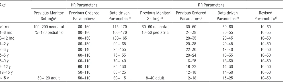
TABLE 1 Data-Driven Vital Sign Parameters for HR and RR Using 10 Age Groups and a Comparison With Former Default Bedside Monitor Presets With 3 Age Groups
Age HR Parameters RR Parameters
Previous Monitor Settingsa
Previous Ordered Parametersb
Data-driven Parametersc
Previous Monitor Settingsa
Previous Ordered Parametersb
Data-driven Parametersc
Revised Parametersd
<1 mo 100–200 neonatal 80–160 115–170 30–60 neonatal 30–60 30–60 10–60
1–6 mo 75–160 pediatric 80–160 105–170 10–50 pediatric 24–38 20–55 10–55
6–12 mo 80–150 100–165 20–35 20–45 10–50
1–2 y 80–150 90–165 20–35 20–45 10–50
2–3 y 80–140 85–155 22–30 18–40 10–50
3–5 y 60–110 75–155 20–24 16–35 10–50
5–9 y 60–110 70–140 16–25 16–30 10–50
9–12 y 60–110 65–130 16–22 14–30 10–50
12–15 y 50–110 60–125 12–18 14–30 10–50
>15 y 50–120 adult 50–110 60–115 8–40 adult 12–18 13–25 10–50
a Preprogrammed within bedside monitors from 2007 to October 2014. b NIH-based vital sign parameters in EHR order sets from 2007 until October 2014.
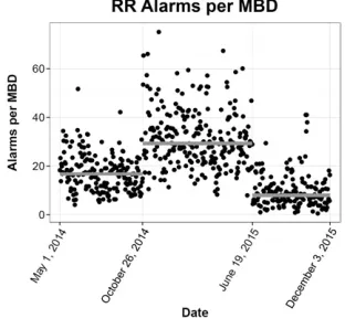
widening RR parameters in phase 2, the frequency of RR alarms fell considerably. Overall, the median number of all monitor alarms per MBD decreased from 52 in the preintervention period to 41 at the end of phase 2, which included the 6 months after the final intervention (P < .001, Fig 1). There was a 17% decrease in median HR alarms (9–7.5 per MBD) and a 53% decrease in RR alarms (16.8–8.0 per MBD). The decrease in alarm frequency for both high and low HR (Fig 2) and high and low RR (Fig 3) were statistically significant (Table 2). Combined, HR and RR alarms fell by 40%. Most of the total cardiorespiratory alarms after phase 2 were from out-of-range SpO2.
Compliance audits that assessed the percentage of patients on continuous monitors with orders accurately corresponding to bedside monitor settings were conducted but not recorded systematically during phase 1. After the completion of phase 2 (from January to March 2016), 6 audits were performed and revealed an average of 80% of bedside monitors were programmed
correctly based on patient age and entered order (range 56–92%). There were zero CRAs, 8 ARCs, and 57 RRT events in the period after the phase 1 intervention through 6 months after the initiation of phase 2 (November 2014 until December 2015). Analysis of HRs and RRs recorded in the EHR within 2 hours before the event when compared with previous bedside monitor default parameters revealed that 18 patients (28%) would have had HR and/or RR out-of-range. When these documented HRs and RRs were compared with intervention parameters after phase 2, 27 patients (42%) had HRs and/ or RRs out-of-range (including all 18 patients who were determined out-of-range with the previous defaults) and an additional 9 patients with HRs
exceeding the new parameters. The 38 patients whose HRs and RRs were within both old and new parameters had other reasons for clinical concern, the most common being low SpO2, acute neurologic events, and hypotension.
DISCUSSION
To our knowledge, this is the first study to demonstrate a significant drop in alarm burden in a pediatric acute care unit after the implementation of HR and RR alarm parameters derived from an inpatient population. Employment of the data-driven HR and iteratively derived RR parameters resulted in a 21% decrease in total alarms from a median of 52 alarms per MBD FIGURE 1
The total alarms (HR, RR, SpO2, and BP) per MBD. A gray line is drawn at the median for each phase of the project, and a red line shows the contributions by HR and RR alarms. Phase 0 (baseline alarm data), phase 1 (initial data-driven HR and RR parameters implementation), and phase 2 (implementation of the same HR parameters and expanded RR parameters) are shown.
FIGURE 2
The HR alarms per MBD. A line is drawn at the median for each phase of the project. Baseline HR data were from May 2014 to October 26, 2014. Phases 1 and 2 both implemented identical data-driven HR parameters.
FIGURE 3
The RR alarms per MBD. A line is drawn at the median for each phase of the project. Phase 0 (baseline), phase 1 (initial data-driven RR parameters implemented), and phase 2 (widening of RR parameters) are shown.
TABLE 2 Comparison of Median Number Alarms With MBD by Alarm Type Before Any
Intervention
After All Interventions Adjusted P (Benjamini-Hochberg) Alarm type
Low HR 1.5 2.2 <.0001
High HR 7.3 4.8 <.0001
Total HR 9.1 7.5 <.0001
Low RR 5.1 2.6 <.0001
High RR 10.0 4.5 <.0001
Total RR 16.8 8.0 <.0001
Totala low alarms 28.3 26.7 .4
Totala high alarms 23.7 13.4 <.0001
Totala alarms 51.5 41.0 <.0001
Low, alarm below set parameter; High, alarm above set parameter.
a Total = HR + RR + SpO
to a median of 41 per MBD. This represents a decrease from 1150 to 840 alarms per 24 hours, or ∼13 fewer alarms in the unit per hour. Our immediate and sustained decrease in HR alarms is consistent with the 12% to 54% decline in out-of-range HR values that was predicted by Bonafide et al12 in
their theoretical model comparing their percentile curves with typical pediatric reference ranges. We did not see this predicted decline in RR alarms during phase 1 because substantial disparities existed between policy and actual nursing practice at the bedside. Although the data-driven RR ranges were wider than the NIH parameters (Table 1), we discovered that the nurses had typically kept the monitor preset RR parameters of 10 to 50 breaths per minute regardless of the order-specified parameters. Thus, despite widening the “notify house officer” orders, the new preprogrammed RR parameters were much narrower than the previous monitor defaults. Reassuringly, there were no patient safety concerns specific to bedside monitor RR parameters during either the preintervention era (2007–2014) or in our safety analysis of our data-driven parameters.13 Our decision
to widen the upper RR limits was further informed by the experience in a pediatric institution in which elevating the upper RR alarm limit significantly (to 200 breaths per minute) contributed to decreased alarm frequency without untoward patient safety consequences.11 Hence,
we felt comfortable implementing widened RR alarm limits in phase 2. The 21% total decrease in alarm burden because of HR, RR, BP, and SpO2 alarms is less than the 43%
decrease in HR and arrhythmia alarms seen in the John Hopkins experience in adult patients16 and
the 56% decrease in HR and RR alarms predicted by Goel et al13
using these parameters compared with the former NIH-based ranges.
However, when HR and RR alarms were isolated from total alarms (specifically, when SpO2, arrhythmia,
and BP alarms are removed), we did witness a 40% decrease (from 25.8 to 15.5 alarms per MBD) in alarm burden, a magnitude that is similar to that predicted by Goel et al13 and is
comparable with the adult experience, 16
both of which excluded SpO2 alarms.
The baseline alarm burden also was already lower than what would have been recorded if the NIH-based orders had been consistently programed at each admission.
In the absence of evidence-based vital sign parameters for the care of hospitalized children, data-driven, age-based HR and RR parameters derived from inpatient populations were a reasonable foundation for a quality-improvement initiative to decrease alarm burden on an acute care unit. Furthermore, iterative modifications to the RR parameters allowed us to safely reduce RR alarm frequency. Similar modifications to the HR parameters were not made because we saw a decline in HR alarm frequency after phase 1; however, this could also be considered in the future. We learned that in conjunction with our single-intervention points (order set implementation and monitor changes), ongoing nursing education and iterative modification of parameters were important to safely decrease alarm burden. We gained a better understanding of local nursing practice and facilitated a better alignment of orders with monitor settings. Schondelmeyer et al17
recently published a study of alarm frequency in a children’s hospital demonstrating that the largest proportion of clinical, nontechnical alarms are due to low SpO2. Similarly,
we found most of our residual alarms after these adjustments to HR and RR were from out-of-range SpO2. Thus,
future alarm reduction efforts may be more impactful if focused on safely reducing saturation alarms.
In addition to demonstrating that iterative adjustments to HR and RR alarm parameters decreases alarm burden in the clinical setting, we have shown that this can be done safely, which is consistent with our retrospective safety analysis.13
Despite widening and shifting HR and RR parameters and employing more specific, preprogramed monitor profiles, 50% more patients who had RRT or ARC events had out-of-range HRs at the time of the event when compared with old, preset parameters. All patients with HRs and RRs in range at the time of clinical concern had other reasons prompting the call for additional resources. This is the first study to address HR- and RR-based alarm burden that has concurrently demonstrated the safety of such an intervention in an inpatient pediatric setting.
There are several limitations to this study. First, most patients admitted to the cardiology acute care ward are transferred from the cardiovascular ICU. However, patients who had spent time in any ICU were excluded in the analysis performed by Goel et al13
REFERENCES
1. The Joint Commission. 2014 National Patient Safety Goal on Clinical Alarm Safety. Oakbrook Terrace, IL: The Joint Commission; 2013
2. The Joint Commission. Sentinel event alert issue 50: medical device alarm safety in hospitals. Available at: https:// www. jointcommission. org/ sea_ issue_ 50/ . Accessed October 12, 2013 3. Alarm-notification problem spotlighted
in Boston globe is all too common.
Health Devices. 2010;39(4):139 4. Gross B, Dahl D, Nielsen L. Physiologic
monitoring alarm load on medical/ surgical floors of a community hospital. Biomed Instrum Technol. 2011;(suppl):29–36
5. Drew BJ, Harris P, Zègre-Hemsey JK, et al. Insights into the problem of alarm fatigue with physiologic monitor devices: a comprehensive observational study of consecutive intensive care unit patients. PLoS One. 2014;9(10):e110274
6. Chambrin MC, Ravaux P, Calvelo-Aros D, Jaborska A, Chopin C, Boniface B. Multicentric study of monitoring alarms in the adult intensive care unit (ICU): a descriptive analysis. Intensive Care Med. 1999;25(12):1360–1366 7. Lawless ST. Crying wolf: false
alarms in a pediatric intensive care unit. Crit Care Med. 1994;22(6): 981–985
8. Görges M, Markewitz BA, Westenskow DR. Improving alarm performance in the medical intensive care unit using delays and clinical context. Anesth Analg. 2009;108(5):1546–1552 9. Bonafide CP, Lin R, Zander M, et al.
Association between exposure to nonactionable physiologic monitor alarms and response time in a children’s hospital. J Hosp Med. 2015;10(6):345–351
10. Paine CW, Goel VV, Ely E, et al. Systematic review of physiologic monitor alarm characteristics and
pragmatic interventions to reduce alarm frequency. J Hosp Med. 2016;11(2):136–144
11. Dandoy CE, Davies SM, Flesch L, et al. A team-based approach to reducing cardiac monitor alarms. Pediatrics. 2014;134(6). Available at: www. pediatrics. org/ cgi/ content/ full/ 134/ 6/ e1686
12. Bonafide CP, Brady PW, Keren R, Conway PH, Marsolo K, Daymont C. Development of heart and respiratory rate percentile curves for hospitalized children. Pediatrics. 2013;131(4). Available at: www. pediatrics. org/ cgi/ content/ full/ 131/ 4/ e1150
13. Goel VV, Poole SF, Longhurst CA, et al. Safety analysis of proposed data-driven physiologic alarm parameters for hospitalized children. J Hosp Med. 2016;11(12):817–823
14. National Institutes of Health. Age-appropriate vital signs. Available at: https:// web. archive. org/ web/ With an average compliance of 80%,
the monitors for these patients at the time of the event may not have been programmed to trigger alarms using the new alarm limits. Also, a cardiorespiratory monitor that produced at least 1 alarm of any type during a 24-hour day counted as an MBD even if that patient was not monitored for the full 24 hours. This would likely bias the results toward the null. Finally, generalizability may be limited, because this was a single time-series study in a quaternary children’s hospital unit that cares predominantly for cardiology patients.
CONCLUSIONS
Implementation of these HR and RR parameters in a children’s hospital acute care unit with predominantly
cardiology patients safely decreased total alarm frequency by 21%. Future opportunities to minimize alarm burden in this population include altering SpO2 alarm ranges, daily
discussions and tailoring settings for each patient to reflect actionable thresholds, and discontinuation of monitoring as soon as medically appropriate.17
ACkNOwLEDGMENTS
We thank all the nurses on the cardiac acute care unit at Lucile Packard Children’s Hospital Stanford, patient care managers Patria Eustaquio, BSN, RN and Piper Church, RN, the unit’s local improvement team, Eric Helfenbein from Phillips, Glen Jones from Stanford Bioengineering and Clinical
Technology, and our hospital’s alarm fatigue task force members (Jonathan Palma, MD; Natalie Pageler, MD; Terry Platchek, MD; and Amy Chapman, MSN, RN, CNS) for their assistance.
ABBREvIATIONS
ARC: acute respiratory code BP: blood pressure
CRA: cardiorespiratory arrest EHR: electronic health record HR: heart rate
MBD: monitored bed day NIH: National Institutes of
Health
RR: respiratory rate RRT: rapid response team SpO2: pulse oxygen saturation
FUNDING: No funding was specifically secured for this study. Ms Poole is supported by the Stanford Biosciences Graduate Program through a Fulbright New Zealand Science and Innovation Graduate Award and through the J.R. Templin Trust Scholarship. These organizations did not have a role in the design, implementation, interpretation, or reporting of this initiative.
20041101222327/ www. cc. nih. gov/ ccc/ pedweb/ pedsstaff/ age. html. Accessed July 26, 2015
15. Ogrinc G, Mooney SE, Estrada C, et al. The SQUIRE (Standards for Quality Improvement Reporting Excellence) guidelines for quality improvement
reporting: explanation and elaboration.
Qual Saf Health Care. 2008;17(suppl 1):i13–i32
16. Graham KC, Cvach M. Monitor alarm fatigue: standardizing use of physiological monitors and decreasing nuisance alarms.
Am J Crit Care. 2010;19(1):28–34, quiz 35
DOI: 10.1542/peds.2016-2458 originally published online July 7, 2017;
2017;140;
Pediatrics
Longhurst, Paul J. Sharek and Veena V. Goel
Alaina K. Kipps, Sarah F. Poole, Cheryl Slaney, Shannon Feehan, Christopher A.
Decrease Alarm Burden
Inpatient-Derived Vital Sign Parameters Implementation: An Initiative to
Services
Updated Information &
http://pediatrics.aappublications.org/content/140/2/e20162458
including high resolution figures, can be found at:
References
http://pediatrics.aappublications.org/content/140/2/e20162458#BIBL
This article cites 14 articles, 3 of which you can access for free at:
Subspecialty Collections
http://www.aappublications.org/cgi/collection/hospital_medicine_sub Hospital Medicine
sub
http://www.aappublications.org/cgi/collection/quality_improvement_ Quality Improvement
_management_sub
http://www.aappublications.org/cgi/collection/administration:practice Administration/Practice Management
following collection(s):
This article, along with others on similar topics, appears in the
Permissions & Licensing
http://www.aappublications.org/site/misc/Permissions.xhtml
in its entirety can be found online at:
Information about reproducing this article in parts (figures, tables) or
Reprints
http://www.aappublications.org/site/misc/reprints.xhtml
DOI: 10.1542/peds.2016-2458 originally published online July 7, 2017;
2017;140;
Pediatrics
Longhurst, Paul J. Sharek and Veena V. Goel
Alaina K. Kipps, Sarah F. Poole, Cheryl Slaney, Shannon Feehan, Christopher A.
Decrease Alarm Burden
Inpatient-Derived Vital Sign Parameters Implementation: An Initiative to
http://pediatrics.aappublications.org/content/140/2/e20162458
located on the World Wide Web at:
The online version of this article, along with updated information and services, is
by the American Academy of Pediatrics. All rights reserved. Print ISSN: 1073-0397.