Research and Reports in Neonatology
Dovepress
R e v i e w
open access to scientific and medical research
Open Access Full Text Article
Neonatal necrotizing enterocolitis
Akhil Maheshwari1
Laura L Corbin2
Robert L Schelonka2
1Division of Neonatology and
Center for Neonatal and Pediatric Gastrointestinal Disease, Department of Pediatrics, University of illinois at Chicago, iL, 2Division of Neonatology,
Department of Pediatrics, Oregon Health and Sciences University, Portland, OR, USA
Correspondence: Akhil Maheshwari Center for Neonatal and Pediatric Gastrointestinal Disease, Children’s Hospital of University of illinois, University of illinois at Chicago, 840 S wood St, CSB 1257, UiC m/c 856, Chicago, iL 60612, USA Tel +1 312 996 4185
Fax +1 312 355 5548 email [email protected]
Abstract: Necrotizing enterocolitis is the most common gastrointestinal emergency in preterm neonates and a major cause of morbidity and mortality in premature infants born before 32 weeks of gestation or with a birth weight less than 1500 g. In this review, we discuss predisposing factors, clinical manifestations, and the quality of evidence for various preventive and therapeutic strategies.
Keywords: necrotizing enterocolitis, inflammation, mucosa, pneumatosis, neonate
Introduction
Necrotizing enterocolitis (NEC), an inflammatory bowel necrosis of infants,1,2 is the
most common gastrointestinal emergency in preterm neonates and a major cause of morbidity and mortality in neonatal intensive care units throughout the world.3,4 In
this review, we discuss pathophysiological factors that may predispose the developing intestine to NEC, describe the clinical manifestations of this disease, and provide a critical appraisal of therapeutic strategies.
Incidence and epidemiology
The incidence of NEC is estimated to be 1–3 per 1000 live births, with more than 90% of all cases occurring in preterm infants.1 NEC occurs in 4%–11% of all premature
infants born with very low birth weight (,1500 g), and the frequency in this subgroup is also inversely related to birth weight and gestational age.5,6 In the National Institute
of Child Health and Development cohort at neonatal research network centers, NEC was recorded in 11.5%, 9%, 6%, and 4% of infants weighing 401–750 g, 751–1000 g, 1001–1250 g, and 1251–1500 g, respectively.7
The incidence of NEC varies significantly between neonatal intensive care units.8–11
Cases occur in each individual neonatal intensive care unit at an “endemic” rate specific for that unit, which may show some seasonal fluctuation and may be punctuated by minor epidemics.7,12–15 Although the reasons for these center differences are unclear,
plausible explanation(s) include biological differences in patient populations and distribution of birth weights, infectious milieu in the neonatal intensive care units, and consistency in labeling of cases that recover without requiring significant medical or surgical intervention.15
Despite improvements in neonatal intensive care and increased overall survival of critically ill premature neonates, mortality rates from NEC can reach 50%.5,16,17 Most
Research and Reports in Neonatology downloaded from https://www.dovepress.com/ by 118.70.13.36 on 27-Aug-2020
For personal use only.
Number of times this article has been viewed
This article was published in the following Dove Press journal: Research and Reports in Neonatology
Dovepress
Maheshwari et al
deaths occur in extremely low birth weight infants, who frequently develop severe disease and require surgery.5
Pathophysiology
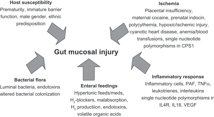
Although the etiopathogenesis of NEC remains unclear, current epidemiological and experimental evidence18,19
identifies several diverse risk factors and supports a multi-factorial model of disease (summarized in Figure 1).
Prematurity is the most important predictor of NEC.
Immaturity of the gastrointestinal tract, particularly in the context of its motility, digestion, perfusion, barrier function, and immune defense, is a major predisposing factor for NEC.20–22 The pathophysiological importance of
prematurity is evident from the near exclusive occurrence of NEC in preterm infants, even though events generally considered to be critical in the pathogenesis of NEC, such as gut mucosal injury, altered barrier function, and bacterial translocation, are recorded frequently in critically ill patients of all ages.23–25
Evidence for genetic predisposition to NEC is modest.
Bhandari et al26 recorded NEC in one or both twins in nine of
63 (14%) pairs of monozygotic twins and in 29 of 189 (15%) pairs of dizygotic twins. After controlling for covariates, genetic factors did not account for any variance in liability for NEC. NEC has been associated with single nucleotide polymorphisms in the interleukin (IL)-4 receptor (+1902G, protective),27 IL-18 (-607A, increased severity),28 vascular
endothelial growth factor (+450C, increased risk),29 and the
carbamoyl-phosphate synthetase 1 genes (T450N, increased
risk).30 In contrast, NEC is not associated with most single
nucleotide polymorphisms that have been linked with Crohn’s disease and/or ulcerative colitis, such as those in the genetic sequences of tumor necrosis factor-alpha (TNF-α), IL-1, IL-4, IL-6, IL-8, and IL-10, CD14, toll-like receptor 4, caspase-recruitment domain 15, and nucleotide-binding oligomerization domain containing 2.31–33
NEC usually occurs in infants who are receiving enteral feedings. Although NEC can occur in neonates who have never been fed, 90%–95% of cases occur in infants with a history of recent volume advancement or reinitiation of enteral feedings.34,35 Besides the risk of direct osmotic
injury to the gut mucosa, feedings may also alter splanchnic blood flow and increase the risk of ischemic injury in underperfused regions by increasing local oxygen needs. In addition, immaturity of motility and digestion in the developing intestine may leave undigested food in the lumen for prolonged periods, promoting bacterial overgrowth and translocation.36 Products of bacterial fermentation, such as
short chain fatty acids, can also injure the immature gut mucosa.37,38
Infants receiving formula feedings are at increased risk of NEC compared with exclusively breastfed neonates.39–46 Formula lacks both cellular as well as soluble
immunoprotective factors, such as IgA and various natural antimicrobials, and also has a propensity to alter the normal postnatal gut bacterial colonization.47–49 Recent studies
indicate that formula feeding in newborn animals may directly induce inflammatory changes in the gut mucosa.50
Host susceptibility
Prematurity, immature barrier function, male gender, ethnic
predisposition
Bacterial flora
Luminal bacteria, endotoxins altered bacterial colonization
Enteral feedings
Hypertonic feeds/meds, H2-blockers, malabsorption,
H2 productiion, endotoxins,
volatile organic acids
Gut mucosal injury
Inflammatory response
Inflammatory cells, PAF, TNFα, leukotrienes, interleukins single nucleotide polymorphisms in
IL4R, IL18, VEGF
Ischemia
Placental insufficiency, maternal cocaine, prenatal indocin, polycythemia, hypoxic/ischemic injury,
cyanotic heart disease, anemia/blood transfusions, single nucleotide
polymorphisms in CPS1
Figure 1 Current epidemiological and experimental information on necrotizing enterocolitis supports a multifactorial model of disease. Clinical and histopathological features
indicate that tissue ischemia, bacterial flora, a dysregulated inflammatory response, and enteral feedings may contribute to the pathogenesis of necrotizing enterocolitis in
premature infants.
Abbreviations: iL, interleukin; veGF, vascular endothelial growth factor; PAF, plasminogen activating factor; TNFα, tumor necrosis factor alpha.
Research and Reports in Neonatology downloaded from https://www.dovepress.com/ by 118.70.13.36 on 27-Aug-2020
Dovepress Neonatal necrotizing enterocolitis
In spite of a large body of data from physiological and retrospective studies, a direct association between specific feeding regimens and/or the rapidity of advancement of feed volumes and NEC has not been conclusively proven.45,51
Several observational studies have suggested that delaying the introduction of enteral feeds beyond the first few days after birth, and using standardized regimes to increase the volume of feeds by less than about 24 mL/kg body weight each day may be associated with a lower risk of NEC.35,52–56
In the National Institute of Child Health and Development neonatal research network, the incidence of NEC was higher at centers where enteral feeding was introduced earlier and feeding volumes advanced rapidly.57 In a recent
retrospective study from a multihospital system,58 fulminant
NEC characterized by massive bowel necrosis and rapid progression to death within 48 hours was associated with advancement of feedings by more than 20 mL/kg/day and/or an increase in concentration of human milk fortifier within 48 hours before developing NEC. However, the association between aggressive enteral feeding and NEC has not been evident in randomized controlled trials comparing slow versus rapid advancement of feedings.51,59–61 A meta-analysis
based on these studies showed that cautiously advanced enteral feedings are not only safe, but may also reduce other morbidities associated with prematurity.62 Similarly,
current trial data do not provide evidence that delayed introduction of progressive enteral feeds reduces the risk of NEC in very low birth weight infants. In a meta-analysis of five randomized controlled trials (600 infants, delayed introduction of feedings defined as later than 5–7 days after birth, and early introduction as less than 4 days after birth), delayed introduction of feedings did not reduce the risk of NEC (relative risk [RR] 0.89, 95% confidence interval [CI] 0.58–1.37) or all-cause mortality (RR 0.93, 95% CI 0.53–1.64). Infants who had delayed introduction of enteral feeds took significantly longer to establish full enteral feeding (reported median difference 3 days).
Mucosal injury may be an early event. Gut epithelial injury is believed to be an early event in NEC. Although the causes of this initial epithelial injury remain unclear, primary apoptotic and autophagic mechanisms have been invoked.63,64
This disruption of the epithelial barrier is presumed to allow bacterial translocation, which in turn triggers a local inflammatory response.63,65,66
Ischemia may play a role in NEC. Coagulative necrosis, which is typically associated with ischemia, is a prominent histopathological finding in NEC.67–69 The predilection
for the ileocecal region, a watershed area supplied by
end-arteries,70 also indicates that ischemia may be an
important pathophysiological event in NEC. Infants with NEC may have decreased endothelial nitric oxide synthase activity and decreased arteriolar nitric oxide production, which can place the developing intestine at a higher risk of ischemic injury.71 However, this association between
hypoxia-ischemia and NEC has not been clearly established in clinical studies in preterm infants.72 Although minor transient
episodes of hypoxia and/or hypotension are not uncommon in premature neonates, major ischemic events are obvious in only a minority of preterm infants with NEC,6,9,72,73 and tend
to occur early in the neonatal period rather than in postnatal weeks 2–4 when NEC occurs.73,74
In full-term infants, NEC tends to occur at an earlier postnatal age than in preterm infants, and is more obviously associated with factors that may conceivably cause splanchnic hypoperfusion. Many term or near-term infants with NEC have a history of placental insufficiency and absence/reversal of end-diastolic blood flow in the umbilical vessels in utero, perinatal asphyxia, polycythemia, episodes of low cardiac output or clinical shock, and congenital cyanotic heart disease.80,83,84
NEC is characterized by a severe, unregulated inflam-matory response. It is characterized by a prominent leu-kocyte infiltrate comprised of activated macrophages and neutrophils.63,65,75 Human and animal studies have
demon-strated increased tissue expression of TNFα and platelet acti-vating factor (PAF), which may propagate ongoing mucosal injury by triggering a cascade of inflammatory mediators, including IL-1, IL-6, IL-8, IL-10, IL-12, and IL-18.36,76–81
Activation of the complement and coagulation cascades, cytokines, reactive oxygen species, and nitric oxide further amplify the mucosal injury.36 In infants with NEC, increased
expression of PAF may be coupled with reduced levels of PAF acetylhydrolase (the enzyme which degrades PAF), further augmenting its local inflammatory effects.36,77,82,83 In
experimental animals, attempts to regulate the inflammatory response by depletion of neutrophils or by using anti-TNFα
antibodies have successfully reduced the severity of tissue damage.84,85
Bacteria play an essential role in the pathogenesis of NEC. Several lines of evidence emphasize the importance of bacterial flora in the pathogenesis of NEC: NEC occurs only after postnatal bacterial colonization of the gastrointestinal tract; intestinal injury prior to colonization may cause strictures or atresia, but not NEC;86 pneumatosis intestinalis,
the pathognomonic finding in NEC, reflects the entrapment of the gaseous products of bacterial fermentation in affected
Research and Reports in Neonatology downloaded from https://www.dovepress.com/ by 118.70.13.36 on 27-Aug-2020
Dovepress
Maheshwari et al
tissues;87 enterally administered aminoglycosides can reduce
the incidence of NEC;88 and NEC-like lesions do not develop
in germ-free animals.16,89 Bacteria play a key role in NEC
by activating the immune system in the mucosa and causing inflammatory injury.66 Bacterial products such as short chain
fatty acids (acetate, butyrate) can also directly damage the epithelial barrier.38,90
Cases of NEC are often temporally and spatially clustered in neonatal intensive care units, suggesting that NEC may be caused by a transmissible agent.91 However,
most studies, whether based on culture techniques or on polymerase chain reaction amplification of 16S ribosomal RNA,92,93 have failed to consistently implicate a single agent.
Cultures of blood and other sterile fluids from infants with NEC usually yield microorganisms that typically colonize critically ill preterm infants and the neonatal intensive care unit microenvironment. Because these microorganisms are not unique to neonatal intensive care units, the interaction of bacteria and bacterial products with the immature intestine tends to receive greater emphasis in the pathogenesis of NEC than the presence of specific bacterial pathogens.65 However,
some recent studies suggest that early duodenal colonization with specific Enterobacteriaceae and Clostridia may predict later development of NEC.93,94 Similarly, in a polymerase
chain reaction-based comparison of fecal microbiota from preterm infants with and without NEC, Wang et al95 found a
marked reduction in bacterial diversity and also an abnormal pattern of bacterial colonization, with a relative abundance of gammaproteobacteria (which include Enterobacteriaceae and Pseudomonadaceae) and reduced numbers of Firmicutes. The oligoclonality of gut microbiota and the disproportionate representation of Gram-negative bacilli may be related to administration of broad-spectrum antibiotics, delayed or interrupted feedings, and exposure to selected multidrug-resistant nursery flora.96,97 Delayed or altered acquisition of
intestinal microbiota by early prolonged antibiotic treatment increases the risk for NEC. This was shown in a recent cohort analysis of 4039 extremely low birth weight infants. Infants who received at least 4 days of initial empirical antibiotic treatment, with sterile body fluid cultures, had an increased risk of NEC with an odds ratio [OR] of 1.34 (95% CI 1.04–1.73).98 The association of specific bacterial groups
with NEC is intriguing and merits further evaluation.
Immaturity of intestinal barrier function may promote bacterial translocation and increase the risk of NEC. Tight junctions and the glycoprotein mucin layer secreted by goblet cells comprise the structural component of the intestinal barrier, whereas IgA, lysozyme, phospholipase A2, and
antimicrobial peptides, such as defensins and cathelicidins, are components of the biochemical barrier.
Immaturity of Paneth cells, specialized crypt cells that produce natural antimicrobials (such as enteric human defensins 5 and 6) and MD2 (a key component of the lipopolysaccharide receptor complex), has been suggested to contribute to the risk of NEC.99–102 Acute NEC is associated
with a low number of Paneth cells, which show weak immunoreactivity or complete absence of lysozyme.103,104
During recovery from NEC, Paneth cell hyperplasia/ metaplasia has been observed with increased expression of enteric defensins.99,101
Secretory IgA (sIgA) antibodies are an important host defense mechanism, preventing luminal antigens and microorganisms from entering the mucosa. In an adult human subject, 70%–80% of all Ig-producing cells in the body are located in the intestinal mucosa and most of these cells produce IgA.105,106 In contrast, neonates lack IgA immunocytes at birth
and the first sIgA may not appear in mucosal secretions until sometime between postnatal week 2 and 8.107 This deficiency
can be partially offset in breastfed infants by sIgA present in colostrum/milk;108 breastfed infants receive about 0.5–1.0 g/
day of antibodies in milk throughout lactation, which is comparable with the 2.5 g daily production of antibodies by a 65 kg adult, and a source of passive immunity against antigens “seen” by the mother-infant dyad.109 In formula-fed premature
infants, the absence of milk-borne sIgA is a significant immunological disadvantage and a likely contributor to the increased risk of NEC.46
Compared with term infants, premature neonates have increased gut mucosal permeability, and this permeability is even greater in infants who subsequently develop NEC.110
Studies on human tissue samples and in rodent models show that the intestinal epithelium is breached early during NEC through apoptosis or necrosis.63 In this process, excessive
nitric oxide production, either directly or through its reactive nitrogen derivative, peroxynitrite, may accentuate epithelial injury through membrane oxidation, induction of apoptosis, and direct mitochondrial damage.111–113 In
preterm infants, a deficiency in the mucus layer may promote bacterial adherence and increase gut mucosal permeability, predisposing to mucosal injury.114 The developmental
deficiency of epithelial trophic factors, such as the epidermal growth factor and heparin-binding epidermal growth factor-like growth factor, may further increase the risk of injury to the epithelial barrier.115–117
Do red blood cell transfusions predispose to NEC? In convalescing preterm infants, red blood cell transfusions have
Research and Reports in Neonatology downloaded from https://www.dovepress.com/ by 118.70.13.36 on 27-Aug-2020
Dovepress Neonatal necrotizing enterocolitis
been temporally associated with NEC.118,119 Red blood cell
transfusions can dampen the normal postprandial increase in mesenteric blood flow in premature infants, particularly in those with a birth weight ,1250 g.120 Immaturity of vascular
auto-regulation in extremely preterm infants is linked to defects in endothelial nitric oxide synthesis68 and could plausibly explain
a higher risk of mucosal injury following transfusions.
Pathology
The disease is commonly localized to the ileocolic region, although the colon may be frequently involved in term infants.121 Some infants with severe, aggressive disease may
develop total gut necrosis (NEC totalis). The four major histo-pathological findings in NEC are coagulative necrosis, bacte-rial overgrowth, pneumatosis intestinalis, and inflammation.
Clinical features
The presenting signs of NEC are protean and may be insidious in onset or sudden and catastrophic. NEC typically presents at 2–4 weeks after birth, although the onset may be as late as 3 months in some infants.16,72,122 The age of onset of NEC
correlates inversely with gestational age at birth in a nonlinear (log-normal) relationship, where infants born at ,28 weeks’ gestation tend to develop NEC at a disproportionately greater postnatal age than their more mature counterparts.123,124
NEC often presents with nonspecific systemic signs, such as tachycardia, apnea, lethargy, and temperature instability.
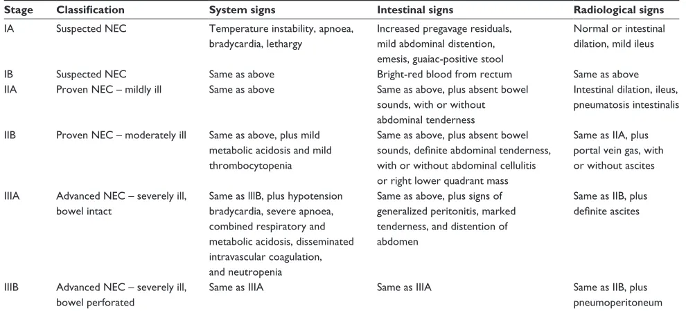
Gastrointestinal signs may include increased prefeed residuals or delayed gastric emptying, emesis, abdominal distention, tenderness, and/or ileus with hypoactive bowel sounds. Grossly bloody stools are seen in approximately 25% of infants. Clinical progression of NEC is commonly staged using the modified Bell’s criteria (Table 1).125 Characteristically, NEC
follows an initial early stage of systemic inflammatory response, followed by a definite stage of localized peritonitis, and finally, an advanced stage of generalized peritonitis.
In a recent study, Clark et al126 described the clinical
characteristics of infants who died of NEC. Compared with 5594 infants who recovered from NEC and were discharged home, there were 1505 infants diagnosed with NEC who died. In multivariate analysis, lower gestational age, lower birth weight, treatment with assisted ventilation on the day of diagnosis of NEC, treatment with vasopressors at the time of diagnosis, and African-American ethnicity were associated with mortality. In another study,58 fulminant
NEC, characterized by massive bowel necrosis and rapid progression to death within 48 hours, was recorded in 7%–10% of all cases and was associated with lower birth weight (1088 ± 545 g versus 1652 ± 817 g), earlier gestational age (27.5 ± 3.3 weeks versus 31.1 ± 4.4 weeks), radiographic evidence of portal venous air, hematocrit ,22%, a history of advancement in feeding volume .20 mL/kg/day, an immature to total neutrophil ratio .0.5, blood lymphocyte count ,4000/µL, and a history of increased concentration of
Table 1 Modified Bell’s staging criteria for necrotizing enterocolitis125
Stage Classification System signs Intestinal signs Radiological signs
iA Suspected NeC Temperature instability, apnoea, bradycardia, lethargy
increased pregavage residuals, mild abdominal distention, emesis, guaiac-positive stool
Normal or intestinal dilation, mild ileus
iB Suspected NeC Same as above Bright-red blood from rectum Same as above
iiA Proven NeC – mildly ill Same as above Same as above, plus absent bowel sounds, with or without abdominal tenderness
intestinal dilation, ileus, pneumatosis intestinalis
iiB Proven NeC – moderately ill Same as above, plus mild metabolic acidosis and mild thrombocytopenia
Same as above, plus absent bowel sounds, definite abdominal tenderness, with or without abdominal cellulitis or right lower quadrant mass
Same as iiA, plus portal vein gas, with or without ascites
iiiA Advanced NeC – severely ill, bowel intact
Same as lllB, plus hypotension bradycardia, severe apnoea, combined respiratory and metabolic acidosis, disseminated intravascular coagulation, and neutropenia
Same as above, plus signs of generalized peritonitis, marked tenderness, and distention of abdomen
Same as iiB, plus definite ascites
iiiB Advanced NeC – severely ill, bowel perforated
Same as iiiA Same as iiiA Same as iiB, plus
pneumoperitoneum Note: This table was published in Pediatric Clinics of North America, vol 33, MC walsh, RM Kliegman, Necrotizing enterocolitis: Treatment based on staging criteria, Pages 179–201, © Copyright elsevier 1986.
Abbreviation: NeC, necrotizing enterocolitis.
Research and Reports in Neonatology downloaded from https://www.dovepress.com/ by 118.70.13.36 on 27-Aug-2020
Dovepress
Maheshwari et al
human milk fortifier within 48 hours before developing NEC. In another study of NEC totalis,127 breast milk feeding was
noted to have a protective effect.
NEC in full-term infants differs from that seen in pre-mature infants. Approximately 10% of infants with NEC are born at term. Unlike preterm infants who develop NEC in the second or third week of life (median 12 days), most term cases are seen within the first week (median 2 days) and often have colonic involvement.121,128,129 NEC in term infants
is usually secondary, associated with conditions such as birth asphyxia, polycythemia, congenital heart disease, rotavirus infections, and Hirschsprung’s disease.121,128–134 Outcomes
are generally better than in preterm neonates, with mortality rates of 0%–13%.121,135,136
Diagnosis
A high index of suspicion in diagnosing at-risk infants is crucial. Most clinical antecedents prior to Bell stage III NEC are nonspecific for gastrointestinal pathology and may not provide sufficient time to the clinician for early institution of treatment measures. In a recent retrospective study, Christensen et al137 reviewed the medical records of
118 infants with stage III NEC. The earliest recognized antecedents of NEC were nonspecific, including apnea/ bradycardia, skin mottling, and irritability, which were first noted at a mean of 2.8 ± 2.1, 4.5 ± 3.1, and 5.4 ± 3.7 hours, respectively, prior to the diagnosis of NEC. The most frequently identified gastrointestinal antecedents were blood in the stools, increased abdominal girth, and elevated prefeeding gastric residuals or emesis, identified 2.0 ± 1.9, 2.8 ± 3.1, and 4.9 ± 4.0 hours before NEC was recognized. No consistent laboratory antecedents were discovered.
Radiographic features remain the mainstay of definitive diagnosis. The pathognomonic sign for NEC is pneumatosis intestinalis (Figure 2).138 These radiolucent shadows have a
bubbly appearance when air is submucosal and become linear when subserosal. Portal venous gas has been associated with a poor prognosis, although this association has recently been questioned.139 Sonographic appearance of portal air is an early
sign. In a prospective cohort study, sonographic portal air had a specificity of 86% for advanced NEC ($stage II), and the sensitivity was lower at 45%.140 It is not known if portal
venous gas visualized by ultrasound or on plain radiographs has the same prognostic significance. Sonographic detection of echoic free fluid and bowel wall thinning may also be more sensitive for intestinal perforation than plain radiography.141
Serial radiographs are invaluable in following the progression of NEC, particularly in the first 48 hours after onset of disease.
Although intestinal perforation may occur within a few hours to as late as 8 days following the onset of NEC,142 more than
two-thirds of all perforations occur within 30–48 hours.143
In some infants who present with bloody stools but minimal systemic signs, pneumatosis may be limited to the colon and may indicate a relatively benign course.144
Most patients with NEC develop leukocytosis and neutrophilia, although neutropenia can occur in advanced disease due to the migration of neutrophils into the peritoneal cavity.145 Blood cultures may grow organisms typically
associated with late-onset sepsis. Thrombocytopenia may occur in stage II and III, and patients with advanced NEC may have evidence of disseminated intravascular coagulation. Breath hydrogen testing was initially heralded as a diagnostic tool, but subsequent studies have shown it as lacking discriminant value.146
The differential diagnosis of NEC includes infections (systemic or intestinal), gastrointestinal obstruction, volvulus, and isolated intestinal perforation. Idiopathic focal intestinal perforations can occur spontaneously or in association with deficiency of the muscularis propria,147 early use of postnatal
corticosteroids alone148 or with indomethacin,149 and with
occult candidal infections.150 Pneumoperitoneum develops
in such patients, but they are usually less ill than those with NEC. Some experts believe that isolated perforations may not be related to NEC.
A B
C D
Figure 2 Abdominal radiographs with characteristic findings of necrotizing
enterocolitis. (A) Diffuse pneumatosis intestinalis with a linear distribution in the left upper and middle quadrants. (B) Characteristic arborization pattern of portal venous gas. (C) Free peritoneal gas with the falciform ligament visible (arrow). (D) Free peritoneal gas seen in a left lateral decubitus view.
Research and Reports in Neonatology downloaded from https://www.dovepress.com/ by 118.70.13.36 on 27-Aug-2020
Dovepress Neonatal necrotizing enterocolitis
Treatment
Medical management
Rapid initiation of therapy is necessary for suspected as well as proven cases of NEC. There is no definitive treatment for established NEC, and therefore treatment is directed at
supportive care and prevention of further injury with cessation of feeding, nasogastric decompression, and administration of intravenous fluids (see Table 2). Infants are usually made nil per os for a variable period of time, depending on the severity of disease. Parenteral antibiotics are widely used for the
Table 2 Treatment and prevention of necrotizing enterocolitis
Therapeutic intervention Current status Evidence level Recommendation level
Treatment Gastric/intestinal
decompression and bowel rest
Provide supportive care and prevent further injury with cessation of feeding, nasogastric decompression, and administration of intravenous fluids. Infants stay nil per os for 3–5 days in stage I, and 10–14 days in stages ii and iii.
iii B
Parenteral antibiotics Broad spectrum antibiotics should be administered based on local antibiotic sensitivity patterns. Anaerobic coverage should be considered in infants with stage iii NeC.
ii-3 C
Primary peritoneal drainage versus exploratory laparotomy
Choices for surgical management in infants with NeC include peritoneal drain placement and exploratory laparotomy. in unstable premature infants with perforated NeC, peritoneal drainage can be cautiously considered as an alternative to exploratory laparotomy, although the best surgical approach in these infants remains unresolved.
i C
Prevention
Antenatal corticosteroids Small beneficial effect of antenatal steroids for reducing risk of NEC. i A Minimal enteral (trophic)
feedings
infants receiving trophic feedings take less time to tolerate full enteral feeds and have a shorter duration of hospital stay, without an effect on the incidence of necrotizing enterocolitis.
i C
Slow advancement of feedings No evidence to suggest that slow advancement of enteral feed volumes reduces the risk of NeC in very low birth weight infants.
i D
Breast milk Although the mechanism of protection is not completely understood, there is strong evidence favoring the use of human milk to reduce the risk of NeC in premature infants.
ii-2 A
Oral immunoglobulins Data from available trials do not support oral administration of immunoglobulin for the prevention of NeC.
i D
enteral antibiotics enteral antibiotic treatment leads to a small reduction in NeC risk; however, increase in antimicrobial-resistant intestinal microbiota precludes routine use of this therapy.
i D
Amino acid supplementation Data are insufficient at present to support supplemental administration of parenteral L-arginine or glutamine to reduce the risk of NeC.
i C
Recombinant cytokines and growth factors
epidermal growth factor is a promising agent in preclinical studies. in early clinical studies, enteral administration of a synthetic amniotic fluid-like solution containing erythropoietin and granulocyte-colony stimulating factor has shown an encouraging safety and efficacy profile.
iii i
Probiotics Probiotics may reduce the risk of severe NeC and related mortality; however, important questions remain regarding optimal choice of agent(s) and dose.
i C
Prebiotics Recent nonhuman animal experimental data suggest that oligofructose prebiotics may be useful in protecting against experimental NeC.
NA* NA*
Notes: *Insufficient human data to determine evidence level or recommendation. Levels of evidence: I, Evidence obtained from at least one properly designed randomized,
controlled trial; ii, evidence obtained from well-designed controlled trials without randomization (ii 1), cohort or case-control analytic studies, (ii 2) evidence obtained from multiple time series with or without the intervention (ii 3); iii, opinions of respected authorities based on clinical experience, descriptive studies, or reports of expert
committees. Levels of recommendations for clinical use: A, Good scientific evidence suggests that the benefits substantially outweigh the potential risks; B, at least fair scientific evidence suggests that the benefits outweigh the potential risks; C, at least fair scientific evidence suggests that there are benefits provided, but the balance between benefits and risks are too close for making general recommendations; D, at least fair scientific evidence suggests that the risks outweigh potential benefits; I, scientific evidence is lacking, of poor quality, or conflicting, such that the risk–benefit balance cannot be assessed.
Abbreviation: NeC, necrotizing enterocolitis.
Research and Reports in Neonatology downloaded from https://www.dovepress.com/ by 118.70.13.36 on 27-Aug-2020
Dovepress
Maheshwari et al
treatment of NEC, but there is surprisingly sparse evidence guiding the choice of antimicrobial agent and duration of therapy. One study comparing alternative treatment regimens that included 90 infants with definite NEC, treated 46 cases with ampicillin and gentamicin, while 44 cases received cefotaxime and vancomycin. Infants $2200 g birthweight had similar outcomes with either regimen. Smaller infants given cefotaxime and vancomycin had a lower risk of culture-positive peritonitis (P= 0.01), and were less likely to die (P= 0.048) or develop thrombocytopenia (P= 0.004). These data suggest that carefully chosen antibiotic regimens can improve the outcome of NEC.151 Antibiotic coverage
for anaerobes should be considered for infants with stage III NEC.
Surgical management
Approximately 20%–40% of patients with pneumatosis intestinalis will require surgical management. Indications for surgery include evidence of perforation seen on abdominal radiographs or positive abdominal paracentesis (stool or organism on Gram stain from peritoneal fluid). Failure of medical management, a single fixed bowel loop on radiographs, abdominal wall erythema, or a palpable mass are all relative indications for surgery. In rare cases, the entire intestine can be involved, precluding surgical intervention. Ideally, surgery should be performed after the development of bowel necrosis, but before perforation and peritonitis occurs.
In unstable premature infants with perforated NEC, peritoneal drainage can be cautiously considered as an alternative to exploratory laparotomy, although the best surgical approach in these infants remains unresolved. In the NECSTEPS trial,152 there was no statistically significant
difference in 90-day survival, dependence on parenteral nutrition, or length of hospital stay in 117 very low birth weight infants randomly assigned to peritoneal drainage or laparotomy. However, other studies have raised important concerns about the routine use of peritoneal drainage. In the NET trial,153 69 extremely low birth weight patients
were randomized to peritoneal drainage or laparotomy, and no significant differences were noted in survival, length of hospital stay, ventilator dependence, or need for parenteral nutrition. However, peritoneal drainage was effective as a definitive treatment in only 4/35 (11%) surviving neonates, and the rest either required a delayed laparotomy (26/34, 74%) or died. In a recent meta-analysis, Rao et al154 reviewed data from the NET and NECSTEPS
trials and detected no significant differences in mortality
within 28 days of peritoneal drainage or laparotomy (28/90 versus 30/95; typical RR 0.99, 95% CI 0.64–1.52; n = 185), mortality by 90 days after the primary procedure (typical RR 1.05, 95% CI 0.71–1.55; n = 185) and the number of infants needing total parenteral nutrition for more than 90 days (typical RR 1.18, 95% CI 0.72–1.95; n = 116). Nearly 50% of infants in the peritoneal drainage group could avoid the need for laparotomy during the study period (44/90 versus 95/96; typical RR 0.49, 95% CI 0.39–0.61; n = 186). One study found that the time to attain full enteral feeds in infants #1000 g was prolonged in the peritoneal drainage group (mean difference 20.77, 95% CI 3.62–37.92).
Although the immediate outcome following peritoneal drainage or laparotomy appears to be similar, there are concerns about the risk of neurodevelopmental impairment following peritoneal drainage. In a recent multicenter trial, peritoneal drainage was associated with increased risk of death or neurodevelopmental impairment.155 A
meta-analysis of three prospective observational studies and two randomized controlled trials suggested a significant excess mortality of 55% associated with peritoneal drainage.156
There is a need for better identification of patients who are less likely to tolerate laparotomy and who may benefit from peritoneal drainage as a temporizing strategy.
Prevention
The existing evidence shows a small beneficial effect of antenatal steroids in reducing the risk of NEC (see Table 2).157
This may be accomplished by accelerating maturation of the gut epithelial barrier and by reducing the overall severity of illness via prevention of lung disease. When analyzed together, eight randomized controlled comparisons of ante-natal corticosteroid administration with placebo or with no treatment, including 1675 infants, showed a risk reduction in NEC of 0.46 (95% CI 0.29–0.74).157 Multiple courses of
antenatal steroids do not appear to reduce the risk of NEC further. In a randomized trial of one to four weekly treat-ments of antenatal steroids or placebo that included 1858 pregnant women, and the outcomes of 2304 infants, the rate of NEC, ie, 1%, was similar in both groups.158
Minimal enteral (trophic) feedings
Initiating feeds by using small amounts of milk or formula may promote the maturation of peristaltic activity and enzymatic systems, release of digestive hormones, and augment intestinal blood flow.3,43,159–162 However, in a
meta-analysis of nine randomized trials including 754
Research and Reports in Neonatology downloaded from https://www.dovepress.com/ by 118.70.13.36 on 27-Aug-2020
Dovepress Neonatal necrotizing enterocolitis
very low birth weight infants,163 early trophic feedings did
not affect feed tolerance, growth rates, or the risk of NEC (RR 1.07, 95% CI 0.67–1.70; risk difference 0.01, 95% CI -0.04–0.05).
Slow advancement of feedings
Meta-analyses of three randomized controlled trials in which a total of 396 infants were included found no significant effects of feeding advancement on the risk of NEC (RR 0.96, 95% CI 0.48–1.92) or all-cause mortality (RR 1.40, 95% CI 0.71–2.80).45 Infants who had slow rates of feed volume
advancement took longer to regain birth weight (median difference 2–5 days) and to establish full enteral feeding (median difference 3–5 days). No statistically significant effect on total duration of hospital stay was detected. The currently available data do not provide evidence that slow advancement of enteral feed volumes reduces the risk of NEC in very low birth weight infants. Of note, few partici-pants were extremely low birth weight or growth restricted, so conclusions about infants at greatest risk for NEC cannot be drawn from the available data.
Breast milk
Experimental and clinical studies show a protective effect of human milk feeds against NEC when compared with formula.39–44,48,164,165 The protective effects of breast milk
against NEC are retained even in pasteurized, banked donor milk. In meta-analysis of data from five randomized trials,46 formula-fed infants were at higher risk of NEC than
infants who received donor milk (RR 2.5, 95% CI 1.2–5.1; risk difference 0.03, 95% CI 0.01–0.06; number needed to harm 33, 95% CI 17–100). More recently, Sullivan et al166
showed that an exclusively human milk-based diet protected extremely premature infants against NEC and surgical NEC when compared with a mother’s milk-based diet that included bovine milk-derived human milk fortifier and preterm formula.
Oral immunoglobulins
Three trials, including a total of 2095 neonates, were reviewed together. Oral administration of IgG or an IgG/IgA combina-tion did not result in a significant reduccombina-tion in incidence of definite NEC (RR 0.84, 95% CI 0.57–1.25), suspected NEC (RR 0.84, 95% CI 0.49–1.46), need for surgery (RR 0.21, 95% CI 0.02–1.75), or death from NEC (RR 1.10, 95% CI 0.47–2.59).49 Based on the available trials, the evidence does
not support the administration of oral immunoglobulin for the prevention of NEC.
enteral antibiotics
To determine the effect of enteral antibiotic prophylaxis and subsequent development of NEC, five randomized controlled trials involving 456 infants were compared. Enteral antibiotic administration resulted in a significant risk reduction for NEC (RR 0.47, CI 0.28–0.78; risk difference -0.10, 0.16 to -0.04); number needed to treat 10 [6–25]). There was a statisti-cally significant reduction in NEC-related deaths (RR 0.32, 0.10–0.96; risk difference -0.07, CI -0.13–0.01) and number needed to treat of 14 (8–100). However, concerns about the development of resistant bacteria remain, and meta-analysis revealed a borderline increase in antimicrobial-resistant intes-tinal microbiota with enteral antibiotic treatment.88
Amino acid supplementation
Nitric oxide augments gastrointestinal perfusion, barrier function, and mucosal repair.167 The supplementation of
L-arginine, a major substrate for nitric oxide production, appears promising in small cohorts in reducing NEC but the data are insufficient at present to support a practice recommendation.168–170 Similarly, glutamine promotes gut
epithelial proliferation and barrier function in animal stud-ies, but a larger multicenter trial of parenteral glutamine supplementation did not show a beneficial effect in reducing the incidence of NEC in preterm infants.171
Probiotics
Probiotics are living microorganisms which, when ingested, can exert a health benefit beyond basic nutrition. Probiotics improve intestinal defense mechanisms, including mucosal IgA secretion, intestinal epithelial cell proliferation, and barrier function, decrease inflammation and epithelial cell apoptosis,172 and may be useful in preventing NEC.173,174
Alfaleh et al175 analyzed 16 eligible trials including
2842 infants.174,176–190 Included trials were highly variable with
regard to enrollment criteria such as birth weight and gesta-tional age, baseline risk of NEC in the control groups, timing, dose, formulation of the probiotics, and feeding regimens. Enteral probiotics significantly reduced the incidence of severe NEC (stage II or more, typical RR 0.35, 95% CI 0.24– 0.52) and mortality (typical RR 0.40, 95% CI 0.27–0.60). Deshpande et al191 selected 11 of these trials174,178–181,184–189
involving 2176 neonates and reported a similar reduction in NEC. The frequency of NEC decreased from 6.56% (71 of 1082) in the control group to 2.37% (26 of 1094) in the probiotics-treated group. Meta-analysis using a fixed-effects model showed reduction in risk of NEC in the probiotics-treated group (RR 0.35, 95% CI 0.23–0.55; P, 0.00001).
Research and Reports in Neonatology downloaded from https://www.dovepress.com/ by 118.70.13.36 on 27-Aug-2020
Dovepress
Maheshwari et al
Only four of these trials reported a significantly higher risk for NEC in the control group.174,179,180,189 The number needed
to treat with probiotics to prevent one case of NEC was 25 (95% CI 17–34). Current evidence indicates that enteral supplementation with probiotics can prevent severe NEC and decrease all-cause mortality in preterm infants. However, further study is needed before routine supplementation using infant formulas with probiotics and also to determine the safety and efficacy of probiotic formulations in extremely low birth weight infants.175,192
Prebiotics
Prebiotics are nondigestible dietary supplements (usually carbohydrates or mucins) which promote proliferation of beneficial commensal bacteria like Lactobacillus and
Bifidobacterium. Recent experimental data suggest that oligofructose prebiotics may be protective against NEC.193–196
Recombinant cytokines
Epidermal growth factor, an important component of gut secretions, human milk, and amniotic fluid, promotes epithelial proliferation, migration, and mucosal repair following injury.197 Oral administration of recombinant
epidermal growth factor protects experimental animals against NEC-like lesions.198 Other studies have similarly evaluated the
role of hematopoietic growth factors, such as erythropoietin and granulocyte-colony stimulating factor. We have recently shown that transforming growth factor-β2 may protect mouse pups against NEC-like injury.66 These cytokines are present in
amniotic fluid and human milk, are swallowed by the fetus in large amounts,199,200 and have a demonstrated an in vitro and
in vivo protective effect on gut mucosa.201–206
Lactoferrin
As an addition to antibiotics, lactoferrin has been considered by some to enhance the response of the immune system when faced with sepsis. Although immune enhancement may play a role in the treatment of NEC, current data do not support the use of lactoferrin as a single agent at this time. Manzoni et al182 randomized 472 very low birth weight
infants to receive either lactoferrin alone or in combination with Lactobacillus rhamnosus GG. Prophylaxis with oral lactoferrin alone did not reduce the incidence of NEC (RR 0.33, 95% CI 0.09–1.17; risk difference -0.04, 95% CI -0.0–0.00), but a significant reduction in NEC was noted when lactoferrin was combined with L. rhamnosus GG (RR 0.05, 95% CI 0.0–0.90; risk difference -0.06, 95% CI -0.10 to -0.02; number needed to treat 17, 95% CI 10–50).
Prognosis
Mortality rates range between 20% and 50%. Approximately 27%–63% of affected infants may require surgery,1,16
and as many as 50% infants may die in the postoperative period.1,15,17 Subacute complications include strictures,
dysmotility, malabsorption, and short gut syndrome.15
Severe NEC has been associated with growth delay that can persist beyond infancy into childhood and poor neurodevelopmental outcome at a corrected gestational age of 18–22 months.207
Summary
Despite advances in the diagnosis and management of many neonatal diseases, NEC remains a devastating condition for many infants. While it is established that very low birth weight infants are at greatest risk for development of NEC, human milk-feeding appears to be the single most effective strategy to reduce, but not eliminate, this disease. Current medical management of NEC is largely supportive and likely does not modify the etiopathogenesis of the disease. Controversies remain regarding optimal surgical management for this condition. Although there are important gaps in our understanding of NEC, future research should focus on prevention of the disease and early recognition that occurs well before the onset of intestinal necrosis.
Acknowledgment
AM is supported by a National Institutes of Health award.
Disclosure
The authors report no conflicts of interest in this work.
References
1. Henry MC, Moss RL. Necrotizing enterocolitis. Annu Rev Med. 2009;60: 111–124.
2. Frost BL, Jilling T, Caplan MS. The importance of pro-inflammatory signaling in neonatal necrotizing enterocolitis. Semin Perinatol. 2008;32(2):100–106.
3. Thompson AM, Bizzarro MJ. Necrotizing enterocolitis in newborns: Pathogenesis, prevention and management. Drugs. 2008;68(9): 1227–1238.
4. Neu J, Walker WA. Necrotizing enterocolitis. N Engl J Med. 2011;364(3): 255–264.
5. Lee JS, Polin RA. Treatment and prevention of necrotizing enterocolitis.
Semin Neonatol. 2003;8(6):449–459.
6. Holman RC, Stoll BJ, Clarke MJ, Glass RI. The epidemiology of necrotizing enterocolitis infant mortality in the United States. Am J
Public Health. 1997;87(12):2026–2031.
7. Guillet R, Stoll BJ, Cotten CM, et al. Association of H2-blocker therapy and higher incidence of necrotizing enterocolitis in very low birth weight infants. Pediatrics. 2006;117(2):e137–e142.
8. Horbar JD, Badger GJ, Carpenter JH, et al. Trends in mortality and morbidity for very low birth weight infants, 1991–1999. Pediatrics. 2002;110(1 Pt 1):143–151.
Research and Reports in Neonatology downloaded from https://www.dovepress.com/ by 118.70.13.36 on 27-Aug-2020
Dovepress Neonatal necrotizing enterocolitis
9. Lemons JA, Bauer CR, Oh W, et al. Very low birth weight outcomes of the National Institute of Child health and human development neonatal research network, January 1995 through December 1996. NICHD Neonatal Research Network. Pediatrics. 2001;107(1):E1.
10. Wiedmeier SE, Henry E, Baer VL, et al. Center differences in NEC within one health-care system may depend on feeding protocol. Am J
Perinatol. 2008;25(1):5–11.
11. Sankaran K, Puckett B, Lee DS, et al. Variations in incidence of necrotizing enterocolitis in Canadian neonatal intensive care units.
J Pediatr Gastroenterol Nutr. 2004;39(4):366–372.
12. Snyder CL, Hall M, Sharma V, St Peter SD. Seasonal variation in the incidence of necrotizing enterocolitis. Pediatr Surg Int. 2010;26(9): 895–898.
13. Guinan M, Schaberg D, Bruhn FW, Richardson CJ, Fox WW. Epidemic occurrence of neonatal necrotizing enterocolitis. Am J Dis Child. 1979; 133(6):594–597.
14. Gerber AR, Hopkins RS, Lauer BA, Curry-Kane AG, Rotbart HA. Increased risk of illness among nursery staff caring for neonates with necrotizing enterocolitis. Pediatr Infect Dis. 1985;4(3):246–249. 15. Stoll BJ. Epidemiology of necrotizing enterocolitis. Clin Perinatol.
1994;21(2):205–218.
16. Lin PW, Stoll BJ. Necrotising enterocolitis. Lancet. 2006;368(9543): 1271–1283.
17. Blakely ML, Lally KP, McDonald S, et al. Postoperative outcomes of extremely low birth-weight infants with necrotizing enterocolitis or isolated intestinal perforation: A prospective cohort study by the NICHD Neonatal Research Network. Ann Surg. 2005;241(6):984–989. 18. Harrell FE. Regression Modeling Strategies with Applications to Linear
Models, Logistic Regression, and Survival Analysis. New York, NY:
Springer-Verlag; 2001.
19. Taylor JM, Ankerst DP, Andridge RR. Validation of biomarker-based risk prediction models. Clin Cancer Res. 2008;14(19):5977–5983. 20. Chandler JC, Hebra A. Necrotizing enterocolitis in infants with very
low birth weight. Semin Pediatr Surg. 2000;9(2):63–72.
21. Snyder CL, Gittes GK, Murphy JP, Sharp RJ, Ashcraft KW, Amoury RA. Survival after necrotizing enterocolitis in infants weighing less than 1,000 g: 25 years’ experience at a single institution. J Pediatr Surg. 1997;32(3):434–437.
22. Rowe MI, Reblock KK, Kurkchubasche AG, Healey PJ. Necrotizing enterocolitis in the extremely low birth weight infant. J Pediatr Surg. 1994;29(8):987–990.
23. Gatt M, Reddy BS, MacFie J. Review article: Bacterial translocation in the critically ill – evidence and methods of prevention. Aliment
Pharmacol Ther. 2007;25(7):741–757.
24. MacFie J, Reddy BS, Gatt M, Jain PK, Sowdi R, Mitchell CJ. Bacterial translocation studied in 927 patients over 13 years. Br J Surg. 2006;93(1):87–93.
25. Stechmiller JK, Treloar D, Allen N. Gut dysfunction in critically ill patients: A review of the literature. Am J Crit Care. 1997;6(3):204–209. 26. Bhandari V, Bizzarro MJ, Shetty A, et al. Familial and genetic
susceptibility to major neonatal morbidities in preterm twins. Pediatrics. 2006;117(6):1901–1906.
27. Treszl A, Heninger E, Kalman A, Schuler A, Tulassay T, Vasarhelyi B. Lower prevalence of IL-4 receptor alpha-chain gene G variant in very-low-birth-weight infants with necrotizing enterocolitis. J Pediatr Surg. 2003;38(9):1374–1378.
28. Heninger E, Treszl A, Kocsis I, Derfalvi B, Tulassay T, Vasarhelyi B. Genetic variants of the interleukin-18 promoter region (-607) influence the course of necrotising enterocolitis in very low birth weight neonates.
Eur J Pediatr. 2002;161(7):410–411.
29. Banyasz I, Bokodi G, Vasarhelyi B, et al. Genetic polymorphisms for vascular endothelial growth factor in perinatal complications. Eur
Cytokine Netw. 2006;17(4):266–270.
30. Moonen RM, Paulussen AD, Souren NY, Kessels AG, Rubio-Gozalbo ME, Villamor E. Carbamoyl phosphate synthetase polymorphisms as a risk factor for necrotizing enterocolitis. Pediatr Res. 2007; 62(2):188–190.
31. Henderson G, Craig S, Baier RJ, Helps N, Brocklehurst P, McGuire W. Cytokine gene polymorphisms in preterm infants with necrotising enterocolitis: Genetic association study. Arch Dis Child Fetal Neonatal Ed. 2009;94(2):F124–F128.
32. Habib Z, Arnaud B, Pascal DL, et al. CARD15/NOD2 is not a predisposing factor for necrotizing enterocolitis. Dig Dis Sci. 2005; 50(9):1684–1687.
33. Szebeni B, Szekeres R, Rusai K, et al. Genetic polymorphisms of CD14, toll-like receptor 4, and caspase-recruitment domain 15 are not associated with necrotizing enterocolitis in very low birth weight infants. J Pediatr Gastroenterol Nutr. 2006;42(1):27–31.
34. Grylack LJ. Neonatal necrotizing enterocolitis revisited. Perinatal
Press. 1986;9:146–148.
35. McKeown RE, Marsh TD, Amarnath U, et al. Role of delayed feeding and of feeding increments in necrotizing enterocolitis. J Pediatr. 1992; 121(5 Pt 1):764–770.
36. Hsueh W, Caplan MS, Qu XW, Tan XD, De Plaen IG, Gonzalez-Crussi F. Neonatal necrotizing enterocolitis: Clinical considerations and pathogenetic concepts. Pediatr Dev Pathol. 2003;6(1):6–23. 37. Di Lorenzo M, Bass J, Krantis A. An intraluminal model of necrotizing
enterocolitis in the developing neonatal piglet. J Pediatr Surg. 1995; 30(8):1138–1142.
38. Nafday SM, Chen W, Peng L, Babyatsky MW, Holzman IR, Lin J. Short-chain fatty acids induce colonic mucosal injury in rats with various postnatal ages. Pediatr Res. 2005;57(2):201–204.
39. Lucas A, Cole TJ. Breast milk and neonatal necrotising enterocolitis.
Lancet. 1990;336(8730):1519–1523.
40. Meinzen-Derr J, Poindexter B, Wrage L, Morrow AL, Stoll B, Donovan EF. Role of human milk in extremely low birth weight infants’ risk of necrotizing enterocolitis or death. J Perinatol. 2009;29(1): 57–62.
41. Sisk PM, Lovelady CA, Dillard RG, Gruber KJ, O’Shea TM. Early human milk feeding is associated with a lower risk of necrotizing enterocolitis in very low birth weight infants. J Perinatol. 2007;27(7): 428–433.
42. Updegrove K. Necrotizing enterocolitis: The evidence for use of human milk in prevention and treatment. J Hum Lact. 2004;20(3):335–339. 43. Sangild PT, Siggers RH, Schmidt M, et al. Diet- and
colonization-dependent intestinal dysfunction predisposes to necrotizing enterocolitis in preterm pigs. Gastroenterology. 2006;130(6):1776–1792. 44. Caplan MS, Amer M, Jilling T. The role of human milk in necrotizing
enterocolitis. Adv Exp Med Biol. 2002;503:83–90.
45. McGuire W, Bombell S. Slow advancement of enteral feed volumes to prevent necrotising enterocolitis in very low birth weight infants.
Cochrane Database Syst Rev. 2008;2:CD001241.
46. Quigley MA, Henderson G, Anthony MY, McGuire W. Formula milk versus donor breast milk for feeding preterm or low birth weight infants.
Cochrane Database Syst Rev. 2007;4:CD002971.
47. Emami CN, Petrosyan M, Giuliani S, et al. Role of the host defense system and intestinal microbial flora in the pathogenesis of necrotizing enterocolitis. Surg Infect (Larchmt). 2009;10(5):407–417.
48. Siggers RH, Siggers J, Thymann T, Boye M, Sangild PT. Nutritional modulation of the gut microbiota and immune system in preterm neonates susceptible to necrotizing enterocolitis. J Nutr Biochem. 2011; 22(6):511–521.
49. Foster J, Cole M. Oral immunoglobulin for preventing necrotizing enterocolitis in preterm and low birth-weight neonates. Cochrane
Database Syst Rev. 2004;1:CD001816.
50. Hang P, Sangild PT, Sit WH, et al. Temporal proteomic analysis of intestine developing necrotizing enterocolitis following enteral formula feeding to preterm pigs. J Proteome Res. 2009;8(1):72–81.
51. Rayyis SF, Ambalavanan N, Wright L, Carlo WA. Randomized trial of “slow” versus “fast” feed advancements on the incidence of necrotizing enterocolitis in very low birth weight infants. J Pediatr. 1999;134(3): 293–297.
52. Brown EG, Sweet AY. Preventing necrotizing enterocolitis in neonates.
JAMA. 1978;240(22):2452–2454.
Research and Reports in Neonatology downloaded from https://www.dovepress.com/ by 118.70.13.36 on 27-Aug-2020
Dovepress
Maheshwari et al
53. Patole S. Safety of enteral feed volumes in neonates at risk for necrotizing enterocolitis: The never-ending story. Pediatrics. 2004;114(1):327. 54. Patole SK, de Klerk N. Impact of standardised feeding regimens on
incidence of neonatal necrotising enterocolitis: A systematic review and meta-analysis of observational studies. Arch Dis Child Fetal Neonatal Ed. 2005;90(2):F147–F151.
55. Premji SS, Chessell L, Paes B, Pinelli J, Jacobson K. A matched cohort study of feeding practice guidelines for infants weighing less than 1,500 g. Adv Neonatal Care. 2002;2(1):27–36.
56. Kuzma-O’Reilly B, Duenas ML, Greecher C, et al. Evaluation, development, and implementation of potentially better practices in neonatal intensive care nutrition. Pediatrics. 2003;111(4 Pt 2): e461–e470.
57. Uauy RD, Fanaroff AA, Korones SB, Phillips EA, Phillips JB, Wright LL. Necrotizing enterocolitis in very low birth weight infants: Biodemographic and clinical correlates. National Institute of Child Health and Human Development Neonatal Research Network. J Pediatr. 1991;119(4):630–638.
58. Lambert DK, Christensen RD, Baer VL, et al. Fulminant necrotizing enterocolitis in a multihospital healthcare system. J Perinatol. May 12, 2011. [Epub ahead of print.]
59. Caple J, Armentrout D, Huseby V, et al. Randomized, controlled trial of slow versus rapid feeding volume advancement in preterm infants.
Pediatrics. 2004;114(6):1597–1600.
60. Salhotra A, Ramji S. Slow versus fast enteral feed advancement in very low birth weight infants: A randomized control trial. Indian Pediatr. 2004;41(5):435–441.
61. Krishnamurthy S, Gupta P, Debnath S, Gomber S. Slow versus rapid enteral feeding advancement in preterm newborn infants 1000–1499 g: A randomized controlled trial. Acta Paediatr. 2010;99(1):42–46. 62. Morgan J, Young L, McGuire W. Slow advancement of enteral feed
volumes to prevent necrotising enterocolitis in very low birth weight infants. Cochrane Database Syst Rev. 2011;3:CD001241.
63. Jilling T, Lu J, Jackson M, Caplan MS. Intestinal epithelial apoptosis initiates gross bowel necrosis in an experimental rat model of neonatal necrotizing enterocolitis. Pediatr Res. 2004;55(4):622–629.
64. Richardson WM, Dai S, Dyer M, et al. Toll like receptor-4 activation links enterocyte autophagy with apoptosis via the stress response gene ATG 16 in the pathogenesis of necrotizing enterocolitis. J Surg Res;158(2):209.
65. Nanthakumar NN, Fusunyan RD, Sanderson I, Walker WA. Inflamma-tion in the developing human intestine: A possible pathophysiologic contribution to necrotizing enterocolitis. Proc Natl Acad Sci U S A. 2000;97(11):6043–6048.
66. Maheshwari A, Kelly DR, Nicola T, et al. TGF-beta(2) suppresses macrophage cytokine production and mucosal inflammatory responses in the developing intestine. Gastroenterology. 2011;140(1):242–253. 67. Nowicki P. Intestinal ischemia and necrotizing enterocolitis. J Pediatr.
1990;117(1 Pt 2):S14–S19.
68. Nowicki PT. Ischemia and necrotizing enterocolitis: Where, when, and how. Semin Pediatr Surg. 2005;14(3):152–158.
69. Nowicki PT, Nankervis CA. The role of the circulation in the pathogenesis of necrotizing enterocolitis. Clin Perinatol. 1994;21(2): 219–234.
70. Rist CB, Watts JC, Lucas RJ. Isolated ischemic necrosis of the cecum in patients with chronic heart disease. Dis Colon Rectum. 1984;27(8): 548–551.
71. Reber KM, Nankervis CA, Nowicki PT. Newborn intestinal circulation. Physiology and pathophysiology. Clin Perinatol. 2002;29(1):23–39. 72. Neu J. The ‘myth’ of asphyxia and hypoxia-ischemia as primary causes
of necrotizing enterocolitis. Biol Neonate. 2005;87(2):97–98. 73. Neu J, Mshvildadze M, Mai V. A roadmap for understanding and
preventing necrotizing enterocolitis. Curr Gastroenterol Rep. 2008; 10(5):450–457.
74. Neu J, Chen M, Beierle E. Intestinal innate immunity: How does it relate to the pathogenesis of necrotizing enterocolitis. Semin Pediatr Surg. 2005;14(3):137–144.
75. Pender SL, Braegger C, Gunther U, Monteleone G, Meuli M, Schuppan G. Matrix metalloproteinases in necrotising enterocolitis.
Pediatr Res. 2003;54(2):160–164.
76. Caplan MS, Sun XM, Hseuh W, Hageman JR. Role of platelet activating factor and tumor necrosis factor-alpha in neonatal necrotizing enterocolitis. J Pediatr. 1990;116(6):960–964.
77. Caplan MS, Simon D, Jilling T. The role of PAF, TLR, and the inflammatory response in neonatal necrotizing enterocolitis. Semin
Pediatr Surg. 2005;14(3):145–151.
78. Viscardi RM, Lyon NH, Sun CC, Hebel JR, Hasday JD. Inflammatory cytokine mRNAs in surgical specimens of necrotizing enterocolitis and normal newborn intestine. Pediatr Pathol Lab Med. 1997;17(4): 547–559.
79. Halpern MD, Holubec H, Dominguez JA, et al. Up-regulation of IL-18 and IL-12 in the ileum of neonatal rats with necrotizing enterocolitis.
Pediatr Res. 2002;51(6):733–739.
80. Halpern MD, Holubec H, Dominguez JA, et al. Hepatic inflammatory mediators contribute to intestinal damage in necrotizing enterocolitis.
Am J Physiol Gastrointest Liver Physiol. 2003;284(4):G695–G6702.
81. Nanthakumar N, Meng D, Goldstein AM, et al. The mechanism of excessive intestinal inflammation in necrotizing enterocolitis: An immature innate immune response. PLoS One. 2011;6(3):e17776. 82. Edelson MB, Bagwell CE, Rozycki HJ. Circulating pro- and
counter-inflammatory cytokine levels and severity in necrotizing enterocolitis.
Pediatrics. 1999;103(4 Pt 1):766–771.
83. Ng PC, Li K, Wong RP, et al. Proinflammatory and anti-inflammatory cytokine responses in preterm infants with systemic infections. Arch
Dis Child Fetal Neonatal Ed. 2003;88(3):F209–F213.
84. Musemeche C, Caplan M, Hsueh W, Sun X, Kelly A. Experimental necrotizing enterocolitis: The role of polymorphonuclear neutrophils.
J Pediatr Surg. 1991;26(9):1047–1049.
85. Halpern MD, Clark JA, Saunders TA, et al. Reduction of experimental necrotizing enterocolitis with anti-TNF-alpha. Am J Physiol Gastrointest
Liver Physiol. 2006;290(4):G757–G764.
86. Hsueh W, Caplan MS, Tan X, MacKendrick W, Gonzalez-Crussi F. Necrotizing enterocolitis of the newborn: Pathogenetic concepts in perspective. Pediatr Dev Pathol. 1998;1(1):2–16.
87. Neu J, Weiss MD. Necrotizing enterocolitis: Pathophysiology and prevention. JPEN J Parenter Enteral Nutr. 1999;23(5 Suppl):S13–S17. 88. Bury RG, Tudehope D. Enteral antibiotics for preventing necrotizing
enterocolitis in low birthweight or preterm infants. Cochrane Database
Syst Rev. 2001;1:CD000405.
89. Chan KL, Ng SP, Chan KW, Wo YH, Tam PK. Pathogenesis of neonatal necrotizing enterocolitis: A study of the role of intraluminal pressure, age and bacterial concentration. Pediatr Surg Int. 2003;19(8):573–577. 90. Lin J, Nafday SM, Chauvin SN, et al. Variable effects of short chain fatty
acids and lactic acid in inducing intestinal mucosal injury in newborn rats. J Pediatr Gastroenterol Nutr. 2002;35(4):545–550.
91. Kliegman RM, Walker WA, Yolken RH. Necrotizing enterocolitis: Research agenda for a disease of unknown etiology and pathogenesis.
Pediatr Res. 1993;34(6):701–708.
92. Millar MR, Linton CJ, Cade A, Glancy D, Hall M, Jalal H. Application of 16S rRNA gene PCR to study bowel flora of preterm infants with and without necrotizing enterocolitis. J Clin Microbiol. 1996;34(10): 2506–2510.
93. Hoy CM, Wood CM, Hawkey PM, Puntis JW. Duodenal microflora in very-low-birth-weight neonates and relation to necrotizing enterocolitis.
J Clin Microbiol. 2000;38(12):4539–4547.
94. de la Cochetiere MF, Piloquet H, des Robert C, Darmaun D, Galmiche JP, Roze JC. Early intestinal bacterial colonization and necrotizing enterocolitis in premature infants: The putative role of Clostridium. Pediatr Res. 2004;56(3):366–370.
95. Wang Y, Hoenig JD, Malin KJ, et al. 16S rRNA gene-based analysis of fecal microbiota from preterm infants with and without necrotizing enterocolitis. ISME J. 2009;3(8):944–954.
96. Van Camp JM, Tomaselli V, Coran AG. Bacterial translocation in the neonate. Curr Opin Pediatr. 1994;6(3):327–333.
Research and Reports in Neonatology downloaded from https://www.dovepress.com/ by 118.70.13.36 on 27-Aug-2020