1535-9778/08/$08.00⫹0 doi:10.1128/EC.00089-08
Copyright © 2008, American Society for Microbiology. All Rights Reserved.
Functional Characterization of a Redundant
Plasmodium
TRAP
Family Invasin, TRAP-Like Protein, by Aldolase Binding and
a Genetic Complementation Test
䌤
Kirsten Heiss,
1Hui Nie,
2Sumit Kumar,
2Thomas M. Daly,
2Lawrence W. Bergman,
2* and Kai Matuschewski
1*
Department of Parasitology, Heidelberg University School of Medicine, 69120 Heidelberg, Germany,1and Center for
Molecular Parasitology, Department of Microbiology and Immunology, Drexel University College of Medicine, Philadelphia, Pennsylvania 191292
Received 11 March 2008/Accepted 14 April 2008
Efficient and specific host cell entry is of exquisite importance for intracellular pathogens. Parasites of the phylumApicomplexaare highly motile and actively enter host cells. These functions are mediated by type I transmembrane invasins of the TRAP family that link an extracellular recognition event to the parasite actin-myosin motor machinery. We systematically tested potential parasite invasins for binding to the actin bridging molecule aldolase and complementation of the vital cytoplasmic domain of the sporozoite invasin TRAP. We show that the ookinete invasin CTRP and a novel, structurally related protein, termed TRAP-like protein (TLP), are functional members of the TRAP family. AlthoughTLP is expressed in invasive stages, targeted gene disruption revealed a nonvital role during life cycle progression. This is the first genetic analysis ofTLP, encoding a redundant TRAP family invasin, in the malaria parasite.
The phylumApicomplexaconsists of unicellular eukaryotes, such asPlasmodiumandToxoplasma gondii, that are obligate intracellular parasites in a wide range of invertebrate and ver-tebrate hosts, including humans. Despite vast differences in host range and target cell specificity, these parasites share a unique mechanism of active actin-dependent motility and host cell entry (18, 27). Gliding locomotion and successful host cell entry through simultaneous formation of a parasitophorous vacuole are rapid processes and are thought to be mechanis-tically coupled (21). Both functions are mediated by the par-asite’s actin-myosin motor machinery (6, 22) and additional proteins, including type I transmembrane proteins of the TRAP/MIC2 family of invasins (12, 29, 30), the invasin bridg-ing protein aldolase (4, 14), and the myosin tail interactbridg-ing protein MTIP (3). According to the current model (1, 26), an extracellular recognition event results in connection of the transmembrane invasins to short actin polymers that in turn are rapidly pulled backwards by immobilized motor myosins.
So far, only TRAP-like invasins have been shown to act directly in parasite locomotion and invasion. In two invasive stages of thePlasmodiumparasite, sporozoites and ookinetes, thrombospondin-related anonymous protein (TRAP) and cir-cumsporozoite- and TRAP-related protein (CTRP), respec-tively, fulfill these functions (5, 29, 30). These proteins share a
unifying primary structure, i.e., combinations of two adhesive modules, the von Willebrand factor A-domain (A domain) (37) and the thrombospondin type I repeat (TSR) (33), in their ectodomains, a transmembrane domain (25) and a cytoplasmic tail domain (CTD) (16). Importantly, the CTD of the sporo-zoite invasin TRAP is essential for gliding motility and cell entry of Plasmodium sporozoites, since a carboxy-terminal truncation ofPlasmodium bergheiTRAP resulted in noninva-sive sporozoites. This finding also permitted a functional assay,
and it was demonstrated that the CTD of the T. gondii
tachyzoite invasin MIC2 could rescue the loss-of-function mu-tant (16). This complementation experiment was the first
di-rect proof for a functional TRAP homolog in T. gondii
tachyzoites. However, this reverse-genetics approach has not been extended to other potential members of the TRAP/MIC2 family yet.
Sporozoites cross various biological barriers and migrate over long distances at a relatively high speed (1 to 3m/s) (7, 21). Similarly, ookinetes traverse barriers, such as the peritro-phic membrane and the mosquito midgut, albeit at a consid-erably lower speed (5m/min) (35). In contrast, merozoites, the invasive stage of the pathogenic red blood cell phase, do not display active locomotion on substrates but employ their motor machinery exclusively for entry into host erythrocytes. In these stages, two TSR-containing transmembrane proteins, termedPlasmodiumthrombospondin-related apical merozoite protein (PTRAMP) (31) and merozoite-specific TRAP ho-molog (MTRAP) (2), have been detected recently. Both pro-teins are reportedly essential for parasite survival (2, 31). MTRAP contains a CTD and interacts with aldolase in vitro, whereas PTRAMP lacks the characteristics of the TRAP fam-ily CTD. A direct function during the merozoite invasion pro-cess has not been demonstrated for any protein yet. Additional invasion-related transmembrane proteins exist; they contain
* Corresponding author. Mailing address for L. W. Bergman: Drexel University College of Medicine, Center for Molecular Parasitology, De-partment of Microbiology and Immunology, Philadelphia, PA 19129. Phone: (215) 991-8376. Fax: (215) 848-2271. E-mail: Lawrence.Bergman @DrexelMed.edu. Mailing address for K. Matuschewski: Heidelberg University School of Medicine, Department of Parasitology, Im Neuenheimer Feld 324, 69120 Heidelberg, Germany. Phone: 49-6221-568284. Fax: 49-6221-564643. E-mail: [email protected] -heidelberg.de.
䌤Published ahead of print on 25 April 2008.
1062
on September 8, 2020 by guest
http://ec.asm.org/
TSR domains only, namely, secreted protein with altered thrombospondin repeat (SPATR) (17) and thrombospondin-related sporozoite protein (TRSP/S21) (15, 19). Both proteins lack the CTD, and none of them have been linked to the parasite actin/myosin motor.
In this study, we established a systematic approach to func-tionally identify TRAP family invasins. Employing two com-plementary assays, in vitro binding to the actin-bridging mol-ecule aldolase and genetic complementation of the TRAP CTD, we tested potential candidates for their capacity to in-teract with the actomyosin motor. We show that the ookinete invasin CTRP and one of the last uncharacterized TRAP-like proteins (TLP) (PFF0800w) are functional members of the TRAP family while EBA175 is not.TLPis expressed in mul-tiple invasive stages but has a redundant role during Plasmo-diumlife cycle progression.
MATERIALS AND METHODS
Plasmodiumlife cycle.P. bergheistrain NK65 was maintained in NMRI mice and Sprague/Dawley rats. For mosquito transmission, mice were assayed for a high proportion of differentiated gametocytes and microgametocyte-stage
para-sites capable of exflagellation.Anopheles stephensimosquitoes were allowed to
blood feed for 15 min on anesthetized mice and maintained under a 14-h light/10-h dark cycle at 75% humidity and 20°C. Mosquitoes were dissected at days 10, 14, and 17 to determine infectivity, midgut sporozoite numbers, and salivary gland sporozoite numbers, respectively. To detect liver-stage parasites in
hepatocytes,⬃3⫻104
Huh7 cells were seeded in eight-well chamber slides and
grown to semiconfluency.P. bergheisporozoites were added, incubated for 90
min at 37°C, and washed off. After 42 to 48 h, liver-stage parasites were visualized
using a primary antibody againstP. bergheiheat shock protein 70 (32). For
analysis of gliding motility, salivary gland sporozoites were deposited on bovine serum albumin-coated glass slides and incubated at 37°C. After fixation with 4% paraformaldehyde, sporozoites and deposited trails were visualized using an
anti-P.bergheiCSP antibody. To determine the prepatent period, 10,000
sporo-zoites were injected intravenously into Sprague/Dawley rats and parasitemia detected by daily examination of Giemsa-stained blood films. For natural trans-mission experiments, young Sprague/Dawley rats were infected by five mosquito bites and parasitemia was examined daily.
PbTRAPcytoplasmic tail swapping.The insertion plasmid used for the tail
swap approach contains the 3⬘untranslated region of PbDHFR(pSE02) (7). For
targeting of PbTRAP, a 5⬘- and 3⬘-truncated sequence of theP. berghei TRAP
open reading frame generated by PCR using the primers TRAP⌬5⬘for (5⬘CG
GAATTCCTTAATGGTCAGGAAATTCTTGACG 3⬘; EcoRI site is
under-lined) and TRAP⌬3⬘rev (5⬘TGCTCTAGATTAGGATCCTGCTATAAAATTA
TAACCAACACC 3⬘; XbaI and BamHI sites are underlined; the in-frame stop
codon is in italics) was cloned into pSE02, resulting in the plasmid pKH01/⌬tail.
This strategy introduces a BamHI cloning site prior to a stop codon for subse-quent cloning of the swap fragments. The following cytoplasmic tail domains
were amplified:P.falciparum TRAP,PfTRAPfor (5⬘CGGGATCCGCAGCAA
CACCCTATGCCGGAGAACC 3⬘; BamHI site is underlined) andPfTRAPrev
(5⬘TGCTCTAGATTAATTCCACTCGTTTTCTTCAGG 3⬘; XbaI site is
un-derlined);P. falciparum CTRP,PfCTRPfor (5⬘ACGGATCCGAGCCTCCTCA
TAGTTCTAATATGG 3⬘; BamHI site is underlined) andPfCTRPrev (5⬘AA
GTCTAGAGAATCAGTTCCACATAGGGTCATCCGCG 3⬘; XbaI site is
underlined);P.berghei TLP,PbTLPfor (5⬘CGGGATCCAAAAACAAACAAA
TAATTCCAACTAGC3⬘; BamHI site is underlined) andPbTLPrev (5⬘TGCT
CTAGATCATTTCCATGGAGAATTGTCATTATAATC 3⬘; XbaI site is
un-derlined), andPfEBA175for (5⬘TATGGATCCTCTGAAGGAGTTATGAAT
GAGAATAATG 3⬘; BamHI site is underlined) andPfEBA175rev (5⬘CTTCT
AGAAAAAAATACATCATATCTTAAATTT 3⬘; XbaI site is underlined). The
obtained PCR fragments were cloned via BamHI/XbaI into pKH01, resulting in
the plasmids pKH02/PfTRAP, pKH03/PbTLP, pKH04/PfEBA175, and pNZ009/
PfCTRP, respectively. Transfection was done as described previously (13) with
SpeI-linearized plasmids. Pyrimethamine-resistant parasite populations were cloned by limiting dilution into 15 NMRI mice. Genotyping of the obtained clonal parasite populations was performed by specific PCRs using the
follow-ing primer combinations:PbTRAPfor (5⬘CCCGGATCCATGAAGCTCTT
AGGAAATAG3⬘) andPbTRAPrev (5⬘CCCGGATCCGTTCCAGTCATTA
TCTTC3⬘) for the PbTRAPwild-type (WT) signal,PbTRAPfor and T7rev (5⬘
GTAATACGACTCACTATAGGGC3⬘), as well as Tgfor (5⬘CCCGCACGGA
CGAATCCAGATGG 3⬘) andPbTRAPrev for the integration-specific PCRs.
For Western blotting, extracts of 100,000 midgut sporozoites were separated on a 10% sodium dodecyl sulfate gel and transferred to a nitrocellulose membrane. TRAP and CSP were detected with polyclonal anti-PbTRAP antisera or a mono-clonal anti-PbCSP antibody (29) and horseradish peroxidase-coupled secondary antibodies.
Recombinant protein expression.The carboxy-terminal domains ofP.
falcip-arumTLP, the PfTLP loss-of-function mutant lacking the penultimate
trypto-phan, PfCTRP, and PfEBA175 were expressed as His-tagged fusion proteins
using the pET28aEscherichia coliexpression vector (Novagen). After induction
using isopropyl--D-thioagalactopyranoside, the protein was purified using
ni-trilotriacetic acid-agarose (Qiagen) under native conditions. The eluted protein was used for aldolase binding assays (see below). The carboxy-terminal 45 amino
acids of WTP. bergheiTRAP and two mutants were similarly expressed as
His-tagged fusions in the vector pET28a and purified as described above. A
glutathioneS-transferase (GST) fusion to full-lengthPlasmodium yoeliialdolase
was obtained from Carlos Buscaglia (New York University) and purified using glutathione agarose.
In vitro aldolase binding assay.Binding assays were performed as previously described (4). Briefly, wells of a 96-well microtiter plate (Maxisorp; Nunc) were coated with recombinant His-tagged fusion proteins that contain different TRAP
tail domains at 4.0g/ml in 0.1 M NaHCO3, pH 9.6, overnight at 4°C. Following
blocking with 5% bovine serum albumin in 10.0 mM imidazole-acetate, 50.0 mM
KCl, 0.2% Tween 20, pH 7.6, biotinylated GST-P. yoeliialdolase was added to
the wells in the buffer above at 1.0g/ml. NeutrAvidin-horseradish peroxidase
(Pierce) was then added in the same buffer at a dilution of 1:2,000. Bound aldolase was quantified with the TMB Slow substrate (Pierce), read at 450 nm
following the addition of 1 M H2SO4. Data are from two independent
experi-ments done in duplicate.
RT-PCR.We isolated poly(A)⫹RNA fromP. falciparum(strains HB3 and
3D7) synchronized blood stages andP. bergheiNK65 salivary gland sporozoites
and gradient purified schizonts using oligo(dT) columns (Invitrogen). Reverse
transcription (RT) was performed using the poly(A)⫹RNA as a template for
first-strand cDNA synthesis (Ambion). Control genomic DNA was isolated from
mixedP. falciparumorP. bergheierythrocytic-stage parasites using silica-gel
columns (Qiagen). cDNA or genomic DNA (1.0l) was used for the PCR
amplifications using gene-specific primer sets.
Gradient purification of parasites and quantitative RT-PCR.After 3 days of infection, when the ascending parasitemia averaged between 15% and 25%,
blood was drawn fromP. yoelii-infected mice and fractionated on
Percoll/Redi-Grad density gradients (Amersham Biosciences). Blood was layered on top of
the gradient and centrifuged for 10 min at 10°C. The five gradient fractions were
removed separately, washed with Hanks balanced salt solution, and treated with HEPES buffer solution, and parasitized cells were collected by centrifugation.
After RNase inhibitor was added, cell pellets were frozen immediately at⫺80°
C until further use. Total RNA was isolated using Trizol and a Qiagen RNAeasy
kit. cDNAs were synthesized from 2g of total RNA isolated from each stage
using the Omniscript cDNA synthesis reaction kit (Qiagen). A reference pool was made by adding equal amounts of total RNA from each fraction prior to
cDNA synthesis. Primer pairs were generated forP. yoelii TLPandMSP1using
the Primer3 software (http://primer3.sourceforge.net; Whitehead Institute for Biomedical Research). All reactions were run in duplicate using an ABI Prism 7700 sequencing detection system, and data were analyzed using the Applied Biosystems Sequence Detector (v.1.7) program. Serial dilutions of input refer-ence pool cDNA were used to generate a standard curve for each target gene. The relative expression levels of each target gene in the five different stages were normalized to the expression level of the reference pool for that particular gene. Bar graphs were plotted as log ratios of amounts of cDNA of each stage to the reference pool.
PbTLPgene targeting.For disruption of PbTLP, two fragments were amplified
using primers TLPrepI_for (5⬘GGGGTACCACAAATTAAAGAACAAATCG
AGGG 3⬘; KpnI site is underlined) and TLPrepI_rev (5⬘CCCAAGCTTGAAT
GGCTCTTAATTTGCCAGTCC 3⬘; HindIII site is underlined) for the 863-bp
5⬘fragment and TLPrepII_for (5⬘CGGAATTCGAGCCGCCTCTATTTAAT
ATTGC 3⬘; EcoRI site is underlined) and TLPrepII_rev (5⬘TCCCCGCGGTG
AACCTCCCAATAGACCCATTCC 3⬘; SacII site is underlined) for the 659-bp
3⬘fragment usingP. bergheigenomic DNA as a template. Both fragments were
cloned into theP. bergheitransfection vector b3D.DTH.D, resulting in the
plasmid pKH20. After transfection, recombinant parasite populations were se-lected using pyrimethamine (13). Clonal parasite lines were obtained by limited dilution into 15 recipient NMRI mice. Genotyping of recombinant parasite
on September 8, 2020 by guest
http://ec.asm.org/
populations was performed by PCR with the following primer combinations:
TgPromrev (5⬘ CGCATTATATGAGTTCATTTTACACAATCC 3⬘) with
PbTLPtestfor (5⬘ TTTTGAGAAGGTATAACCCATATTCC 3⬘) (test1) and
Tgfor and PbTLPtestrev (5⬘ TCCCCGCGGAACATCCATATTAAATAAC
ATCG 3⬘) (test2) for successful gene replacement andPbTLPfor_1 (5⬘CGGG
ATCCTAGGTGGTTCTACTAAGG 3⬘) andPbTLPrev_1 (5⬘TGCACTGCAG
TCAATTTTGATCTTTATAATTTTC 3⬘) for the PbTLPWT signal.
For RT-PCR analysis, poly(A)⫹RNA from gradient-purified schizonts of WT
and knockout parasites was isolated and used for cDNA synthesis. For detection
ofTLPtranscripts, two different primer sets were used:PbTLPfor_2 (5⬘CGCG
GATCCCTATTTGATAATATCGATACAGACCC 3⬘) and PbTLPrev_2 (5⬘
TGCTCTAGAAATCTATATCCTTTTTGTCATCCAC 3⬘) and PbTLPfor_2
andPbTLPrev_1. PbMyoA-and PbMSP1-specific primer sets were used as
tran-script controls.
Nucleotide sequence accession number.The nucleotide sequence reported in this paper has been submitted to the GenBank database with the accession number AY484471.
RESULTS
Preselection of parasite invasins by in vitro aldolase bind-ing.We initiated our analysis by prescreening the cytoplasmic CTDs of candidate transmembrane proteins for in vitro bind-ing to aldolase. We coated microtiter plates with recombinant His-tagged polypeptides that correspond to the CTDs of the sporozoite invasin PbTRAP (29), the ookinete-specific protein PfCTRP (5, 30), the candidate merozoite invasin PfEBA-175 (10), and a previously uncharacterized potential PfTRAP-like protein (PFF0800w), termed TRAP-like protein (PfTLP).
Immobilized proteins were tested for binding of aP.yoelii aldolase GST fusion protein (Fig. 1). As negative controls, we included TRAP mutants that lack the penultimate tryptophan or the carboxy-terminal acidic cluster, resulting in nonproduc-tive motility in vivo (16). In good agreement with published data (4, 14), we detected binding of the PbTRAP CTD to aldolase, which was strictly dependent on the presence of the key residues (Fig. 1). Importantly, the CTDs of PfCTRP and PfTLP interacted with aldolase to a similar extent, suggesting that both proteins may function in TRAP-mediated processes. In contrast, the PfEBA175 CTD, which is acidic in nature but lacks the key tryptophan residue (10), failed to bind aldolase. Failure of PfEBA175 to bind to aldolase further substantiates
that the observed in vitro interactions are specific and reflect a shared property of TRAP proteins.
To confirm the relevance of the PfTLP/aldolase interaction, we tested a mutant form of TLP that contained an alanine in place of the penultimate tryptophan (Fig. 1). As expected, aldolase binding was reduced to background levels. Together, these findings suggest that CTRP and TLP might interact with the parasite actomyosin motor machinery.
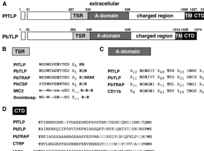
TRAP-like protein.TLP is a type I transmembrane protein (Fig. 2A) that contains in its ectodomain one TSR (Fig. 2B) and an A domain (Fig. 2C) in the reverse order compared to TRAP. Of note, the key residues of A domains are also present in the amino-terminal portion between the signal sequence and the TSR, indicating a potential additional binding motif (data not shown). In its transmembrane domain, TLP contains the signature for a potential rhomboid cleavage site (2, 34), indi-cating that TLP may be processed similarly to TRAP and MIC2. The carboxy-terminal domain of TLP contains the pe-nultimate tryptophan and scattered negatively charged resi-dues (Fig. 2D). In the case of TRAP, both signatures were previously shown to drive sporozoite motility (16). Similarity of the domain architecture and the in vitro aldolase binding sug-gested that TLP belongs to the TRAP family of parasite inva-sins.
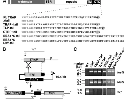
Genetic complementation of the TRAP tail. We next em-ployed a reverse-genetics approach with the rodent malaria model parasiteP. bergheiin order to test whether the candidate proteins can be functionally grouped into the class of TRAP family invasins, as has been shown previously for MIC2 (16). Similar to this strategy, we designed a set of TRAP CTD swap mutants based on the corresponding regions that we tested in the aldolase assay (Fig. 3A). We included the CTDs of PfTRAP, PfCTRP, PbTLP, and PfEBA175. In addition, we generated a potential gain-of-function mutant of PfEBA175 that contains a penultimate tryptophan in order to test whether the acidic carboxy-terminal residues of EBA175 can at least partially interact with the motor machinery.
The TRAP tail fusion genes were introduced into WT par-asites by insertional replacement (Fig. 3B). The targeting plas-mids are predicted to insert by a single-crossover event, result-ing in a 5⬘ functional copy that contains the desired TRAP
fusion protein under the control of the endogenous TRAP
promoter and a 3⬘ copy that lacks the promoter and start codon and hence is not expressed. The successful integration events were confirmed in the clonal parasite populations by insertion-specific PCR amplification (Fig. 3C). The
clonal-swap parasite populations were transmitted to Anopheles
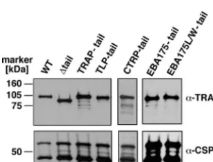
stephensi mosquitoes and assessed for their capacity to form oocysts and viable midgut sporozoites (Table 1). As expected, no differences were observed between the swap parasites. Functional production of midgut sporozoites permitted testing for proper expression of the PbTRAP fusion proteins in West-ern blot analysis (Fig. 4). All parasites produced comparable amounts of the PbTRAP protein.
Phenotypic analysis of the TRAP swap sporozoites enabled us to identify TRAP family members based on functional cri-teria (Table 1). As expected, the mutant that contained a CTD truncation lacks the capacities to invade mosquito salivary glands and to induce a patent blood-stage infection in rats (16).
Replacement of the P. berghei TRAP CTD with the
corre-FIG. 1. In vitro aldolase binding assay to identify TRAP family invasins. Enzyme-linked immunosorbent assay plates coated with His-tagged fusion proteins of either PbTRAP, two PbTRAP loss-of-func-tion mutants lacking the charged residues (trap-acid) or the penulti-mate tryptophan (trap-w/a), PfCTRP, PfTLP, a PfTLP loss-of-function mutant lacking the penultimate tryptophan (tlp-w/a), or PfEBA175 were incubated with a biotinylated GST-P. yoeliialdolase fusion pro-tein. Bound aldolase was quantified in an avidin-substrate assay.
on September 8, 2020 by guest
http://ec.asm.org/
spondingP. falciparumTRAP region resulted in viable sporo-zoites that invade salivary glands comparably to the WT. When tested for infectivity of the mammalian host in vitro or in vivo, PfTRAPsporozoites are as infectious as the WT.
As predicted, the PfCTRPparasites were infectious in the mammalian host in numbers comparable to those of the PfTRAP parasites. Although infectivity of mosquito salivary glands was reduced in PfCTRPparasites, they were evidently capable of invasion (Table 1). Similarly, PbTLP parasites showed a reduced rate of invasion of salivary glands. While liver-stage development in vitro was intermediate between WT parasites and a negative control with a large deletion of the major portion of the tail (⌬tail parasites), inoculation with PbTLPswap mutants consistently resulted in substantial num-bers of mature liver-stage parasites and patent animals when injected in vivo (Table 1).
Notably, both versions of the PfEBA175 CTDs failed to complement the PbTRAP deletion. This finding excludes a potential role of EBA175 in a TRAP-related step during inva-sion by providing a direct link to the parasite motor. Most importantly, the failure of the PfEBA175L/W mutant (contain-ing an additional penultimate tryptophan) to complement the PbTRAP CTD indicates that presence of a penultimate
tryp-tophan and a carboxy-terminal acidic cluster, while necessary, is not sufficient for a direct function in parasite motility. We
conclude that the CTDs ofCTRPandTLPcomplementTRAP
functions, albeit not as well as TRAP. Therefore, both proteins are functional members of the TRAP family in addition to their overall structural relationship.
TLPis expressed in blood-stage parasites and sporozoites.
The functional characterization ofTLPas a third TRAP family member inPlasmodiumprompted us to determine its expres-sion in thePlasmodiumlife cycle. We first tested expression of P. berghei TLPin sporozoites and late-blood-stage parasites by RT-PCR (Fig. 5A). In contrast to PbTRAP(29) and PbCTRP (5, 30), PbTLP is expressed in multiple stages, indicating a shared function between sporozoites and merozoites. We next examined the expression profiling of the P. yoelii ortholog during erythrocytic schizogony by quantitative real-time RT-PCR (Fig. 5B). PyTLPis apparently under stage-specific ex-pression control and highly upregulated in schizonts, the stage preceding infectious merozoites.
To exclude potential minor contamination with sexual blood-stage parasites, we extended our expression analysis to P. falciparumstrains (strains HB3 and 3D7) that both lost their ability to form gametocytes in vitro. We synchronizedP. fal-FIG. 2. Schematic diagram of thePlasmodiumTLP. (A) Representation of the primary structure ofP. falciparumTLP (PFF0800w) andP. berghei TLP (AY484471). Displayed are the extracellular TSR, followed by an A domain, the transmembrane span (TM), and the CTD. (B) Comparison of the TSRs in selected TSR-containing proteins. Shown are the conserved tryptophans, the central dicysteine motif, and the cluster of positive residues. In addition to the TRAP family members TLP and TRAP, TSRs ofP. bergheicircumsporozoite protein (PbCSP),T. gondiimicronemal protein 2 (MIC2), and human thrombospondin (thrombosp.) are shown. (C) Comparison of MIDAS motifs in the A-domain-containing proteins TLP, TRAP, and the integrin CD11b. Invariant residues of the MIDAS are highlighted in bold. (D) TLP has a TRAP family-like cytoplasmic domain (CTD). An amino acid alignment of the putative CTDs ofP. falciparumandP. bergheiTLP,P. bergheiTRAP,P. falciparumCTRP, andT. gondiiMIC2 are shown. The strictly conserved juxtamembrane tyrosine and penultimate tryptophan residues are shown in black, the carboxy-terminal acidic residues in gray.
on September 8, 2020 by guest
http://ec.asm.org/
ciparumparasites and generated total cDNA of highly purified ring, trophozoite, and schizont stages (Fig. 5C). Similar to that of transcripts that function in merozoite invasion (PfAMA1 and PfMSP7[23]), PfTLPexpression commences in schizonts, corroborating our expression data from the rodent parasites. Together our data demonstrate expression ofTLPin multiple invasive stages of rodent and humanPlasmodiumparasites and specific upregulation prior to merozoite invasion.
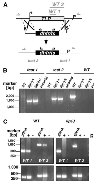
TLPis dispensable forPlasmodiumlife cycle progression.To test whether TLP is important for asexual replication ofP. berghei, we first targeted the PbTLPgene with an integration vector that disrupts the gene locus via a single-crossover event (data not shown). Several attempts to disrupt the gene were not successful, while an integration control that recovered the WTTLPcopy yielded recombinant parasites (data not shown). To distinguish between an essential function and difficulties in targeting the gene, we constructed a replacement vector
con-taining the PbTLP5⬘and 3⬘untranslated regions that flank the positive selection marker cassette (Fig. 6A). Upon a double-crossover event, this vector is predicted to delete the entire PbTLPlocus. After transfection and continuous selection with the antifolate pyrimethamine, we obtained a parental popula-tion that was used for single parasite cloning. Genotyping of two independent clonal parasite lines verified the correct gene replacement event (Fig. 6B). To further confirm the absence of TLPtranscripts in thetlp⫺parasites, we performed RT-PCR of cDNAs generated from blood-stage mutant and WT para-site poly(A)⫹RNA (Fig. 6C). As predicted, noTLPtranscripts were detected in the knockout parasites, while it was readily detectable in WT parasites. The successful generation ofTLP -deficient parasites demonstrates that this gene is not essential during the pathogenic blood-stage cycle in vivo.
We extended our in vivo analysis to the entire parasite life cycle and tested sporozoite formation and maturation in the
FIG. 3. Functional identification of TRAP family members inPlasmodium. (A) Schematic representation of the TRAP tail swap experiments. Shown are the cytoplasmic tails ofP. bergheiTRAP, a negative control with a large deletion of the major portion of the tail (⌬tail), a positive control containing the tail of theP. falciparumTRAP ortholog (TRAP-tail), and the replacement of the TRAP tail with the corresponding region of PbTLP (TLP-tail), PfCTRP (CTRP-tail), PfEBA 175 (EBA175-tail), or a mutant version of PfEBA175 that contains an additional penultimate tryptophan (EBA175L/W-tail). Carboxy-terminal negatively charged residues are shown in bold, and the penultimate tryptophan is boxed. (B) Generation of theTRAPtail mutations by insertional replacement. The WTTRAPgenomic locus is targeted with a SpeI-linearized insertion plasmid containing a 5⬘truncation of theTRAPopen reading frame, the corresponding tail swaps, the 3⬘untranslated region ofDHFR/TS, and thedhfr/tspositive selectable marker. Upon a single-crossover event, the region of homology is duplicated, resulting in a 5⬘copy with the functionalTRAPswap mutant and a nonfunctional 3⬘mutant that lacks the promoter and the start codon. Integration-specific test primer combinations are indicated by arrows, and expected fragments are shown as lines. (C) The successful integration event in the resistant parasite population is confirmed by insertion-specific primer combinations.
on September 8, 2020 by guest
http://ec.asm.org/
Anophelesvector and transmission to the mammalian host (Ta-ble 2). Natural transmission experiments by blood-feeding of tlp⫺-parasite-infectedAnophelesmosquitoes on malaria-naive rats revealed normal completion of the life cycle, indistiguish-able from that of WT parasites. Quantification of sporozoite numbers in midgut-associated oocysts and salivary glands, the final target organ in the mosquito, yielded similar numbers for tlp⫺ and WT parasites. Mature, salivary gland-associated sporozoites displayed continuous gliding locomotion, albeit at a lower frequency, and full in vitro and in vivo infectivity in cultured hepatoma cells and when injected intravenously into rats, respectively. Together, these findings show thatTLPdoes not play an essential role at any stage of the malaria parasite life cycle.
DISCUSSION
The most important physiological function of TRAP family invasins is to drive parasite motility and host cell entry (16). The identification of the merozoite invasin that links the acto-myosin motor machinery to the merozoite surface ligands would set the stage for novel intervention strategies that spe-cifically target merozoite entry into host erythrocytes. Toward the identification of this missing link in thePlasmodiumlife
cycle, we combined two approaches, in vitro binding to aldolase (14) and genetic complementation of the PbTRAPtail (16), which together permit the systematic identification of candidate proteins that link an extracellular recognition event to the intracellular parasite motor complex.
Examination of the P. falciparumgenome database (9) re-vealed the presence of a previously unidentified TRAP-like protein in addition to the sporozoite and ookinete invasins
TRAP and CTRP, respectively. While the Plasmodium
ge-nomes harbors numerous proteins that contain throm-bospondin repeats, such as SPATR (17), TRSP (15, 19), PTRAMP (31), and MTRAP (2), only TLP shares all unifying structural features of TRAP family invasins, i.e., the combined presence of A domains and TSRs in their ectodomain and a conserved cytoplasmic tail domain (Fig. 2A). The TSR con-tains the typical amino-terminal WSxW tetrapeptide and carboxy-terminal basic residues that are separated by two in-variant cysteines (Fig. 2B). A domains contain metal ion-de-pendent adhesion sites (MIDAS) that are implicated in ligand binding (20, 24). Of the invariant residues that coordinate the central divalent cation, the two flanking aspartates and the central threonine are conserved (Fig. 2C). Notably, only one of generally two serines is present in the amino-terminal DxSxS sequence. We also noticed the presence of a second region between amino acids 48 and 267 of PfTLP that might consti-tute an unconventional A domain. Biochemical approaches are required to test the adhesive properties of this region in com-parison to the conventional A domain. In addition to the two well-characterized adhesion modules, TLP contains a sizeable portion, termed “charged region,” between the A domain and the type I transmembrane span that displays an unusually high content of lysine, arginine, glutamic acid, and aspartic acid residues. The carboxy-terminal domain of TLP contains the penultimate tryptophan and a cluster of negatively charged residues (Fig. 2D), which, in the case of TRAP, drive parasite motility (16).
By two independent assays, in vitro aldolase binding of the recombinant CTD and a reverse-genetics strategy, we func-tionally identified TLP as a thirdPlasmodiummember of this protein family. The CTD of TLP carries key residues that are
FIG. 4. Western blot analysis of TRAP tail swap parasites. Midgut sporozoite extracts from 100,000 WT or mutant sporozoites were sep-arated on a 10% SDS gel and probed with polyclonal anti-PbTRAP-repeat serum (␣-TRAP) and monoclonal anti-PbCSP antibodies.
TABLE 1. Phenotypes of TRAP tail swap parasites
Genotype No. of midgut sporozoites/
infected mosquitoa
No. of salivary gland sporozoites/
infected mosquitoa
No. of liver-stage
parasitesb
No. of animals infected (no. positive/no. injected)
Prepatent period,
daysc
WT 24,870 (⫾17,373) 12,271 (⫾5,816) 259 (⫾48) 6/6 3.5
⌬tail 23,514 (⫾6,828) 1,353 (⫾428) 0.9 (⫾0.7) 0/6d
PfTRAP 34,064 (⫾15,707) 16,037 (⫾7,114) 229 (⫾75) 8/8e (4.3)
PfCTRP 23,283 (⫾9,163) 5,872 (⫾2,081) 151 (⫾92) 8/8f (4.2)
PbTLP 24,845 (⫾14,047) 5,314 (⫾3,038) 34.5 (⫾5) 5/5g (4.5)
PfEBA175L 31,715 (⫾18,805) 2,943 (⫾1,256) 0.2 (⫾0.4) 1/10h (6.5)
PfEBA175LW 31,645 (⫾9,622) 3,074 (⫾1,339) 0 0/8d
a
Values are means, with SDs given in parentheses.
b
Liver-stage parasites are total mature exoerythrocytic forms visualized 48 h after incubation of 10,000 salivary gland sporozoites with subconfluent cultured
hepatocytes. Values are means⫾SDs.
c
Prepatent period is the time until the first detection of an erythrocytic-stage parasite in Giemsa-stained blood smears after intravenous injection of 10,000 salivary gland sporozoites. Parentheses indicate that a proportion of parasites contained mixed populations.
d
One animal became patent and was subsequently shown to have completely reverted to the WT locus.
e
One animal contained a mixed population of clonal PfTRAPand residual WT parasites.
f
Two animals contained a mixed population of clonal PfCTRPand residual WT parasites.
g
Three animals contained a mixed population of clonal PbTLPand residual WT parasites.
h
The one animal contained the clonal parasite.
on September 8, 2020 by guest
http://ec.asm.org/
shared by members of the TRAP family. This region can par-tially complementTRAPfunctions during sporozoite invasion of mosquito salivary glands and hepatocytes. Similarly, the CTD of the ookinete invasin CTRP can functionally replace the TRAP tail. Functional assignment of CTRP to the TRAP family was predicted, sincectrp⫺ookinetes no longer traverse the mosquito midgut and do not display productive motility (5, 30). It will be interesting to test whether MTRAP, which was previously shown to likely exert a vital function during in vitro
FIG. 5. Expression ofTLPin invasive stages. (A) RT-PCR from poly(A)⫹RNA of gradient-purified schizonts and salivary gland sporo-zoites ofP. bergheiusing primers specific for PbTLPand, as a positive control,P. bergheimerozoite capping protein 1 (PbMCP1). RT, reverse transcriptase; SPZ., sporozoites; BS, blood stages. (B) Quantitative real-time RT-PCR ofP. yoelii MSP1(white boxes) and PyTLP(gray boxes) using RNA from synchronized ring stages (Rings), early tro-phozoites (8 h) (Early T.), midstage trotro-phozoites (12 h) (Mid T.), late trophozoites (16 h) (Late T.), and schizont (Schiz.) as a template. Change in gene expression levels is shown as mean values (⫾standard deviations) of stage-specific signal divided by the mean signal of the pooled RNA samples (pool). (C) RT-PCR from poly(A)⫹RNA of synchronizedP. falciparumblood stages (ring stages, trophozoites, and schizonts) with two pairs of gene-specific primers for PfTLPand prim-ers for PfAMA1 (apical membrane antigen 1), PfMSP7 (merozoite surface protein 7), and PfACT1(actin 1) as controls.
FIG. 6. Targeted disruption of PbTLP. (A) Replacement strategy to generatetlp⫺parasites. The WTTLPgenomic locus is targeted with a KpnI/SacII-linearized replacement plasmid (pREP) containing 5⬘ and 3⬘untranslated regions adjacent to theTLPopen reading frame and thedhfr/tspositive selectable marker. Upon a double-crossover event, the open reading frame is replaced by the selectable marker. Replacement-specific test and WT primer combinations are indicated by arrows and expected fragments as lines. (B) Replacement-specific PCR analysis. The successful replacement event is verified by primer combination (test 1 and test 2) that can amplify only a signal from the
REPlocus. Absence of the WT signal fromtlp⫺parasites confirms the purity of the clonal population. (C) Absence ofTLPtranscripts intlp⫺
parasites. cDNA from WT ortlp⫺late-blood-stage parasites was am-plified in the presence (⫹) or absence (⫺) of reverse transcriptase (RT) with twoTLP-specific primer combinations (WT1 and WT2). As loading controls, RT-PCRs with myosin A (MyoA)- and merozoite surface protein 1 (MSP1)-specific primers were added. gDNA, wild-type genomic DNA.
on September 8, 2020 by guest
http://ec.asm.org/
growth ofP. falciparumblood stages (2), can complement the TRAP tail. To date, the bridging protein of the motor machin-ery used by the malaria merozoite to propel itself into the host erythrocyte remains unknown, although biochemical evi-dence suggests a potential, specific role for MTRAP in this process (2).
In this study, we provide evidence from independent ap-proaches that TLP is a member of the growing family of TRAP invasins. Although the precise role of aldolase in bridging the CTDs of invasins to microfilaments remains to be determined, in vitro binding to aldolase is a hallmark of members of the TRAP family and a valuable predictor for a role in motility and invasion (4, 14). That efficient interaction of the CTD of TLP with aldolase is relevant in vivo is corroborated by our finding that this region can partially complementTRAPfunctions in sporozoites. Previously it was shown that the TRAP CTD sub-stitutes for the unrelated cytoplasmic domain of EBA-175, one of several members of the erythrocyte binding ligand family (10). Using the TRAP tail swap approach, we establish that EBA-175 does not complement TRAP functions. This finding contrasts with the reverse experiment, i.e., complementation of the EBA175 carboxy-terminal domain by TRAP (10), and raises the interesting possibility that TLP and/or MTRAP and EBA-175 act in concert, probably through TLP/MTRAP-de-pendent recruitment of EBA-175 to the invasion machinery. We can now test this hypothesis by studying biochemical in-teractions between the proteins and by genetic replacement of the cytoplasmic tail encoded byEBA-175with the correspond-ing regions ofTLPand/orMTRAP. Similarly, another mero-zoite ligand, termed PTRAMP, may play a yet-undefined role during invasion (31). This protein contains neither an A do-main nor the CTD of the TRAP family of invasins, and it presumably plays a vital role in blood stages (31).
Based on our data, we propose that TLP interacts with the actin-bridging molecule aldolase in invading-parasite stages and plays a redundant role in linking target cells and parasite ligands to the actin-myosin motor machinery. In analogy to TRAP, the prototype of this family of proteins, TLP secretion may be precisely regulated and may occur only after initial target cell contact (8). The presence ofTLPin all stages tested suggests a conserved, albeit not essential, function that is shared in all life cycle stages. Our expression data are further
supported by the presence of PyTLPin a sporozoite expressed-sequence-tag library (17) and a cDNA library generated from axenic liver stages (36). Indeed, abundant expression of known merozoite ligands in sporozoites has been described for AMA1 and EBA175 (11, 28), suggesting that merozoites and sporo-zoites, which both invade via simultaneous formation of a parasitophorous vacuole, share multiple surface ligands.
Our finding that a previously unrecognized TRAP family member performs a redundant role in parasite life cycle pro-gression suggests that parasite motility and host cell entry are driven by a more complex and partially redundant group of transmembrane and surface proteins than previously antici-pated.
ACKNOWLEDGMENTS
We particularly acknowledge Victor Nussenzweig (NYU) and Louis Miller (NIH) for continuous critical discussions. We thank Na Zhou, Andreas Kunze, Ann-Kristin Mu¨ller, and Markus Ganter for expert assistance.
This work was supported by grants from the NIH (AI48226) to L.W.B. and the research focus “Tropical Medicine Heidelberg,” a junior grant (no. 190/2002) from the Medical Faculty of Heidelberg University, the European Commission (BioMalPar, no. 23), the Joachim Siebeneicher Foundation, and the Chica and Heinz Schaller Foundation to K.M.
REFERENCES
1.Baum, J., A. T. Papenfuss, B. Baum, T. P. Speed, and A. F. Cowman.2006. Regulation of apicomplexan actin-based motility. Nat. Rev. Microbiol.
4:621–628.
2.Baum, J., D. Richard, J. Healer, M. Rug, Z. Krnajski, T.-W. Gilberger, J. L. Green, A. A. Holder, and A. F. Cowman.2006. A conserved molecular motor drives cell invasion and gliding motility across malaria life cycle stages and
other apicomplexan parasites. J. Biol. Chem.281:5197–5208.
3.Bergman, L. W., K. Kaiser, H. Fujioka, I. Coppens, T. M. Daly, S. Fox, K. Matuschewski, V. Nussenzweig, and S. H. I. Kappe.2003. Myosin A tail domain interacting protein (MTIP) localizes to the inner membrane complex ofPlasmodiumsporozoites. J. Cell Sci.116:39–49.
4.Buscaglia, C. A., I. Coppens, W. G. Hol, and V. Nussenzweig.2003. Sites of interaction between aldolase and thrombospondin-related anonymous
pro-tein inPlasmodium.Mol. Biol. Cell14:4947–4957.
5.Dessens, J. T., A. L. Beetsma, G. Dimopoulos, K. Wengelnik, A. Crisanti, F. C. Kafatos, and R. E. Sinden. 1999. CTRP is essential for mosquito
infection by malaria ookinetes. EMBO J.18:6221–6227.
6.Dobrowolski, J. M., and L. D. Sibley.1996.Toxoplasmainvasion of
mam-malian cells is powered by the actin cytoskeleton of the parasite. Cell84:
933–939.
7.Frevert, U., S. Engelmann, S. Zougbe´de´, J. Stange, B. Ng, K. Matuschewski, L. Liebes, and H. Yee.2005. Intravital observation ofPlasmodium berghei
sporozoite infection of the liver. PLoS Biol.3:e192.
TABLE 2. Phenotypes oftlp⫺parasites
Genotype No. of mg sporozoites/
infected mosquitoa No. of sg sporozoites/
infected mosquitob Gliding motilityc No. of liver-stage
parasitesd
No. of animals infected (no.
patent/no.
infected)e
Prepatent periodf
(days)
i.v. By bite i.v. By bite
WT 35,079 (⫾674) 7,970 (⫾5,473) ⫹⫹ 434 (⫾134) 2/2 1/1 4.25 4.5
tlp⫺ 26,997 (⫾22,724) 7,438 (⫾4,631) ⫹ 428 (⫾118) 6/6 2/2 4.3 5.0
a
mg, midgut. Values are means, with SDs given in parentheses.
b
sg, salivary gland. Values are means, with SDs given in parentheses.
c
Gliding motility was visualized by CSP labeling of salivary gland sporozoites on glass slides, and motile sporozoites were counted using a fluorescence microscope.
⫹⫹, continuous, multiple trails for more than 30% of sporozoites;⫹, continuous, multiple trails for at least 10% of sporozoites.
d
Liver-stage parasites are total mature exoerythrocytic forms (mean⫾SD) visualized 48 h after incubation of 10,000 salivary gland sporozoites with subconfluent
cultured hepatocytes.
e
i.v., intravenous injection; by bite, infectious mosquito bites.
f
Prepatent period is the time until the first detection of an erythrocytic-stage parasite in Giemsa-stained blood smears after intravenous (i.v.) injection of 10,000 salivary gland sporozoites or 5 infectious mosquito bites (by bite).
on September 8, 2020 by guest
http://ec.asm.org/
8.Gantt, S., C. Persson, K. Rose, A. J. Birkett, R. Abagayan, and V. Nussen-zweig.2000. Antibodies against TRAP do not inhibitPlasmodiumsporozoite
infectivityin vivo. Infect. Immun.68:3667–3673.
9.Gardner, M. J., N. Hall, E. Fung, O. White, M. Berriman, R. W. Hyman, J. M. Carlton, A. Pain, K. E. Nelson, S. Bowman, I. T. Paulsen, K. James, J. A. Eisen, K. Rutherford, S. L. Salzberg, A. Craig, S. Kyes, M. S. Chan, V. Nene, S. J. Shallom, B. Suh, J. Peterson, S. Anguoli, M. Pertea, J. Allen, J. Selengut, D. Haft, M. W. Mather, A. B. Vaidya, D. M. Martin, A. H. Fair-lamb, M. J. Fraunholz, D. S. Roos, S. A. Ralph, G. I. McFadden, L. M. Cummings, G. M. Subramanian, C. Mungall, J. C. Venter, D. Carucci, S. L. Hoffman, C. Newbold, R. W. Davis, C. M. Fraser, and B. Barrell.2002.
Genome sequence of the human malaria parasitePlasmodium falciparum.
Nature419:498–511.
10.Gilberger, T.-W., J. K. Thompson, M. B. Reed, R. T. Good, and A. F. Cowman.2003. The cytoplasmic domain of thePlasmodium falciparum li-gand EBA-175 is essential for invasion but not protein trafficking. J. Cell
Biol.162:317–327.
11.Gru¨ner, A. C., K. Brahimi, F. Letourneur, L. Renia, W. Eling, G. Snounou, and P. Druilhe. 2001. Expression of erythrocyte-binding antigen 175 in
sporozoites and liver stages ofPlasmodium falciparum.J. Infect. Dis.184:
892–897.
12.Huynh, M. H., and V. B. Carruthers.2006.ToxoplasmaMIC2 is a major
determinant of invasion and virulence. PLoS Pathog.2:e84.
13.Janse, C. J., B. Franke-Fayard, G. R. Mair, J. Ramesar, C. Thiel, S. Engelmann, K. Matuschewski, G. J. van Gemert, R. W. Sauerwein, and A. P. Waters.2006. High efficiency transfection ofPlasmodium bergheifacilitates
novel selection procedures. Mol. Biochem. Parasitol.145:60–70.
14.Jewett, T. J., and L. D. Sibley.2003. Aldolase forms a bridge between cell surface adhesins and the actin cytoskeleton in apicomplexan parasites. Mol.
Cell11:885–894.
15.Kaiser, K., K. Matuschewski, N. Camargo, J. Ross, and S. H. Kappe.2004.
Differential transcriptome profiling identifiesPlasmodiumgenes encoding
pre-erythrocytic stage-specific proteins. Mol. Microbiol.51:1221–1232.
16.Kappe, S., T. Bruderer, S. Gantt, H. Fujioka, V. Nussenzweig, and R. Me´nard.1999. Conservation of a gliding motility and cell invasion machinery
in apicomplexan parasites. J. Cell Biol.147:937–943.
17.Kappe, S. H. I., M. J. Gardner, S. M. Brown, J. Ross, K. Matuschewski, J. M. Ribeiro, J. H. Adams, J. Quakenbusch, J. Cho, D. J. Carucci, S. L. Hoffman, and V. Nussenzweig.2001. Exploring the transcriptome of the malaria
sporo-zoite stage. Proc. Natl. Acad. Sci. USA98:9895–9900.
18.Kappe, S. H. I., C. A. Buscaglia, L. W. Bergman, I. Coppens, and V. Nus-senzweig.2004. Apicomplexan gliding motility and host cell invasion:
over-hauling the motor model. Trends Parasitol.20:13–16.
19.Labaied, M., N. Camargo, and S. H. Kappe.2007. Depletion ofPlasmodium bergheithrombospondin-related sporozoite protein reveals a role in host cell
entry by sporozoites. Mol. Biochem. Parasitol.153:158–166.
20.Lee, J.-O., P. Rieu, M. A. Arnout, and R. Liddington.1995. Crystal structure of the A domain from the alpha subunit of integrin CR3 (CD11b/CD18).
Cell80:631–638.
21.Matuschewski, K., and H. Schu¨ler.2008. Actin/myosin-based gliding motility
in apicomplexan parasites, p. 110–120.InB. Burleigh and D. Soldati-Favre
(ed.), Molecular mechanisms of parasite invasion. Springer, Heidelberg, Germany.
22.Meissner, M., D. Schlu¨ter, and D. Soldati.2002. Role ofToxoplasma gondii
myosin A in powering parasite gliding and host cell invasion. Science298:
837–840.
23.Mello, K., T. M. Daly, C. A. Long, J. M. Burns, and L. W. Bergman.2004. Members of the merozoite surface protein 7 family with similar expression
patterns differ in ability to protect againstPlasmodium yoeliimalaria. Infect.
Immun.72:1010–1018.
24.Michishita, M., V. Videm, and M. A. Arnaout.1993. A novel divalent cation-binding site in the A domain of the b2 integrin CR3 (CD11b/CD18) is
essential for ligand binding. Cell72:857–867.
25.Opitz, C., M. Di Christina, M. Reiss, T. Ruppert, A. Crisanti, and D. Soldati.
2002. Intramembrane cleavage of microneme proteins at the surface of the
apicomplexan parasiteToxoplasma gondii.EMBO J.21:1577–1585.
26.Schu¨ler, H., and K. Matuschewski.2006.Plasmodiummotility: actin not
actin’ like actin. Trends Parasitol.22:146–147.
27.Sibley, L. D.2004. Intracellular parasite invasion strategies. Science304:248– 253.
28.Silvie, O., J.-F. Franetich, S. Charrin, M. S. Mueller, A. Siau, M. Bodescot, E. Rubinstein, L. Hannoun, Y. Charoenvit, C. H. Kocken, A. W. Thomas, G. J. van Gemert, R. W. Sauerwein, M. J. Blackman, R. F. Anders, G. Pluschke, and D. Mazier.2004. A role for apical membrane antigen 1 during
invasion of hepatocytes by Plasmodium falciparum sporozoites. J. Biol.
Chem.279:9490–9496.
29.Sultan, A. A., V. Thathy, U. Frevert, K. J. Robson, A. Crisanti, V. Nussen-zweig, R. S. NussenNussen-zweig, and R. Me´nard.1997. TRAP is necessary for
gliding motility and infectivity ofPlasmodiumsporozoites. Cell90:511–522.
30.Templeton, T. J., D. C. Kaslow, and D. A. Fidock.2000. Developmental
arrest of the human malaria parasite Plasmodium falciparumwithin the
mosquito midgut via CTRP gene disruption. Mol. Microbiol.36:1–9.
31.Thompson, J., R. E. Cooke, S. Moore, L. F. Anderson, C. J. Janse, and A. P. Waters.2004. PTRAMP; a conservedPlasmodiumthrombospondin-related
apical merozoite protein. Mol. Biochem. Parasitol.134:225–232.
32.Tsuji, M., D. Mattei, R. S. Nussenzweig, D. Eichinger, and F. Zavala.1994. Demonstration of heat-shock protein 70 in the sporozoite stage of malaria
parasites. Parasitol. Res.80:16–21.
33.Tucker, R. P.2004. The thrombospondin type 1 repeat superfamily. Int.
J. Biochem. Cell. Biol.36:969–974.
34.Urban, S., and M. Freeman.2003. Substrate specificity of rhomboid in-tramembrane proteases is governed by helix-breaking residues in the
sub-strate transmembrane domain. Mol. Cell11:1425–1434.
35.Vlachou, D., T. Zimmermann, R. Cantera, C. J. Janse, A. P. Waters, and F. Kafatos.2004. Real-time, in vivo analysis of malaria ookinete locomotion
and mosquito midgut invasion. Cell. Microbiol.6:671–685.
36.Wang, Q., S. Brown, D. S. Roos, V. Nussenzweig, and P. Bhanot.2004.
Transcriptome of axenic liver stages ofPlasmodium yoelii.Mol. Biochem.
Parasitol.137:161–168.
37.Whittaker, C. A., and R. O. Hynes.2002. Distribution and evolution of von Willebrand/integrin A domains: widely dispersed domains with roles in cell
adhesion and elsewhere. Mol. Biol. Cell13:3369–3387.