R E S E A R C H
Open Access
PrP
Sc
spreading patterns in the brain of sheep
linked to different prion types
Wiebke M Wemheuer
1, Sylvie L Benestad
2, Arne Wrede
1, Wilhelm E Wemheuer
3, Bertram Brenig
3, Bjørn Bratberg
2,
Walter J Schulz-Schaeffer
1*Abstract
Scrapie in sheep and goats has been known for more than 250 years and belongs nowadays to the so-called prion diseases that also include e.g. bovine spongiform encephalopathy in cattle (BSE) and Creutzfeldt-Jakob disease in humans. According to the prion hypothesis, the pathological isoform (PrPSc) of the cellular prion protein (PrPc) comprises the essential, if not exclusive, component of the transmissible agent. Currently, two types of scrapie disease are known - classical and atypical/Nor98 scrapie. In the present study we examine 24 cases of classical and 25 cases of atypical/Nor98 scrapie with the sensitive PET blot method and validate the results with conventional immunohistochemistry. The sequential detection of PrPScaggregates in the CNS of classical scrapie sheep implies that after neuroinvasion a spread from spinal cord and obex to the cerebellum, diencephalon and frontal cortex via the rostral brainstem takes place. We categorize the spread of PrPScinto four stages: the CNS entry stage, the brainstem stage, the cruciate sulcus stage and finally the basal ganglia stage. Such a sequential development of PrPScwas not detectable upon analysis of the present atypical/Nor98 scrapie cases. PrPScdistribution in one case of atypical/Nor98 scrapie in a presumably early disease phase suggests that the spread of PrPScaggregates starts in the di- or telencephalon. In addition to the spontaneous generation of PrPSc, an uptake of the infectious agent into the brain, that bypasses the brainstem and starts its accumulation in the thalamus, needs to be taken into consideration for atypical/Nor98 scrapie.
Introduction
Scrapie in sheep and goats, which has been reported for more than 250 years [1], belongs to the transmissible spongiform encephalopathies (TSEs) - also known as prion diseases. This group of fatal diseases includes bovine spongiform encephalopathy (BSE) in cattle, chronic wasting disease (CWD) in deer and Creutzfeldt-Jakob disease (CJD) in humans. TSEs are characterized by the accumulation of protein aggregates, which are relatively stable against proteolysis. According to the prion hypothesis, a misfolded protein is the relevant part of the infectious agent [2]. It is widely accepted that this“proteinaceous infectious particle”is the patho-logical isoform of the physiopatho-logical prion protein (PrPc) which is encoded by a cellular gene [3]. Recently, it has been shown that infectivity can be generated from a
synthetic misfolded form of the prion protein [4]. Depending on the kind of prion disease, the pathological prion protein (PrPSc) is detectable solely in the central nervous system (CNS) or may also be found in other tis-sues, especially in those of the lymphoreticular system (LRS) [5].
In the worldwide population of small ruminants, BSE and scrapie are considered to be the relevant TSEs affect-ing sheep and goats. Scrapie, however, is not a homoge-nous disease form, as demonstrated by the existence of several strains upon transmission to rodents [6] and the peculiar molecular properties of the sheep-passaged scra-pie isolate CH1641 [7,8]. The discovery of a novel type of scrapie in Norway in 1998 (Nor98) that was clearly dis-tinguishable from all previously reported forms of scrapie [9], and that was soon after detected in several other countries, added to the diversity of this TSE [10]. In our present work we concentrate on scrapie field cases that include cases of“classical”scrapie as well as“atypical"/ Nor98 scrapie. Obvious differences exist between the two scrapie forms with regard to the epidemiology of the * Correspondence: [email protected]
1Prion and Dementia Research Unit, Department of Neuropathology,
University Medical Center, Georg-August University, Robert-Koch Str. 40, 37075 Goettingen, Germany
Full list of author information is available at the end of the article
disease and the properties of the proteinaceous particle. The latter include Western blot profiles and the stability against denaturation and proteases [11-13]. The two forms of sheep scrapie also differ with regard to the gen-otypes affected. Amino acids at codon 136 (A/V), 154 (H/R) and 171 (H/Q/R) are considered to markedly influ-ence susceptibility to classical scrapie; the most suscepti-ble alleles are V136R154Q171(VRQ) and A136R154Q171
(ARQ), while the A136R154R171allele (ARR) seems to
con-fer a certain resistance against the disease [14,15]. Atypi-cal/Nor98 scrapie affects a number of genotypes, including the ARR allele, and animals with the AHQ allele or a Phenylalanin (F) instead of Leucin (L) at codon 141 in the ARQ allele are proportionally overrepresented [16-18].
The results of a number of case reports and studies have shown that the deposition form and distribution of PrPSc aggregates in atypical/Nor89 scrapie sheep are clearly distinct from classical scrapie; immunohisto-chemical methods and recently the sensitive PET blot method have been used for the detection of PrPScin the ovine brain [9,19-23]. Formerly, the PET blot had only been used for the sensitive detection of PrPScin extra-cerebral organs of classical scrapie sheep [24-27]. Sur-prisingly, the anatomical distribution of PrPSc in the ovine brain found in the literature is more thoroughly documented for atypical/Nor98 scrapie than for classical scrapie. Although the pathogenesis of classical scrapie is well-studied [28,29], detailed descriptions on how the infectious agent spreads once it has reached the brain seem to be lacking for both scrapie types. For classical scrapie, numerous reports exist on the different forms of PrPScthat can be found in the brain tissue and the presence of PrPSc aggregates in peripheral neural and non-neural tissues - at least in sheep carrying suscepti-ble PrP genotypes. Also, the entry of the infectious agent into the CNS has been described thoroughly for field classical scrapie infections and has been shown to agree with the oral infection of sheep with BSE and scrapie as well as the oral infection of rodent models infected with scrapie [29-32]. The infectious agent apparently enters the CNS via the intermediolateral col-umn of the thoracic spinal cord (Th8 - Th10 in natural
scrapie infection) and the dorsal motor nucleus of the vagus nerve (DMNV) in the brainstem. Unfortunately, reports on the spread of ovine PrPScfrom the brainstem into the brain are usually not very detailed. In atypical/ Nor98 scrapie, most of the PrPScload in affected sheep is found in the cerebellum and cerebrum. It still needs to be determined whether this novel disease is a spora-dic prion disease or not. If sheep could acquire the dis-ease from their environment, where would the infectious agent enter the CNS? The pattern of PrPSc deposition is apparently reproduced when atypical/
Nor98 scrapie is transmitted from one sheep to another via intracerebral inoculation [33].
In this study the PrPScdeposition pattern in the CNS of 24 classical and 25 atypical/Nor98 field scrapie sheep was determined using the sensitive and specific PET blot method. Different amounts of PrPScin the CNS of classical scrapie have been assigned to different stages of PrPScspread into the brain, depending on the affected neuroanatomical structures.
Materials and methods
Material
The brains and, if available, the spinal cords as well as lymphatic tissue (tonsils and/or retropharyngeal lymph nodes) were collected from 49 scrapie field cases and 6 further sheep from scrapie-free flocks as controls. Scra-pie positivity was diagnosed either ante mortem by ton-sil biopsy or post mortem using the respective methods stipulated by the EU VO999/2001 at that time (samples were collected during a time span of 12 years). The scrapie-positive group included 19 German and 5 Nor-wegian sheep diagnosed with classical scrapie and 24 Norwegian atypical/Nor98 scrapie cases, plus one German atypical/Nor98 case. The control group was made up of six German sheep derived from scrapie-free flocks. The PrP genotypes were determined either by PCR and melting curve analysis [34] or by automated sequencing as described previously [9]. Further informa-tion on the individual animals including age, breed, gen-otype, presence of clinical signs and availability of LRS and spinal cord is listed in Table 1.
Depending on the circumstances under which the samples were collected, the post mortem times of tissues varied between 2 h and 4 days. Usually one half of the brain/tonsil/lymph node was fixed in 4% buffered for-maldehyde, cut into slices and embedded in paraffin within five to seven days, while the other half was frozen and stored at -80 °C.
Histopathology
One to threeμm-thick CNS/lymphatic tissue sections were cut, collected on silane-coated glass slides and stained with haematoxylin and eosin (H&E). Brain sec-tions were also stained with Luxol Fast Blue then coun-terstained by periodic acid Schiff` reagent (LFB/PAS) for the orientation and discrimination of neuronal nuclei and neural tracts.
PET blot
Table 1 Genotype, age, breed, the presence of clinical signs and the availability of lymphatic tissue and spinal cord of the individual sheep
Genotype Breed Age in
months
Lymphatic tissue available
Spinal cord available
Clinical signs present
Classical scrapie cases
ARQ/ARQ G.M./B.M. crossbreed ~42 yes yes no
ARQ/ARQ G.M./B.M. crossbreed ~24 yes yes no
ARQ/ARQ G.M./B.M. crossbreed >48 yes yes no
ARQ/ARQ Black headed Mutton ~72 yes yes no
ARQ/ARQ German Merino ~60 yes yes yes
ARQ/ARQ German Merino >48 yes yes yes
ARQ/ARQ G.M./B.M. crossbreed >48 yes yes yes
ARQ/ARQ G.M./B.M. crossbreed unknown yes no unknown
ARQ/ARQ G.M./B.M. crossbreed ~48 yes no unknown
ARQ/ARQ Black headed Mutton unknown yes no unknown
ARQ/ARQ B.M./Mountain sheep crossbreed 27 yes yes yes
ARQ/ARQ G.M./B.M./Mountain sheep crossbreed
25 yes yes yes
ARQ/ARQ B.M./Mountain sheep crossbreed unknown yes yes yes
ARQ/ARQ B.M./Mountain sheep crossbreed 29 yes yes no
ARQ/ARQ B.M./Mountain sheep crossbreed 38 yes yes yes
VRQ/ARQ Texel unknown yes no unknown
VRQ/ARQ Norwegian pelt sheep ~24 yes no unknown
VRQ/ARQ Steigar sheep ~42 no no unknown
VRQ/ARQ Texel unknown yes no unknown
VRQ/ARQ Steigar sheep unknown no no unknown
VRQ/ARQ Texel/Mountain sheep crossbreed 32 yes yes yes
VRQ/ARH Texel 30 yes yes no
VRQ/ARH Steigar sheep unknown yes no unknown
VRQ/ARH Texel unknown yes no unknown
Atypical/ Nor98 scrapie cases
AFRQ/ARQ Spæl sheep ~78 no no uncertain
AFRQ/AHQ Suffolk/Rygja/Steigar crossbreed ~72 no yes yes
ARQ/AHQ German Merino unknown yes no unknown
AHQ/AHQ Steigar sheep ~48 yes yes yes
AHQ/AHQ Spæl sheep ~72 yes no yes
AHQ/AHQ Norwegian white sheep ~84 yes no yes
AHQ/AHQ Spæl sheep ~42 yes yes uncertain
AHQ/AHQ Spæl sheep ~48 yes no yes
AHQ/AHQ Spæl sheep ~72 no no yes
AHQ/ARH Norwegian white sheep ~120 yes no yes
AHQ/AFRQ Dala sheep ~84 no yes yes
AHQ/AFRQ Steigar sheep ~60 no no yes
AFRQ/ AFRQ
Norwegian white sheep ~60 yes no uncertain
AFRQ/ AFRQ
classical and atypical/Nor98 sheep scrapie [23]. In brief, immunolabeling of PrPScwas performed after a 1-3 μm tissue section had been placed on a nitrocellulose mem-brane (0.45 μm, Bio-Rad, Hercules, CA, USA) which was then deparaffinized and rehydrated. This was fol-lowed by treatment with proteinase K (250 μg/mL; Sigma-Aldrich, MO, USA) overnight at 56 °C and the decontamination of the membranes in 4 M guanidine thiocyanate (GdnSCN) for 30 min. Membranes were blocked with 0.2% casein in PBS containing 1% Tween before the primary antibody (mAbP4) was applied 1:5000 in TBST. An alkaline phosphatase-coupled goat-anti-mouse antibody (Dako, Glostrup, Denmark) and the formazan-reaction with NBT/BCIP were used to visualize the result. Thorough rinsing of the membranes with TBST was required between the different steps.
Immunohistochemistry
Tissue sections on silane-coated glass slides were stained with one of the primary mAbs P4, L42 (R-Biopharm, Darmstadt, Germany), F89/160.1.5 (Veterinary Medical Research and Developement, Pullman, WA, USA), and 12F10 (kindly provided by W Bodemer and D Motzkus, German Primate Center), which were used 1:500 in combination with an alkaline phosphatase-coupled goat anti-mouse antibody (Dako) and neufuchsine as chro-mogene as described previously [23]. Alternatively, a
commercially available kit from Dako (Envision AEC, Glostrup, Denmark) was applied by using mAb F89/ 160.1.5 at a dilution of 1:2000 in combination with the mAb 2G11 (1:200, kindly provided by J Grosclaude (INRA, Jouy-en-Josas, France)).
Examination and evaluation of immunolabelled sections
From each sheep all available sections of the CNS and the LRS were examined with the PET blot, and the intensity of the PrPSc staining as well as the forms and distribution of the PrPScdeposition were evaluated. The presence of PrPSc deposits and the deposition forms in CNS and LRS sections were verified by immunohisto-chemistry. This was usually done using either mAb P4 (German cases) or mAb F89/160.1.5 in combination with mAb 2G11 (Norwegian cases), but if considered necessary immunohistochemistry was repeated with further antibodies as stated above. The intensity of PrPScdeposits in the PET blots was evaluated on a scale of 0.5 to 4 (0 = no PrPScdeposits visible; 0.5 = very little indefinable deposits; 1 = very little distinct PrPSc depos-its; 1.5 = little distinct PrPSc deposits; 2 = moderate PrPScdeposits, all deposition forms well distinguishable; 2.5 = moderate to pronounced PrPSc deposits, all deposition forms well distinguishable; 3 = pronounced PrPSc deposits, deposition forms partly interfere with each other; 3.5 = pronounced PrPScdeposits, deposition Table 1 Genotype, age, breed, the presence of clinical signs and the availability of lymphatic tissue and spinal cord of the individual sheep(Continued)
AFRQ/ AFRQ
Norwegian white sheep ~78 yes no yes
AFRQ/ AFRQ
Rygja/Dala crossbreed ~84 yes no uncertain
AFRQ/ AFRQ
Dala sheep ~108 yes no no
AHQ/ARR Spæl sheep ~72 no no yes
AHQ/ARR Norwegian white sheep ~96 yes no no
AHQ/ARR Dala sheep ~72 yes no uncertain
AHQ/ARR Dala sheep ~84 no yes no
ARR/AFRQ Norwegian white sheep ~96 no no unknown
ARR/AFRQ Norwegian white sheep ~66 yes no no
ARR/ARR Steigar ~84 no yes no
unknown Rygja/Dala crossbreed unknown no no no
Control cases
ARQ/ARQ Skudde unknown yes no no
ARR/ARH Leine 30 yes no no
ARR/ARR German Merino ~132 yes no no
ARR/ARR Leine ~60 yes no no
ARR/ARR Leine ~96 yes no no
ARR/ARR Leine >48 yes no no
forms interfere with each other; 4: maximal PrPSc depos-its, deposition forms interfere with each other). The value system of the scale itself was established and agreed on by two independent persons that routinely evaluate PET blots.
Western blot analysis
Ten percent tissue homogenates (wt/vol) were either prepared in PBS containing 0.5% desoxycholic acid sodium salt (DOC) using glass grinding tubes and pes-tles or 20% homogenates were obtained by the standard sampling procedure of the TeSeE Western Blot Kit (Bio-Rad, Hercules, CA, USA).
Twenty percent homogenates were processed using the TeSeE sheep/goat Western Blot Kit according to the manufacturer’s instructions. The antibody P4 was added at a dilution of 1:1000 to the primary antibody of the kit. Ten percent homogenates were subjected to a differ-ent protocol using homemade 15% acrylamid gels, a 0.45μm nitrocellulose (NC) membrane (Bio-Rad) for semi-dry blotting and mAbP4 (1:2000). The membrane was treated with 4 M GdnSCN and blocked with 0.2% casein in PBS including 1% Tween for 30 min respec-tively before the primary antibody was applied overnight at 4 °C. An HRP-conjugated goat anti-mouse antibody (Dako, Carpintera, CA, USA) and Super Signal Femto West Maximum Sensitivity Substrate (Perbio, Erembo-degem, Belgium) were used to visualize the result on x-ray film. The molecular size of PrPSc was compared only within one system.
Results
Western blot
In all sheep that had been classified as atypical/Nor98 scrapie cases, the characteristic small fragment of 11-12 kDa [9] was present in CNS tissue samples after pro-teinase K digestion. Hereof the typical triplet pattern of 18-30 kDa in all classical scrapie sheep was clearly dis-tinguishable. We usually used CNS tissue for Western blotting to determine the molecular profile. Only in one sheep with classical scrapie PrPSc were amounts in the brainstem so minimal that lymphatic tissue was needed to perform a valid Western blot. To ensure that Wes-tern blot PrPSc patterns of different tissues were com-parable in one sheep, lymphatic tissues of further sheep with classical scrapie (from this study) were examined as well.
PET blot and immunohistochemistry
Disease-associated prion protein could be identified in the CNS of all scrapie sheep with the PET blot and there was no PrPScdetectable in the tissues of the nega-tive control group. As previously described, immunohis-tochemical methods were able to confirm the presence
of PrPSc deposits in all sheep except for one atypical/ Nor98 case, despite using of a panel of antibodies [23].
Immunolabeling with the PET blot method allowed the identification of a number of deposition forms of PrPScin the CNS and all were confirmed by immuno-histochemical methods.
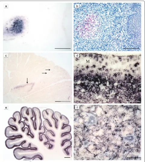
As described before [23] PrPSc was detectable in the LRS tissue of all classical scrapie sheep where it was present, but in none of the atypical/Nor98 scrapie ani-mals with available LRS tissue could PrPScbe found (for availability of lymphatic tissue see Table 1). Figure 1a/b shows PET blot and immunohistochemical staining of the PrPSc aggregates in the follicle of a tonsil derived from a classical scrapie case.
Deposition forms
Intra- and perineuronal PrPSc aggregates were found with the PET blot solely in classical scrapie (Figure 1f), as were subpial, subenpendymal, and perivascular depos-its. Extra neuronal PrPSc aggregates in the brains of sheep affected by classical scrapie often had a ramified appearance and were found in grey and white matter structures (Figure 1d and 1f). They were addressed as glia-associated PrPSc aggregates and found to be rela-tively conspicuous in the cerebellar molecular layer where they took a stellate form [36] (Figure 1d).
In contrast, PrPSc aggregates found in the white matter of atypical/Nor98 scrapie sheep were always well-defined granules that varied a bit in size and were occasionally arranged like pearls on a string. The latter deposition form could also be observed in classical cases, but here also linear PrPScwas sometimes present. PrPScdeposits in the grey matter of atypical/Nor98 scra-pie cases generally showed a fine granular pattern, also termed “synaptic/reticular” in human TSEs rather than
“fine granular” [12,37] (see Figure 1c). In some atypical/ Nor98 scrapie cases, larger plaque-like aggregates could be seen in the substantia nigra, basal ganglia, thalamic nuclei and white matter. However, a differentiation between real plaques (amyloid) and plaque-like deposits is not possible with immunohistochemical detection methods or with the PET blot method as demonstrated before [38].
A discrimination between globular and punctuate deposits in the white matter of atypical/Nor98 cases [11,21] was irreproducible with the PET blot, which is why the term“granular” for the PrPScdeposits that were present in the white matter was chosen. Punctuate PrPSc deposits, comprising smaller aggregates than granular PrPScdeposits but more defined than the reticular PrPSc aggregates, were detected in the grey matter of classical scrapie sheep.
Figure 1Characteristic PrPScdeposition patterns in classical and atypical/Nor98 scrapie:The same lymph follicle in the tonsil of a sheep with classical scrapie is shown stained either with the PET blot method (a) or conventional immunohistochemistry (b) (both mAb P4, bars = 250μm). In the cervical spinal cord segment of a sheep with atypical/Nor98 scrapie (c) stained with the PET blot method (mAb P4, bar = 800 μm) synaptic PrPScaggregates are present in the substantia gelatinosa (vertical arrow) and granular PrPScin the corticospinal tract (horizontal arrows). In the cerebellar cortex of a classical scrapie sheep stained with the PET blot (d) complex PrPScdeposits are visible. Glia-associated PrPSc
deposits take a stellate form in the molecular layer (mAb P4, bar = 150μm; M = molecular layer, P = layer of Purkinje cells, G = granular cell layer). In the majority of the atypical/Nor98 scrapie sheep the cerebellar cortices show a more intense staining of the molecular layer than the granular layer (e) (PET blot, mAb P4; bar 600μm). Intraneuronal, perineuronal and glia-associated PrPScaggregates (f) in the reticular formation of
Distribution of PrPScin the CNS
Sequential appearance of PrPScdistribution in the CNS of classical scrapie sheep To determine the sequential appearance of PrPSc in the CNS, all field cases of classical scrapie were subjected to a thorough examination regarding the anatomical structures affected by PrPSc deposition. In the following, all cases were
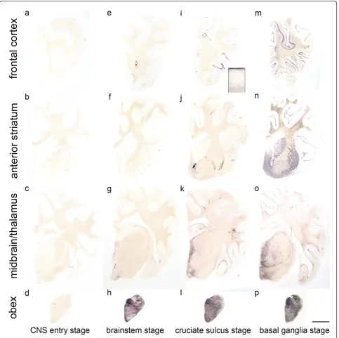
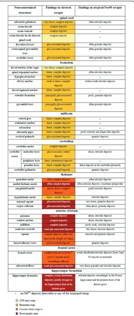
arranged according to the amount of PrPSc they had accumulated in total, and the occurrence of PrPSc in a panel of 127 neuroanatomical loci was compared between the cases. From this evaluation arose a classifi-cation of the classical scrapie cases into four stages of PrPScspread in the CNS (see Figures 2, 3, 4 and 5). Cri-teria for these turned out to be certain neuroanatomical
Figure 2Classification of the PrPSc spread during disease development in classical scrapie.The examined classical scrapie cases classified into four stages of PrP spread according to certain affected neuroanatomical sites (PET blots, mAb P4). In theCNS entry stage (a - d)only discrete PrPScdeposits are visible in the obex region, while in thebrainstem stage (e - h)PrPScaggregates are clearly visible in the brainstem
structures whose involvement marked a stage, meaning that the respective structure accumulated PrPScaggregates (with a minimal score of 1) in all animals belonging to this stage and the following stage/stages. They are described in detail below and visualized in Figures 3 and 4.
Figure 3 Progression of classical scrapie in the brain shown for certain affected neuroanatomical sites. The colour code agrees with the one in Figure 5.
Figure 4Accumulation of PrPScin different brain regions during disease progression:The four stages of the examined classical scrapie cases are depicted in four overlying graphs that illustrate how PrPScaggregates (PET blot method, mAb P4) are increasingly accumulated in the brains from caudal (left) to rostral (right). Evaluation of PrPScintensity was performed on a scale from 0.5 - 4 (see material and methods) and shown for the following brain areas: 1 dorsal motor nucleus of the vagus nerve (DMNV), 2 inferior olive, 3 dorsal tegmental nucleus, 4 cerebellar molecular layer, 5 cerebellar granular layer, 6 cerebral peduncle, 7 central grey (mesencephalon), 8 caudate nucleus, 9 ventral pallidum, 10 rostral commissure, 11 cruciate sulcus, 12 frontal white matter.
Figure 5Form and appearance of PrPScdeposits in classical and atypical/Nor98 scrapie sheep presented for representative CNS regions. As a sequential development of PrPScdistribution
could not be observed upon analysis of the present atypical/Nor98 scrapie cases, no coding colours were used for the results of this scrapie type in contrast to classical scrapie sheep. PrPScdeposits are
CNS entry stage:One sheep showed only few discrete PrPSc deposits in the brain that were restricted to the dorsal motor nucleus of the vagus nerve (DMNV), the solitary tract nucleus and the spinal trigeminal tract in the brainstem. Further PrPSc aggregates could be detected in the substantia intermedialis lateralis and centralis of the thoracic spinal cord. This first stage, where PrPScis detectable only in these CNS areas, can be considered the“CNS entry stage” in accordance with studies of other authors who have monitored the ascen-sion of PrPScfrom the intestines to the CNS [29,30].
Brainstem stage:In the second stage, all segments of the spinal cord and all nuclei of the obex region accu-mulate PrPScwhich also disseminates to the more ros-tral parts of the medulla; this may therefore be called
“brainstem stage”. In the caudal medulla the cellulae marginales and substantia gelatinosa of the spinal tri-geminal tract nucleus show a very intense staining. The mesencephalon and thalamus display discrete PrPSc deposits which are generally found to be subpial and/or perivascular while the mamillary body, habenular nuclei and the hypothalamic nuclei accumulate substantial amounts of PrPSc. The cerebellar nuclei accumulate PrPScif the rostral medulla is largely involved and focal deposits of PrPScare visible in the cerebellar cortex.
Cruciate sulcus stage: During the next stage, the mesencephalon, amygdaloid nuclei, septal nuclei, optic tract, cerebral peduncle, hippocampus formation, frontal cortex and subcortical white matter are increasingly affected. Regarding the frontal cortex, it is notably the sulcus cruciatus - and in a number of cases only this part of the cortex - that accumulates PrPScin its deeper cortical layers (see Figure 2i). This stage is therefore designated“cruciate sulcus stage”. PrPScdeposits in the cerebellar cortex are not yet evenly distributed.
Basal ganglia stage:In the final stage, PrPScdeposits can be seen also in the medial thalamic nuclei (medio-dorsal, ventrolateral, ventral posterior and anterior group), the corpora geniculata and the basal ganglia. A positive staining for PrPScin the latter determines a classical case in our definition for the “basal ganglia stage”. The white matter also displays remarkable amounts of PrPSc, which are strongly linked to perivas-cular distribution.
All stages are depicted in Figures 2, 3 and 4 and the stage at which PrPScreaches a respective neuroanatomi-cal site is indicated in Figure 5 using a colour code. In the sheep examined in this study we could not find any influence of the different genotypes on the neuroanato-mical distribution of PrPScaggregates.
Comparison of PrPScdeposition patterns in classical and atypical/Nor98 scrapiePrPScdeposits in atypical/ Nor98 scrapie cases were examined and evaluated in the same way as with the classical field cases.
In contrast to the classical scrapie cases, differentiating distribution/spread stages of PrPScin the CNS was not fea-sible with the atypical/Nor98 scrapie cases. In Figure 6, the same brain sections that illustrate the different stages of classical scrapie in Figure 2 are depicted for a case of aty-pical/Nor98 scrapie. In all atyaty-pical/Nor98 scrapie sheep, where brainstem material was available (n= 15) apart from one (see below), PrPScaggregates were detectable in the rhombencephalon and mesencephalon. Regularly affected neuroanatomical structures were the spinal tri-geminal nucleus, reticular formation, pyramid, pontine fibres, substantia nigra and cerebral peduncle. In the spinal cord the corticospinal tract and substantia gelatinosa accu-mulated PrPScin most cases (Figure 1c). Certain grey mat-ter structures such as the DMNV, hypoglossal nucleus, dorsal tegmental nucleus, oculomotor nucleus, red nucleus
and central grey of the mesencephalon, never displayed any PrPScin the examined atypical/Nor98 scrapie cases. These listed neuroanatomical sites, however, accumulated large amounts of PrPScin the respective stage of PrPSc dis-tribution in the CNS of classical scrapie sheep as explained above (Figures 2, 3, 4 and 5). There were no PrPSc aggre-gates detectable in the cerebellar nuclei of the examined atypical/Nor98 scrapie cases, in contrast to the classical scrapie cases as described above. The synaptic or reticular PrPScstaining pattern in the cerebellar cortex of atypical/ Nor98 scrapie sheep was in most cases more intense in the molecular than in the granular layer (Figure 1e). Intra-and extracellular complex PrPScaggregates in the cerebel-lar cortex of classical scrapie sheep were predominantly present in the granular layer and surrounding the Purkinje cells; the molecular layer displayed mainly glia-associated PrPScdeposits that took a stellate form (Figure 1d). The cerebellar peduncles and white matter of the cerebellum
itself showed PrPScaggregates for both scrapie types. In the diencephalon of most atypical/Nor98 scrapie sheep, the corpora geniculata, medial thalamic nuclei and reticu-lar nucleus accumulated PrPScaggregates. In all atypical/ Nor98 cases where the anterior striatum could be exam-ined (n= 14), PrPScdeposits were also present in the cau-date nucleus and putamen. The white matter of diencephalon and telencephalon showed PrPScdeposits in both types of sheep scrapie. In atypical/Nor98 scrapie, these were mainly confined to the subcortical fibres and certain white matter tracts, e.g. the corpus callosum or the commissura rostralis (Figure 7d, arrow), while the distri-bution in classical scrapie was more disseminated.
There was one case in which PrPSc deposits were detectable with the PET blot only in the supratentorial (cerebral) brain structures and to a very small degree in the cerebellar cortex. The brainstem, including midbrain and spinal cord, were completely spared in this case,
which was eventually considered to represent an early stage of atypical/Nor98 scrapie [23].
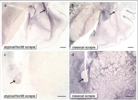
In Figure 7 the contrasts in PrPScintensity existing in the grey and white matter between the two types of scrapie are demonstrated in a case of atypical/Nor98 scrapie and a classical scrapie case of the“basal ganglia stage": in classical scrapie it is the centromedial amygda-loid nuclei (Figure 7b) as well as the septal nuclei and basal ganglia (Figure 7d) that show substantially more PrPScthan the external capsule (Figure 7b, arrow) and the rostral commissure (Figure 7d, arrow). In atypical/ Nor98 scrapie, this principle turns out to be exactly the opposite, with the external capsule (Figure 7a, arrow) and the rostral commissure (Figure 7c, arrow) accumu-lating rather intense PrPSc deposits in contrast to the adjacent grey matter.
The lateral olfactory tract displayed PrPScaggregates in both scrapie types with the respective PrPScdeposition patterns described above. Yet, the Islands of Calleja -clusters of neuronal granular cells in the olfactory tuber-cle - showed dense PrPSc deposits solely in classical scrapie cases and were completely devoid of PrPScin aty-pical/Nor98 scrapie sheep. Regarding the hippocampus formation in classical scrapie cases, there was usually a more intense staining of the hippocampus and the fissura hippocampi compared to the dentate gyrus. In contrast to the atypical/Nor98 scrapie cases, there was no obvious accentuation of any layers. Atypical/Nor98 scrapie sheep showed a rather intense PrPScstaining of the granular layer of the dentate gyrus, the fissura hippocampi and the interconnective fibres between hippocampus and alveus (similar to the subcortical white matter) in comparison to the adjacent layers. The pyramid layer of the hippocam-pus appeared to be completely devoid of PrPScdeposits. The intensity of PrPSc staining in a single case was usually in agreement with the intensity of PrPScdeposits that could be found in the cerebral cortex of both scrapie types. As mentioned above, the complex PrPScaggregates in classical scrapie were mainly confined to the deeper cortical layers (laminae V and VI) while reticular/synaptic PrPScdeposits in the cortices of atypical/Nor98 scrapie sheep were distributed more evenly, although an accent-uation of laminae I and IV could be noted in some cases. Like in classical scrapie, differences regarding the dis-tribution of PrPSc deposition could not be related to genotypes.
Discussion
In this study 24 cases of classical and 25 cases of atypi-cal/Nor98 scrapie cases were examined with the PET blot method, focusing on the similarities and differences in the distribution of PrPScdeposits that were detectable with this method. Recently the PET blot has been shown to provide a sensitive and specific detection of
PrPScin both types of sheep scrapie in the same manner as had been previously shown for human, bovine and rodent neuronal and non-neuronal tissues [30,35,38-41]. The high sensitivity of this method allows PrPScdeposits to be detected even in FFI patients where conventional immunohistochemistry fails to detect them, and con-trasts with Western blotting, which requires up to 1 g of tissue equivalent [42]. The PET blot provides, apart from its sensitivity and specificity, a good overview of where to find PrPSc in a brain section (Figure 2), as no counterstaining is necessary. The fine resolution of the immunolabeling gives a good impression of the struc-tures that accumulate PrPSc(Figure 1f), but the general delineation of the single cell is better with immunohis-tochemistry, which is why these two methods comple-ment each other in a sensible way.
Neuroinvasion and spread of PrPScin the ovine brain
spread via sympathetic nerve fibres of the Plexus ner-vorum perivascularis. Haematogenous neuroinvasion has also been discussed with regard to the circumventricular organs (CVOs) due to the fact that these are usually affected in scrapie-infected sheep and that they are not protected by the BBB [46]. The possibility that the CVOs might be in contact with PrPScfrom the blood during the pathogenesis of the disease cannot be excluded, but our results argue against a major involve-ment of the CVOs in neuroinvasion. In a very early case the DMNV was affected, but the area postrema and further CVOs were devoid of PrPSc (Figure 2d). This agrees again with the results obtained for the oral infec-tion of hamsters with scrapie [30].
In contrast to the classical scrapie cases, a sequential development of PrPScdistribution cannot be seen upon analysis of the present atypical/Nor98 scrapie cases. PrPScdistribution in one sheep of presumably early dis-ease phase suggests that the aggregation of PrPSchas its origin in the di- or telencephalon. A spontaneous gen-esis of misfolded PrP could arise in the cerebral cortex. On the other hand, an ascending spread of the infec-tious agent that bypasses the brainstem and enters the CNS via sensible nerve fibres should be taken into con-sideration, e.g. proprioceptive fibres [47] or the spi-nothalamic tract. This would lead to further spreading of PrPScfrom the thalamic nuclei to the cerebellar and cerebral cortex and from these to the brainstem and spinal cord, e.g. via the corticospinal tract.
Where does the spread of Nor98- PrPScstart in the brain? It has been speculated by Nentwig et al. [48] that the PrPSc deposits and histopathologic lesions in atypical/ Nor98 scrapie possibly evolve from the cerebrum to the cerebellum and the brainstem, but according to their examination of six sheep brains, this concept would not explain the PrPScdistribution in one sheep where they found PrPScmainly in the cerebellum. However, immu-nohistochemistry - as used by these authors - is some-times not able to detect the fine reticular deposits, e.g. seen in the cortex of Creutzfeldt-Jakob disease type 1, especially in the rare VV1 subtype [49]. The sensitive PET blot method, in contrast, is able to visualize these reticular deposits [38]. PrPSc deposits in one case described by Nentwig et al. could have therefore simply been missed in the cortex by immunohistochemistry. If this proved to be correct, according to the argumenta-tion of Nentwig et al., PrPScdeposition and histopatho-logic lesions could indeed evolve from the cerebrum into the cerebellum and the brainstem. The 15 whole brains of atypical/Nor98 scrapie sheep examined by Moore et al. [21] should accordingly represent more or less the final stage of disease, as PrPSccan be generally found in all parts of the brain, including the brainstem.
In other reports on the occurrence of atypical/Nor98 scrapie, cases have been described in which no PrPSc was detectable by immunohistochemistry in the obex region at all, but in the cerebellum and cerebrum [50-54]. If the misfolding of PrPcin atypical/Nor98 scra-pie does really start in the cerebrum it is obvious why early stages are not present in the worldwide pool of preserved atypical/Nor98 brains, as only the sampling of brainstem and cerebellum is compulsory in small rumi-nants according to EU regulations. Thus the question of whether PrPSc accumulation might start sporadically in the cerebrum - and if so, at one or more sites at the same time? - cannot be resolved by this or any other current study using field cases of atypical/Nor98 scrapie. This situation is comparable to the one with CJD type 1, where a spontaneous misfolding of PrPcin the cere-bral cortex and a caudal spread from there is assumed, but not proven [55]. The incidence of atypical/Nor98 in sheep is higher than that of CJD in humans [17]. A case control study of atypical/Nor98 scrapie has shown that animal movement does not seem to be a factor for the transmission of atypical/Nor98 scrapie between flocks; thus if sheep were to acquire this prion disease from their environment, its contagiousness would indeed be very low [56]. It has been speculated that this might be due to the relatively low protease stability, which could also explain the lack of intracellular PrPScdeposits [33].
There are certainly small differences between the PrPScdistribution detected by Moore et al. [21] in their described atypical/Nor98 scrapie cases and the ones revealed here by the PET blot method. For instance, in the present atypical/Nor98 scrapie material, PrPSc was never detectable in the cerebellar nuclei. Also, the affected parts of the hippocampus appear to be different. This might be due to differences in the treatment of tis-sue, the methods and/or differences in the antibodies used (mAb2G11 versus mAbP4). As previously reported, perineuronal staining has also been detected for the sub-stantia nigra in some atypical/Nor98 scrapie sheep using immunohistochemistry [11], whereas in our study only plaque-like PrPScdeposits could be seen in this neuroa-natomical structure. Similarly, neuronal deposits could be found in many affected sites of classical scrapie, but in contrast to previous publications [22], neither PET blot nor immunohistochemistry revealed PrPSc in the Purkinje cells of the cerebellum. It is known that espe-cially intraneuronal immunoreactivity needs to be inter-preted with caution [10]. However, the congruence between previous reports on PrPSc
deposition patterns and the present results is obvious.
Conclusion
Nor98 scrapie cases using the sensitive and specific PET blot method. We were able show a sequential appear-ance of PrPScaggregates in the CNS of sheep with clas-sical scrapie, but not in atypical/Nor98 scrapie. The four emerging stages of spread in classical scrapie were defined by the accumulation of PrPScin certain neuroa-natomical structures. These structures accumulated PrPScaggregates in all animals belonging to this stage and the following stage/stages. Further conclusions drawn from this study regarding atypical/Nor98 scrapie might help in future to elucidate its origin and poten-tially related prion disease types like Creutzfeldt-Jakob disease type 1.
Acknowledgements
We would like to thank Tatjana Pfander, Nadine Rupprecht and Kerstin Brekerbohm for their skilful technical assistance. The work was supported by the VolkswagenStiftung (grants ZN 1294 and ZN 2168 to W.J.S.S).
Author details
1Prion and Dementia Research Unit, Department of Neuropathology,
University Medical Center, Georg-August University, Robert-Koch Str. 40, 37075 Goettingen, Germany.2Norwegian Veterinary Institute, P.O. Box 750
Sentrum, 0106 Oslo, Norway.3Institute of Veterinary Medicine, Faculty for
Agricultural Sciences, Georg-August University, Burckhardweg 2, 37075 Goettingen, Germany.
Authors’contributions
WMW carried out the PET blot studies, participated in
immunohistochemistry, Western blot, tissue acquisition and the design of the study and drafted the manuscript. SLB participated in
immunohistochemistry, Western blot, tissue acquisition and design of the study and co-edited the manuscript. AW, WEW, BeB and BjB participated in tissue acquisition and diagnosing the cases. WJSS conceived the study, participated in its design and coordination and co-edited the manuscript. All authors read and approved the final manuscript.
Competing interests
The authors declare that they have no competing interests.
Received: 20 September 2010 Accepted: 15 February 2011 Published: 15 February 2011
References
1. McGowan JP:Scrapie in sheep.Scottish J Agric1922,5:365-375. 2. Prusiner SB:Novel proteinaceous infectious particles cause scrapie.
Science1982,216:136-144.
3. Oesch B, Westaway D, Walchli M, McKinley MP, Kent SB, Aebersold R, Barry RA, Tempst P, Teplow DB, Hood LE, Prusiner SB, Weissmann C:A cellular gene encodes scrapie PrP 27-30 protein.Cell1985,40:735-746. 4. Wang F, Wang X, Yuan CG, Ma J:Generating a prion with bacterially
expressed recombinant prion protein.Science2010,327:1132-1135. 5. Bendheim PE, Brown HR, Rudelli RD, Scala LJ, Goller NL, Wen GY,
Kascsak RJ, Cashman NR, Bolton DC:Nearly ubiquitous tissue distribution of the scrapie agent precursor protein.Neurology1992,42:149-156. 6. Bruce ME, Boyle A, Cousens S, McConnell I, Foster J, Goldmann W, Fraser H:
Strain characterization of natural sheep scrapie and comparison with BSE.J Gen Virol2002,83:695-704.
7. Hope J, Wood SC, Birkett CR, Chong A, Bruce ME, Cairns D, Goldmann W, Hunter N, Bostock CJ:Molecular analysis of ovine prion protein identifies similarities between BSE and an experimental isolate of natural scrapie, CH1641.J Gen Virol1999,80:1-4.
8. Stack MJ, Chaplin MJ, Clark J:Differentiation of prion protein glycoforms from naturally occurring sheep scrapie, sheep-passaged scrapie strains (CH1641 and SSBP1), bovine spongiform encephalopathy (BSE) cases and Romney and Cheviot breed sheep experimentally inoculated with
BSE using two monoclonal antibodies.Acta Neuropathol2002, 104:279-286.
9. Benestad SL, Sarradin P, Thu B, Schönheit J, Tranulis MA, Bratberg B:Cases of scrapie with unusual features in Norway and designation of a new type, Nor98.Vet Rec2003,153:202-208.
10. EFSA:Opinion of the Scientific Panel on Biological Hazards on classification of atypical Transmissible Spongiform Encephalopathy (TSE) cases in Small Ruminants.The EFSA Journal2005,276:1-30.
11. Benestad SL, Arsac JN, Goldmann W, Nöremark M:Atypical/Nor98 scrapie: properties of the agent, genetics, and epidemiology.Vet Res2008,39:19. 12. Wemheuer WM, Benestad SL, Wrede A, Schulze-Sturm U, Wemheuer WE,
Hahmann U, Gawinecka J, Schütz E, Zerr I, Brenig B, Bratberg B, Andreoletti O, Schulz-Schaeffer WJ:Similarities between forms of sheep scrapie and Creutzfeldt-Jakob disease are encoded by distinct prion types.Am J Pathol2009,175:2566-2573.
13. Simon S, Nugier J, Morel N, Boutal H, Créminon C, Benestad SL, Andréoletti O, Lantier F, Bilheude JM, Feyssaguet M, Biacabe AG, Baron T, Grassi J:Rapid typing of transmissible spongiform encephalopathy strains with differential ELISA.Emerg Infect Dis2008,14:608-616. 14. Belt PB, Muileman IH, Schreuder BE, Bos-de Ruijter J, Gielkens AL, Smits MA:
Identification of five allelic variants of the sheep PrP gene and their association with natural scrapie.J Gen Virol1995,76:509-517. 15. Hunter N:PrP genetics in sheep and the applications for scrapie and
BSE.Trends Microbiol1997,5:331-334.
16. Lühken G, Buschmann A, Groschup MH, Erhardt G:Prion protein allele A136 H154Q171 is associated with high susceptibility to scrapie in purebred and crossbred German Merinoland sheep.Arch Virol2004,149:1571-1580. 17. Lühken G, Buschmann A, Brandt H, Eiden M, Groschup MH, Erhardt G:
Epidemiological and genetical differences between classical and atypical scrapie cases.Vet Res2007,38:65-80.
18. Moum T, Olsaker I, Hopp P, Moldal T, Valheim M, Moum T, Benestad SL: Polymorphisms at codons 141 and 154 in the ovine prion protein gene are associated with scrapie Nor98 cases.J Gen Virol2005,86:231-235. 19. González L, Martin S, Begara-McGorum I, Hunter N, Houston F, Simmons M,
Jeffrey M:Effects of agent strain and host genotype on PrP accumulation in the brain of sheep naturally and experimentally affected with scrapie.J Comp Pathol2002,126:17-29.
20. Miller JM, Jenny AL, Taylor WD, Marsh RF, Rubenstein R, Race RE: Immunohistochemical detection of prion protein in sheep with scrapie.J Vet Diagn Invest1993,5:309-316.
21. Moore SJ, Simmons M, Chaplin M, Spiropoulos J:Neuroanatomical distribution of abnormal prion protein in naturally occurring atypical scrapie cases in Great Britain.Acta Neuropathol2008,116:547-559. 22. van Keulen LJ, Schreuder BE, Meloen RH, Poelen-van den Berg M,
Mooij-Harkes G, Vromans ME, Langeveld JP:Immunohistochemical detection and localization of prion protein in brain tissue of sheep with natural scrapie.Vet Pathol1995,32:299-308.
23. Wemheuer WM, Benestad SL, Wrede A, Wemheuer WE, Brenig B, Bratberg B, Schulz-Schaeffer WJ:Detection of classical and atypical/Nor98 scrapie by the paraffin-embedded tissue blot method.Vet Rec2009, 164:677-681.
24. Andréoletti O, Simon S, Lacroux C, Morel N, Tabouret G, Chabert A, Lugan S, Corbière F, Ferre P, Foucras G, Laude H, Eychenne F, Grassi J, Schelcher F:PrPSc accumulation in myocytes from sheep incubating natural scrapie.Nat Med2004,10:591-593.
25. Lacroux C, Corbière F, Tabouret G, Lugan S, Costes P, Mathey J, Delmas JM, Weisbecker JL, Foucras G, Cassard H, Elsen JM, Schelcher F, Andreoletti O: Dynamics and genetics of PrPSc placental accumulation in sheep.J Gen Virol2007,88:1056-1061.
26. Ligios C, Sigurdson CJ, Santucciu C, Carcassola G, Manco G, Basagni M, Maestrale C, Cancedda MG, Madau L, Aguzzi A:PrPSc in mammary glands of sheep affected by scrapie and mastitis.Nat Med2005,11:1137-1138. 27. Thomzig A, Schulz-Schaeffer W, Wrede A, Wemheuer W, Brenig B, Kratzel C,
Lemmer K, Beekes M:Accumulation of pathological prion protein PrPSc in the skin of animals with experimental and natural scrapie.PLoS Pathog2007,3:e66.
28. Ersdal C, Ulvund MJ, Espenes A, Benestad SL, Sarradin P, Landsverk T: Mapping PrPSc propagation in experimental and natural scrapie in sheep with different PrP genotypes.Vet Pathol2005,42:258-274. 29. van Keulen LJ, Schreuder BE, Vromans ME, Langeveld JP, Smits MA:
30. McBride PA, Schulz-Schaeffer WJ, Donaldson M, Bruce M, Diringer H, Kretzschmar HA, Beekes M:Early spread of scrapie from the
gastrointestinal tract to the central nervous system involves autonomic fibers of the splanchnic and vagus nerves.J Virol2001,75:9320-9327. 31. van Keulen LJ, Vromans ME, Dolstra CH, Bossers A, Van Zijderveld FG:
Pathogenesis of bovine spongiform encephalopathy in sheep.Arch Virol 2008,153:445-453.
32. Ryder SJ, Dexter GE, Heasman L, Warner R, Moore SJ:Accumulation and dissemination of prion protein in experimental sheep scrapie in the natural host.BMC Vet Res2009,5:9.
33. Simmons MM, Konold T, Simmons HA, Spencer YI, Lockey R, Spiropoulos J, Everitt S, Clifford D:Experimental transmission of atypical scrapie to sheep.BMC Vet Res2007,3:20.
34. Schütz E, Scharfenstein M, Brenig B:Genotyping of ovine prion protein gene (PRNP) variants by PCR with melting curve analysis.Clin Chem 2006,52:1426-1429.
35. Schulz-Schaeffer WJ, Fatzer R, Vandevelde M, Kretzschmar HA:Detection of PrP(Sc) in subclinical BSE with the paraffin-embedded tissue (PET) blot. Arch Virol Suppl2000, 173-180.
36. González L, Martin S, Jeffrey M:Distinct profiles of PrP(d) immunoreactivity in the brain of scrapie- and BSE-infected sheep: implications for differential cell targeting and PrP processing.J Gen Virol 2003,84:1339-1350.
37. Kitamoto T, Tateishi J:Human prion diseases with variant prion protein. Philos Trans R Soc Lond B Biol Sci1994,343:391-398.
38. Schulz-Schaeffer WJ, Tschöke S, Kranefuss N, Dröse W, Hause-Reitner D, Giese A, Groschup MH, Kretzschmar HA:The paraffin-embedded tissue blot detects PrP(Sc) early in the incubation time in prion diseases.Am J Pathol2000,156:51-56.
39. Lezmi S, Bencsik A, Baron T:PET-blot Analysis Contributes to BSE Strain Recognition in C57Bl/6 Mice.J Histochem Cytochem2006,54:1087-1094. 40. Peden AH, Ritchie DL, Head MW, Ironside JW:Detection and localization
of PrPSc in the skeletal muscle of patients with variant, iatrogenic, and sporadic forms of Creutzfeldt-Jakob disease.Am J Pathol2006, 168:927-935.
41. Thomzig A, Kratzel C, Lenz G, Krüger D, Beekes M:Widespread PrPSc accumulation in muscles of hamsters orally infected with scrapie.EMBO Rep2003,4:530-533.
42. Reder AT, Mednick AS, Brown P, Spire JP, Van Cauter E, Wollmann RL, Cervenakova L, Goldfarb LG, Garay A, Ovsiew F, Gajdusek DC, Roos RP: Clinical and genetic studies of fatal familial insomnia.Neurology1995, 45:1068-1075.
43. Schulz-Schaeffer W, McBride PA, Beekes M, Kretzschmar HA:Spread of PrPSc in orally infected animals during incubation time of prion disease. XIV International Conference of Neuropathology2000, 663.
44. Jeffrey M, Goodsir CM, Holliman A, Higgins RJ, Bruce ME, McBride PA, Fraser JR:Determination of the frequency and distribution of vascular and parenchymal amyloid with polyclonal and N-terminal-specific PrP antibodies in scrapie-affected sheep and mice.Vet Rec1998,142:534-537. 45. Delattre JY, Krol G, Thaler HT, Posner JB:Distribution of brain metastases.
Arch Neurol1988,45:741-744.
46. Sisó S, Jeffrey M, González L:Neuroinvasion in sheep transmissible spongiform encephalopathies: the role of the haematogenous route. Neuropathol Appl Neurobiol2009,35:232-246.
47. Rüb U, Schultz C, Del TK, Gierga K, Reifenberger G, de Vos RA, Seifried C, Braak H, Auburger G:Anatomically based guidelines for systematic investigation of the central somatosensory system and their application to a spinocerebellar ataxia type 2 (SCA2) patient.Neuropathol Appl Neurobiol2003,29:418-433.
48. Nentwig A, Oevermann A, Heim D, Botteron C, Zellweger K, Drogemuller C, Zurbriggen A, Seuberlich T:Diversity in Neuroanatomical Distribution of Abnormal Prion Protein in Atypical Scrapie.PLoS Pathog2007,3:e82. 49. Parchi P, Giese A, Capellari S, Brown P, Schulz-Schaeffer W, Windl O, Zerr I,
Budka H, Kopp N, Piccardo P, Poser S, Rojiani A, Streichemberger N, Julien J, Vital C, Ghetti B, Gambetti P, Kretzschmar H:Classification of sporadic Creutzfeldt-Jakob disease based on molecular and phenotypic analysis of 300 subjects.Ann Neurol1999,46:224-233.
50. Buschmann A, Biacabe AG, Ziegler U, Bencsik A, Madec JY, Erhardt G, Lühken G, Baron T, Groschup MH:Atypical scrapie cases in Germany and France are identified by discrepant reaction patterns in BSE rapid tests.J Virol Methods2004,117:27-36.
51. Buschmann A, Lühken G, Schultz J, Erhardt G, Groschup MH:Neuronal accumulation of abnormal prion protein in sheep carrying a scrapie-resistant genotype (PrPARR/ARR).J Gen Virol2004,85:2727-2733. 52. Gavier-Widen D, Nöremark M, Benestad S, Simmons M, Renstrom L,
Bratberg B, Elvander M, af Segerstad CH:Recognition of the Nor98 variant of scrapie in the Swedish sheep population.J Vet Diagn Invest2004, 16:562-567.
53. Onnasch H, Gunn HM, Bradshaw BJ, Benestad SL, Bassett HF:Two Irish cases of scrapie resembling Nor98.Vet Rec2004,155:636-637. 54. Orge L, Galo A, Machado C, Lima C, Ochoa C, Silva J, Ramos M, Simas JP:
Identification of putative atypical scrapie in sheep in Portugal.J Gen Virol 2004,85:3487-3491.
55. Heye N, Cervos-Navarro J:Focal involvement and lateralization in Creutzfeldt-Jakob disease: correlation of clinical,
electroencephalographic and neuropathological findings.Eur Neurol 1992,32:289-292.
56. Hopp P, Omer MK, Heier BT:A case-control study of scrapie Nor98 in Norwegian sheep flocks.J Gen Virol2006,87:3729-3736.
doi:10.1186/1297-9716-42-32
Cite this article as:Wemheueret al.:PrPScspreading patterns in the
brain of sheep linked to different prion types.Veterinary Research2011
42:32.
Submit your next manuscript to BioMed Central and take full advantage of:
• Convenient online submission
• Thorough peer review
• No space constraints or color figure charges
• Immediate publication on acceptance
• Inclusion in PubMed, CAS, Scopus and Google Scholar
• Research which is freely available for redistribution