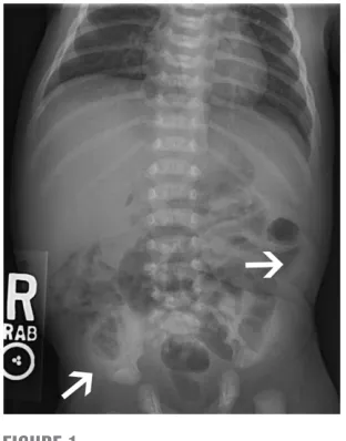
Bloody Stools in a 3-Day-Old Term Infant
Full text
Figure
Related documents
In this section, the load cycle data analysis process is discussed and a representative load cycle for a single cell is presented that is incorporated in the
Abbreviations: aFP, α -fetoprotein; alB, albumin; alP, alkaline phosphatase; alT, alanine transaminase; asT, aspartate aminotransferase; Bclc, Barcelona clinic for liver cancer;
between Fn level and chemotherapy benefits in stage III/IV patients, a subgroup analysis was carried out based on Fn level and we found that patients receiving chemotherapy
They argue that leverage is an instrument that is very sensitive to changes in corporate value determined by capital structure (Modigliani & Miller, 1958). The higher the
Continuous patient monitoring can be an extension to the ―Elderly Family Member Monitoring‖ application; this application, however, requires the medical services
The new development in the product is ensured by innovation in the same product or new ideas implementing with using technology for attracting more consumers in
This article addresses issues of first professional, in-service and continuing education and training with particular reference to requirements reported by a large group of
We would like to acknowledge National Medical Research Center, Al Zawiyah-Libya, for generous help by providing us with analysis instrument and technical support and Division
