Abstract: -Tight sandstone samples from Fuyu oil layer in Daan oilfield of Songliao Basin were mainly studied in this paper. A 3D digital core model with real pore throat structure was constructed by combining Tomography with advanced "Maxima-Ball". The research showed that the tight siltstone samples had bad pore connectivity. The storage space was mainly composed of intragranular dissolution pores. Image computed porosity was 0.5 %, with an average pore radius of 1.89 um and an average throat radius of 1.01 um. Tomography relative to conventional core testing method had advantages such as results intuitive, rich data and non-destructive to sample. With the further development of computer technology, nondestructive testing technology will become an important technology involved in the oil field exploration and development.
Keywords: - Tight sandstone ; Nondestructive testing technology; Maxima-Ball; Pore structure
I.
INTRODUCTION
Industrial nondestructive testing technology in our country has played a very important role in industry [1].According to industry detection and scientific research, and the application of measurement in recent years,
such as structure of nondestructive testing ,defect analysis and quality assurance and control, nondestructive testing technology in the increasingly become a powerful X-ray tomography imaging detection tools, and its application in characterization of reservoir pore structure is becoming more and more widely [2-3], X-ray CT imaging technology has the advantage of rapidly, all-round, and a wide range of rock sample scanning imaging [4-5]
, the digital core can be more intuitive to study the porosity of the reservoir rock characteristics [ 6-8 ], its application in the direction of the evaluation of pore structure will be more broad. This paper takes Daan oilfield in southern Songliao Basin dense sandstone samples as the research object, using X-ray CT imaging technology has realized the pore network model extraction and quantitative characterization of the pore parameters, the visualization of microscopic pore structure laid a good foundation for the multi-scale pore structure study.
II.
MICRO
CT
SCAN
Fig.1 MicroXCT-200 Fig.2 X-ray layout system schematic diagram
Fig.3 Rock sample Fig.4 2D images of CT scanning(resolution 1.0664um)
III.
2D
IMAGE
ANAIYSIS
AND
PROCESSING
Using Avizo 8.0 software image segmentation techniques, we get reconstruced 2D gray image of binarization segmentation, the bright part of CT images is high density material, the dark part is considered as pore structure, the pore area is red rendering (fig. 5a), divided into pore and granular matrix, the seepage simulation can be used in pore network modeling and segmentation image (fig. 5b).
Fig. 6 3D stereogram of core Fig. 7 Pore connectivity rendering
V.
ESTABLISHMENT
OF
3D
PORE
NETWORK
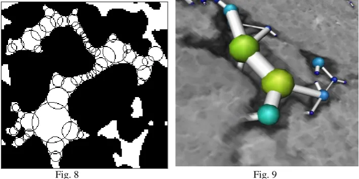
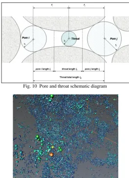
Using the method of " Maxima-Ball" to get pore network structure of extraction and modeling, we can not only improve the speed of network extraction, and also ensures the accuracy of the pore distribution and connectivity features. " Maxima-Ball " method is a series of different size of the spheres filling into the pore space of 3D core image, there is a connection relationship according to the radius between filling various sizes of the ball . The core internal pore structure will be sent to you by overlapped and contained ball string to represent (fig.8. Pore network structure of pore and throat is established through the ball string to find local smallest ball l between the two big ball, thus forming the “pore-throat-pore” matching relationship (fig.9 and fig.10), and eventually the whole ball string structure can be simplified to pore network structure model (fig.11).
Fig. 8 Fig. 9
Fig. 10 Pore and throat schematic diagram
Fig. 11 3D pore network structure model (ball for pore, tube for throat)
VI.
THE
RESULTS
AND
ANAILSIS
Fig. 14 Types of reservoir space under 2D gray scale images (resolution 1.0664 um)
VII.
THE
EXPEREMENTAL
RESULTS
AND
ANALYSIS
1 .In this paper, using X-ray CT imaging technology combined with advanced " Maxima-Ball" method,we established the micro scale real 3D pore model of pore throat structure, CT scanning experiment results show that the samples of argillaceous siltstone has poor connectivity, reservoir space is mainly intergranular dissolved pore.
2 .Compared with the conventional X-ray CT imaging technology core testing method ,the result of X-ray CT imaging technology is intuitive, simple and feasible, the technique of microscopic pore throat based on CT scanning laid a foundation for further study of multi-scale dense sandstone pore structure characteristic.
REFERENCES
[1] Gong-Tian Shen. China the progress of nondestructive testing and evaluation technology [J].Nondestructive Testing , 2008, 11:2-6.
[2] Bin Bai, Ru-Kai Zhu, Songtao Wu, et al. Multi-scale method of Nano(Micro)-CT study on modifications inside natural building stones by means of high resolution X-ray CT [J]. Petroleum Exploration and Development. 2013(03): 329-333.
[3] Bijeljic B, Muggeridge A H, Blunt M J. Pore-scale modeling of longitudinal dispersion[J]. Water Resources Research. 2004, 40(11):417-427.
[4] Sakwat A A D. Nanoscnaale X-raydi imaging. Nature Photonics. 2010, 4(267): 840-848. [5] Attwood D. Microscopy: Nanotomography comes of age. Nature. 2006, 442(7103): 642-643.
[6] Xiang-Jun Liu , Hong-Lin Zhu, Li-Xi Liang. Digital Rock Physics of Sandstone Based on Micro CT Technology [J]. Chinese Journal Of Geophysics 2014 (4) : 1133-1140.
[7] Na Su, Yong-Gang Duan, Chun-Sheng Yu. Reconstruction of Microscopic Pore Structure in Low Permeability Gas Reservoirs by Micro-CT Scanning [J]. Oil & Gas Geology. 2011 (5) : 792-796.
[8] Le Qu, Wei Sun, Huan-Hong Du, etc al. Characterization Technique of Pore Structure by 3D Digital Core Based on CT Scanning and Its Application: An Example from Sangonghe Formation of 116 Well Field in Mobei Oilfield [J]. Modern Geology. 2014 (01) : 190-196.