Adaptive Segmentation of Cells and Particles in
Fluorescent Microscope Image
M. Kanchana H. Bharani
Professor Assistant Professor
Department of Information Technology Department of Information Technology
Karpaga Vinayaga College of Engineering & Technology, Chinna Kolambakkam, Madurantakam Taluk,
Kanchipuram-603308, Tamil Nadu, India
Karpaga Vinayaga College of Engineering & Technology, Chinna Kolambakkam, Madurantakam Taluk,
Kanchipuram-603308, Tamil Nadu, India
Abstract
Understanding the mechanisms of cell motility and their regulation is an important challenge in biomedical research. The ability of cells to exert forces on their environment and alter their shape as they move is essential to various biological processes such as the immune response, embryonic development, or tumor genesis .Recent technological advances in con-focal fluorescence microscopy have given researchers the opportunity to investigate these complex processes in vivo. However, they also lead to a tremendous increase in the amount of image data acquired during the studies. Therefore, the analysis of time-lapse experiments relies increasingly on automated image processing techniques. Namely, there is a high demand for fast and robust methods to help biologists to quantitatively analyze time-lapse image data. The potential of the proposed tracking scheme and the advantages and disadvantages of both frameworks are demonstrated on 2-D and 3-D time-lapse series of rat adipose-derived mesenchymal stem cells and human lung squamous cell carcinoma cells, respectively. The crucial tasks are, in particular, segmenting, tracking, and evaluating movement tracks and morphological changes of cells, sub-cellular components and other particles.
Keywords: Fluorescent Microscope Image, Adaptive Segmentation
_______________________________________________________________________________________________________
I.
I
NTRODUCTIONWe present a fast and robust approach to tracking the evolving shape of whole fluorescent cells in time-lapse series. The proposed tracking scheme involves two steps. First, coherence-enhancing diffusion filtering is applied on each frame to reduce the amount of noise and enhance flow-like structures. Second, the cell boundaries are detected by minimizing the Chan- Vese model in the fast level set-like and graph cut frameworks. To allow simultaneous tracking of multiple cells over time, both frameworks have been integrated with a topological prior exploiting the object indication function. The potential of the proposed tracking scheme and the advantages and disadvantages of both frameworks are demonstrated on 2D and 3D time-lapse series of rat adipose-derived mesenchymal stem cells and human lung squamous cell carcinoma cells, respectively. In biomedical experiments sub-cellular particles of interest are fluorescently labelled in different chromatographic bands yielding multi-channel images which are subsequently analyzed by automatic image analysis techniques. Unfortunately explicit labelling of the complete cell area is typically not possible and enforces to extract it from one of the available channels originally intended for detection of other particles. In our experimental data each image contains three channels. In the nucleus channel of the images nuclei manifest themselves by more or less homogeneously textured bright blob.
II.
R
ELATED WORKSSegmentation of cells and detection of particles in fluorescently labelled microscopy images are instances of general problem image analysis. Due to the special characteristics of these images, adaptations are required and have been proposed. In (Dzyubachyk et al., 2007) and (Dzyubachyk et al., 2008) a level-set based approach for segmentation and tracking of cells is proposed. For initial segmentation in the first time frame, the fitting term of the classical Chan-Vese model (Chan and Vese, 2001) is replaced with a Gaussian likelihood for the intensity values with unknown variance.
proposed by (Dufour et al., 2008). They combine wavelet-based particle detection adopting the approach of (Olivo- Marin, 2002) with active contour-based cell area segmentation. Unfortunately their snake energy is not transferable to our domain as the cells in our scenario show quite in homogeneous and grainy intensity distributions which do not allow for easy modelling within snake energy functional.
III.
P
ROPOSED METHODThe proposed tracking scheme, which involves two steps, first, a coherence enhancing diffusion filter is applied on each frame to reduce the amount of noise and enhance flow-like structures. Second, the cell boundaries are detected by minimizing the Chan-Vese model. Active contours can either be modelled implicitly adopting level-set approaches or explicitly in terms of parametric contour models, i.e. snakes. While the numerical tractability of level-set approaches is often superior compared to snake optimization techniques, topology preservation is usually easier to guarantee by snake approaches as topological stability is inherent in the model theory underlying snakes.
In our scenario the number of objects to segment is known in advance, hence the topology of the desired segmentation result is known. For this reason we prefer explicit active contour models for this application. In explicit approaches the contour of an object is defined as a function c : R → R2 which maps a parameter value s ∈ [0,1] defined along a given contour c to points (x,y) in 2-D space. For object segmentation an energy functional over the contour function c(s) is defined. The contour is evolved towards a local minimum of the energy, which gives the final segmentation result.
Modules Description A.
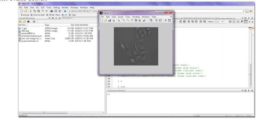
Preprocessing 1)
Frames are allocated from given input video Align the width and height of video
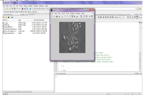
To convert color frame into gray frame Coherence Enhancing Diffusion Filter 2)
A mathematically well-founded method based on the solution of a nonlinear anisotropic diffusion equation. This spatial filter can enhance flow-like structures while reducing the amount of noise.
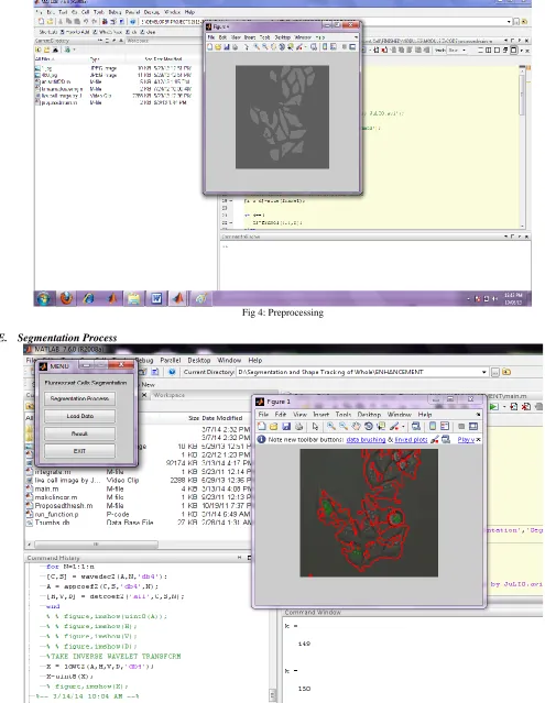
Chan-Vese model 3)
The number of cells in the first frame is not known in advance, the cell boundaries are detected using the Chan-Vese model without any topological constraint. Both frameworks are initialized using a fully automated weighted 2means clustering that corresponds to the minimization of equation without the regularization term. Finally binary output image established as the cells to be tracked.
IV.
O
UTPUT ANALYSISColor Frame Convert A.
Adaptive Histogram Equalization B.
Fig. 2: Histogram Increase
Level of Adapthiseq C.
Preprocessing Module D.
Fig 4: Preprocessing
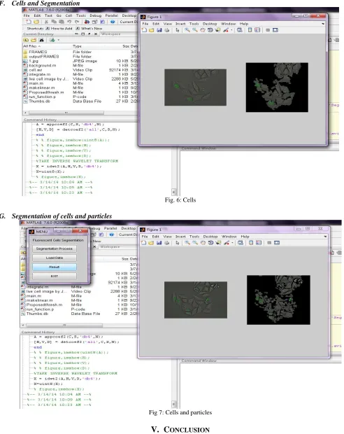
Cells and Segmentation F.
Fig. 6: Cells
Segmentation of cells and particles G.
Fig 7: Cells and particles
V.
C
ONCLUSIONtracking scheme, which involves two steps, first, a coherence enhancing diffusion filter is applied on each frame to reduce the amount of noise and enhance flow-like structures. Second, the cell boundaries are detected by minimizing the Chan-Vese model
R
EFERENCES[1] C. Zimmer, B. Zhang, A. Dufour, A. Th´ebaud, S. Berlemont, V. Meas- Yedid, and J.-C. Olivo-Marin, “On the digital trail of mobile cells,” IEEE Signal Processing Magazine, vol. 23, no. 3, pp. 54–62, 2006.
[2] R. Ananthakrishnan and A. Ehrlicher, “The forces behind cell movement,” International Journal of Biological Sciences, vol. 3, no. 5, pp. 303–317, 2007. [3] R. Fern´andez-Gonz´alez, A. Mu˜noz-Barrutia, M. H. Barcellos-Hof, and C. Ortiz-de-Sol´orzano, “Quantitative in vivo microscopy: the return from the
„omics‟,” Current Opinion in Biotechnology, vol. 17, no. 5, pp. 501– 510, 2006.
[4] C. Vonesch, F. Aguet, J.-L. Vonesch, and M. Unser, “The colored revolution of bio imaging,” IEEE Signal Processing Magazine, vol. 23, no. 3, pp. 20–31, 2006.
[5] E. Meijering, O. Dzyubachyk, I. Smal, and W. A. Cappellen, “Tracking in cell and developmental biology,” Seminars in Cell and Developmental Biology, vol. 20, no. 8, pp. 894–902, 2009.
[6] P. Sarder and A. Nehorai, “Deconvolution methods for 3-D fluorescence microscopy images,” IEEE Signal Processing Magazine, vol. 23, no. 3, pp. 32–45, 2006.
[7] J. Weickert, A. Bruhn, T. Brox, and N. Papenberg, “A survey on variational optic flow methods for small displacements,” in Mathematical Models for Registration and Applications to Medical Imaging. Springer- Verlag, 2006, pp. 103–136.
[8] P. Matula, P. Matula, M. Kozubek, and V. Dvoˇr´ak, “Fast point-based 3D alignment of live cells,” IEEE Transactions on Image Processing, vol. 15, no. 8, pp. 2388–2396, 2006.
[9] K. Li, M. Chen, T. Kanade, E. D. Miller, L. E. Weiss, and P. G. Campbella, “Cell population tracking and lineage construction with spatiotemporal context,” Medical Image Analysis, vol. 12, no. 5, pp. 546–566, 2008.
[10] O. Al-Kofahi, R. J. Radke, S. K. Goderie, Q. Shen, S. Temple, and B. Roysam, “Automated cell lineage construction: A rapid method to analyze clonal development established with murine neural progenitor cells,” Cell Cycle, vol. 5, no. 3, pp. 327–335, 2006.
[11] N. Harder, F. Mora-Berm´udez, W. J. Godinez, J. Ellenberg, R. Eils, and K. Rohr, “Automated analysis of the mitotic phases of human cells in 3D fluorescence microscopy,” in Medical Image Computing and Computer- Assisted Intervention, 2006, pp. 840–848.
[12] F. Li, X. Zhou, J. Ma, and S. T. C. Wong, “Multiple nuclei tracking using integer programming for quantitative cancer cell cycle analysis,” IEEE Transactions on Medical Imaging, vol. 29, no. 1, pp. 96–105, 2010.
[13] D. Padfield, J. Rittscher, and B. Roysam, “Coupled minimum-cost flow cell tracking for high-throughput quantitative analysis,” Medical Image Analysis, vol. 15, no. 1, pp. 650–668, 2011.
[14] O. Debeir, P. V. Ham, R. Kiss, and C. Decaestecker, “Tracking of migrating cells under phase-contrast video microscopy with combined mean-shift processes,” IEEE Transactions on Medical Imaging, vol. 24, no. 6, pp. 697–711, 2005.