International Journal of Pharmaceutical Research & Allied Sciences, 2019, 8(4):97-104
Research Article
CODEN(USA) : IJPRPM
ISSN : 2277-3657
Regional Left Ventricular Function Analysis By 128-Row Multi-Detector
Computed Tomography in Patients with Coronary Artery Disease
Hanan A. Bahaaeldin1, Ibrahim A. Libda1, Ahmed A. El Sammak1, Ekhlas M. Hussien2, Farida M. El Fawal1
1Department of Radiology, Faculty of Medicine, Zagazig University, Zagazig, Egypt 2Department of Cardiology, Faculty of Medicine, Zagazig University, Zagazig, Egypt.
Email: dr.flower35 @ yahoo.com
ABSTRACT
Purpose: The aim of this study was to estimate the role of multidetector computed tomography (MDCT) in the evaluation of LV regional wall motion abnormalities (RWMA) in subjects complaining of coronary artery heart disease (CAD) and to compare MDCT data with two dimension standard echocardiography (2DSE) as the standard reference. Patients and Methods: Sixty subjects with supposed coronary artery heart disease were submitted to retrospective gating contrast-enhanced MDCT. 10 phases of the cardiac cycle were performed to detect end-systolic and end-diastolic phases at LV short-axis view. LV Regional wall motion was assessed qualitatively (visually in cine-mode) and quantitatively (measuring the percentage of systolic wall thickening on static end-diastolic and end-systolic images) on cardiac short-axis view and long-axis views using a 17-segment model. 2DSE was performed within two weeks before MDCT. Results: Good segmental agreement was found between echocardiography and MDCT (k=0.7; p < 0.001), MDCT detected 720 (98.7%) of 729 segments that showed normal motility, 172 (74.7%) of 230 segments showed hypokinesia and 49 (80.3%) of 61 segments showed akinesia or dyskinesia. Regarding the diagnostic performance, the sensitivity, specificity, and accuracy of MDCT reached 80.4%, 97.4%, and 93.5%, respectively, assuming 2DSE as the gold standard. Conclusion: Evaluation of regional left ventricular function by using MDCT is a precise method, with good agreement with 2D ECG.
Key words: Coronary artery disease, regional left ventricular function, MDCT.
INTRODUCTION
PATIENTS AND METHODS
Population and Study Design
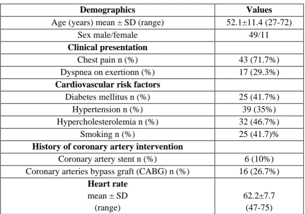
This investigation was conducted at Zagazig University Hospitals, Radiology Department, from February 2017 to February 2019. Sixty patients from the cardiology outpatient clinic with either known or suspected coronary artery disease were referred for MDCT coronary angiography; they were 49 males and 11 females, age ranged from 27 to 72 years, mean age of 52±11 years old. The average heart rate was 62 beats/min. The patients had chest pain (71.7%) and/or dyspnea on exertion (29.3%). The patients' data are summarized in Table 1. Patients exclusion criteria were: renal insufficiency (serum creatinine<1.5), morbid obesity, arrhythmias and pulmonary diseases that hinder breath-holding during MDCT acquisition.
Table 1. Patients’ data
Demographics Values
Age (years) mean ± SD (range) 52.1±11.4 (27-72)
Sex male/female 49/11
Clinical presentation
Chest pain n (%) 43 (71.7%)
Dyspnea on exertionn (%) 17 (29.3%)
Cardiovascular risk factors
Diabetes mellitus n (%) 25 (41.7%)
Hypertension n (%) 39 (35%)
Hypercholesterolemia n (%) 32 (46.7%)
Smoking n (%) 25 (41.7)%
History of coronary artery intervention
Coronary artery stent n (%) 6 (10%)
Coronary arteries bypass graft (CABG) n (%) 16 (26.7%)
Heart rate
mean ± SD (range)
62.2±7.7 (47-75)
All patients signed a written informed consent and filled a written survey including demographic and clinical data. The consent forms and echocardiography and CT protocols, which were utilized in the present investigation were approved by the Institutional Review Board (IRB) of Zagazig University.
To determine coronary artery disease and assess left ventricular regional function, all patients underwent contrast-enhanced retrospective ECG gated coronary CT angiography (CCTA) and 2D echocardiography performed within two weeks before CCTA.
Protocol of CCTA Patient preparation
All patients were premedicated with 50 mg/day metoprolol one day before MDCT examination. The patients with a pulse exceeding of 70 bpm were given another 50mg of oral metoprolol 30min before the exam in order to reduce cardiac motion artifacts. To accomplish coronary vasodilatation, sublingual nitroglycerin was given as the patient lying on the table.
MDCT image acquisition
All CT angiographic examinations were performed using Philips Ingenuity core 128 TM v3.5.7.25001 (Philips healthcare systems, Netherlands) in Zagazig University hospital. The following parameters were used: 16×0.75 mm detector collimation, 0.39 s rotation time, the pitch of 0.2-0.3, increment 0.5mm and reconstruction slice width 0.6mm, the tube current was 300 ± 40 mA at 120-140 kV. Scanning direction; craniocaudal. Mean scan time was 12± 1.5 seconds, and the total time for the examination was less than 10 minutes.
MDCT image analysis
Images were reconstructed at ten phases: 0, 30, 40, 45, 50, 60, 70, 75, 80, & 90 % of the R–R interval. Philips Extended intellispace™ portal Workstation post-processed the images of all ten phases. LV regional wall motion was assessed visually (in cine-mode) and the degree of systolic wall thickening was quantified (on static end-diastolic and end-systolic images) on cardiac short-axis view and long-axis views (four-chamber, three-chamber, and two-chamber views) using a 17-segment model. [11] The left ventricular wall motion was considered normal if the left ventricular wall thickness increased by more than 40% during systole. Hypokinesia was considered when the left ventricular wall thickness increased by less than 30%, and akinesia if there was wall thickening of less than 10% . [5] The outward motion of myocardium during systole with underlying wall thinning was considered as dyskinesia (Fig 1). A three-point scoring system was used to determine the wall motion of each segment 1: normal, 2: hypokinetic, or 3: dyskinetic or akinetic. [7, 8]
Segmental agreement between echocardiography and MDCT was calculated for actual scores for 17 segments. It was repeated using binary values (normal and abnormal) to calculate the diagnostic performance. To correct for the possibility of misregistration between segments, a second approach divided each left ventricle into 3 coronary artery territories, according to American Heart Association recommendations, [11] so that the anterior wall and anterior septum represented the left anterior descending artery, the inferior wall and inferior septum represented the right coronary artery, and the posterior and lateral walls represented the left circumflex artery. If ≥2 segments in a particular territory were abnormal, the region was considered abnormal. The average time spent on functional analysis at the workstation was 15 minutes.
Echocardiography
Transthoracic Echocardiography (TTE) is assumed to be the gold standard. An altrasound machine (Vivid-7; GE-Vingmed, Milwaukee Wis) was used to examine all of the participants. Images were acquired in apical 2- and 4-chamber views and standard parasternal using a 3.5 MHz transducer by a 15-year experienced echocardiologist, blinded to clinical data and MDCT findings. To evaluate regional LV function, short-axis slices (at the mitral valve, papillary muscle, and apical levels), apical 4- and 2-chamber and long-axis views were recorded. Each segment was scored as 1: normokinesia, 2: hypokinesia, and 3: akinesia or dyskinesia using 17 segments model.
Statistical analysis:
Continuous data are expressed as mean±standard deviation (SD). Segmental agreement between echocardiography and MDCT was calculated for actual scores for 17 segments according to the American Heart Association using Cohen’s kappa coefficient. It was repeated by using binary values (abnormal and normal) and for each coronary artery territory. Assuming echocardiography to be the gold standard, the specificity and sensitivity of MDCT were calculated to detect an abnormal segment for each coronary artery territory individually and for all segments.
RESULTS
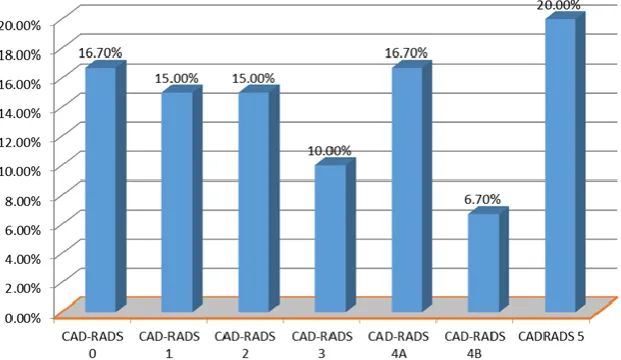
The current study enrolled 60 patients (18.3% were female and 81.7% were males) with known or suspected CAD. 16.7% of the studied patients (10/60) were categorized as CAD-RADS 0, while 36.7% (n=22) of them underwent coronary artery revascularization procedures either CABG (26.7%) or stenting (10%). The CADRADS categorization of the studied group is illustrated in fig 2.
In this study, there was an overall good agreement between MDCT and 2DSE in detecting an abnormal regional wall motion. According to 3 point scoring system; there was a good agreement between ECHO and MDCT in the detection of regional wall motion abnormality using (κ=0.7; p < 0.001). Among the totally examined 1020 segments, MDCT detected 720 (98.7%) of 729 segments that showed normal motility by 2D ECHO examination, 172 (74.7%) of 230 segments showed hypokinesia by 2D ECHO examination and 49 (80.3%) of 61 segments showed akinesia or dyskinesia by 2D ECHO examination (Table 2).
Using a binary analysis (normal or abnormal); Among our 1020 examined segments, 729 segments were normal, and 291 segments were abnormal by echocardiography and on MDCT 782 segments were normal and 238 segments were abnormal. There was an excellent agreement between MDCT and echocardiography for binary scores (κ=0.8; p < 0.001) (Table 3). With respect to ECG as a gold method, MDCT recorded a high sensitivity, specificity, and accuracy reached 80.4%, 97.4%, and 93.5% respectively, in the present study (Table 4).
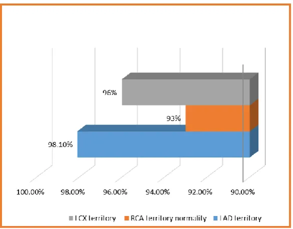
Figure 3: Bar chart for comparing the diagnostic accuracy of MDCT in the detection of coronary artery territory wall motion abnormalities relative to 2DSE.
Table 2. Agreement between echocardiography and MDCT in detection of regional wall motion abnormality (RWMA) regarding 1 to 3 scores:
All segments
ECHO
Kappa
agreement p-value
1 No. (%)
2 No. (%)
3 No. (%)
MDCT
1 (777)
720 (98.7%)
53 (23%)
4 (6.5%)
0.7 0.001**
2 (188 )
8 (1.09%)
172 (74.7%)
8 (13.1%) 3
(55)
1 (.137%)
5 (2.1%)
49 (80.3%)
Total (1020) 729 230 61
Table 3. Agreement between ECHO and MDCT in the detection of regional wall motion abnormality for binary scores:
ECHO
Kappa
agreement p-value
Normal No. (729) %
Abnormal No. %(291)
MDCT regional wall
motion
Normal
(777 ) 720 98.7% 57 19.5%
0.8 0.001**
Abnormal
(243) 9.0 1.3% 23480.4%
Table 4. Diagnostic accuracy of MDCT in the detection of regional left ventricular functional comparing to echocardiography:
Sensitivity Specificity PVP PVN Accuracy
Binary score 80.4% 97.4% 96.2% 92.6% 93.5%
LAD territory normality 100.0% 93.4% 97.4% 100.0% 98.1%
RCA territory normality 100.0% 78.6% 90.6% 100.0% 93.0%
LCX territory normality 100.0% 81.8% 95.1% 100.0% 96.0%
DISCUSSION
Many indices of LV diastolic functions are efficiently measured with 2DSE like E to A wave ratio, deceleration time and relaxation time. Additionally, 2DSE can evaluate the function of cardiac valves and other cardiovascular structures. [13, 14] Notably, echocardiography is a fundamentally real-time imaging method and is not limited by arrhythmias. Though, echocardiography has its limitations in the application in diagnosis related to patient conditions such as chronic lung disease, previous cardiothoracic surgery, and obesity, which restrict optimal visualization of cardiac chambers due to poor acoustic windows. Many investigators reported that good visualization of the endocardium can be affected by the operator-dependent variables such as gain settings and depth of scanning. [12, 14-16]
In spite of using MDCT in the diagnosis of coronary artery diseases, it is improbable to substitute echocardiography for the valuation of regional LV function, but it is characterized by its capacity to assess ventricular function and perfusion in addition to the coronary arteries in a scan. In addition, MDCT can detect the anatomical relationship between the coronary artery and its supplements to the myocardium through its excellent ability to visualize wall thickening and thickness, but its progressive resolution is less than that of echocardiography. Therefore, it is very important to know how MDCT parallels echocardiography in its capacity to evaluate regional myocardial function. [7]
The present study demonstrated a general good covenant between MDCT and 2D ECHO in detecting an abnormal regional wall motion. According to the 3-point scoring system, there was good agreement between ECHO and MDCT in detecting regional wall motion abnormality using (k=0.7; p<0.001). The best agreement was in detection of normal then akinetic segments with less ability in the detection of hypokinetic segments. There was an excellent arrangement between MDCT and echocardiography (k=0.8; p<0.001) by applying a binary statistical analysis (normal or abnormal). Regarding echocardiography as the gold standard, MDCT had an accuracy of 93.5%.
Analysis depending on the coronary artery territories detected that the diagnostic accuracy of MDCT for LAD and LCX regions (98% and 96%) was better than RCA regions (93.1%). The possible reason may be attributed to breathing artifacts due to the proximity of the inferior wall to the diaphragm. This was in agreement with a study done by Lessick et al.[7]
Several previous studies established a good association between MDCT and 2DSE in the evaluation of LV regional wall motion abnormality. [2, 7, 8, 17-20] Butler et al. (2006) and Ko et al. (2010) demonstrated that wall motion assessment by 64-slice MDCT agreed well with 2D-TTE (75% agreement; κ=0.61) and (94% agreement; k=0.7) respectively, which was similar to results in a study by Henneman et al., who found in his study a covenant in 96% of the ventricular segments (κ=0.82). Slam et al. also proved a good agreement between 16-row MDCT and echocardiography (κ=0.78). Lessick et al. found in his study that the sensitivity and specificity were averaged 89% and 96%, respectively compared to echocardiography as the method. [2, 7, 8, 17-19] and this was consistent with our results. Ko et al. stated that the score of MDCT abnormal segments is higher than 2D-ECHO; the sensitivity was very high (97%), but specificity was slightly decreased (82%) in detecting regional dysfunction, which is disagreed with our results. The relatively high observer variability can explain the results owing to the impaired temporal resolution of MDCT in comparison to echocardiography. [8] The main limitation in the evaluation of regional wall motion abnormality is the limited temporal resolution of MDCT. [8] Further advances in the temporal resolution of MDCT would be helpful to equal results from competitive imaging modalities such as CMR. Another problem in interpreting the regional wall motion abnormalities was its dependence on a visual interpretation that is different between radiologists. [16] We tried to overcome this problem, by using a quantitative method for the evaluation of RWMA by measuring LV myocardial thickness at end-systolic and end-diastolic phases and calculation of the degree of systolic thickening.
Finally, the major drawback of retrospective ECG-gated coronary CT angiography is the higher radiation exposure. [21] Newly, prospective ECG-gated method had been advanced and the dose of radiation is decreased effectively. It offers important information such as anatomical features of heart, the pericardium, and the size of cardiac chambers. Inappropriately, it is not applied to estimate LV or RV performance; accordingly, ECG was used for a single cardiac phase. [22] For reduction of a dose, there is another efficient method represented in the ECG table dependent tube up-to-date modulation, by which the dose can be reduced up to half as nearly 25% of the maximum tube current is used through systole matched to diastole, but this will affect the assessment of RWMA and LVESV. [15, 23, 24].
Therefore, in spite of these outcomes and the integral benefits of MDCT than 2D-echocardiography, the need for intravenous contrast stain and the exposure to radiation are limited the widespread use of this method as a tool to solely evaluate LV performance. While attention must be put into consideration when involving the right coronary artery region and the extent of the fault, for the reason that MDCT tends to miscalculate the number of abnormal segments compared to echocardiography. Further investigations should be performed to estimate the use of a greater temporal resolution other than the role of standardized quantification of regional function.
Conflict of Interest: The authors declared no conflict of interest.
Financial Disclosures: No financial disclosures.
Abbreviations:
2DSE: Two dimensional standard echocardiography, CAD: Coronary artery disease, CCTA: coronary CT angiography, CMR: Cardiac magnetic resonance imaging, LAD: left anterior descending artery, LCX: Left Circumflex artery, LVESV: Left ventricular end-systolic volume, MDCT: Multi-detector computed tomography, RCA: Right coronary artery, RWMA: regional wall motion abnormalities, TTE: Trans-thoracic Echocardiography
REFERENCES
1. Finn JP, Nael K, Deshpande V, Ratib O, Laub G. Cardiac MR imaging: state of the technology. Radiology. 2006 Nov;241(2):338-54.
2. Henneman MM, Schuijf JD, Jukema JW, Holman ER, Lamb HJ, De Roos A, van der Wall EE, Bax JJ. Assessment of global and regional left ventricular function and volumes with 64-slice MSCT: a comparison with 2D echocardiography. Journal of nuclear cardiology. 2006 Jul 1;13(4):480-7.
3. Sami SM, Elfawal SK, Abdelgawad MS, Zidan MA, Zaki AM, Mowaki AF. MDCT in the study of left ventricular function compared with MRI in patients with myocardial ischemia. The Egyptian Journal of Radiology and Nuclear Medicine. 2018 Mar 1;49(1):29-41.
4. Ghorbani A, Shirzadpour E, Kaffashian MR, Mohamadpour M, Seifinejad Y, Amraei M. Anti-Atherosclerotic Effects of the Hydroalcoholic Extract of Crocus sativus L. (saffron) Petals on Hypercholesterolemic Rats. International Journal of Pharmaceutical and Phytopharmacological Research, 2018 ;8(6) :99-104.
5. Dahmardeh H, Sedaghat M. Epidemiological Investigation of Cardiovascular Diseases in Khatamol Anbia Hospital in Zahedan City in 2016. Pharmacophore, 2017;8(6),60-3.
6. Areshidze DA, Mischenko DV, Makartseva LA, Kucher SA, Kozlova MA, Timchenko LD, Rzhepakovsky IV, Nagdalian AA, Pushkin SV. Some Functional Measures of the Organism of Rats at Modeling of Ischemic Heart Disease in Two Different Ways. Entomology and Applied Science Letters. 2018 Jan 1;5(4):19-29.
7. Lessick J, Mutlak D, Rispler S, Ghersin E, Dragu R, Litmanovich D, Engel A, Reisner SA, Agmon Y. Comparison of multidetector computed tomography versus echocardiography for assessing regional left ventricular function. The American journal of cardiology. 2005 Oct 1;96(7):1011-5.
8. Ko SM, Kim YJ, Park JH, Choi NM. Assessment of left ventricular ejection fraction and regional wall motion with 64-slice multidetector CT: a comparison with two-dimensional transthoracic echocardiography. The British journal of radiology. 2010 Jan;83(985):28-34.
9. Sucang L, Weinert L, Mor-Avi V, Neil J, Ebner C, Steringer-Mascherbauer R, Schmidt F, Schummers G, Lang RM, Nesser HJ. Quantitative assessment of left ventricular size and function: Side-by-side comparison of three dimensional echocardiography and computed tomography against magnetic resonance reference. In Journal of the American College of Cardiology 2006 Feb 21 (Vol. 47, No. 4, pp. 139A-139A). 360 Park Ave South, New York, NY 10010-1710 USA: Elsevier Science Inc.
10. Maffei E, Messalli G, Martini C, Nieman K, Catalano O, Rossi A, Seitun S, Guaricci AI, Tedeschi C, Mollet NR, Cademartiri F. Left and right ventricle assessment with Cardiac CT: validation study vs. Cardiac MR. European radiology. 2012 May 1;22(5):1041-9.
12. Bansal D, Singh RM, Sarkar M, Sureddi R, Mcbreen KC, Griffis T, Sinha A, Mehta JL. Assessment of left ventricular function: comparison of cardiac multidetector-row computed tomography with two-dimension standard echocardiography for assessment of left ventricular function. The international journal of cardiovascular imaging. 2008 Mar 1;24(3):317-25.
13. Singh RM, Singh BM, Mehta JL. Role of cardiac CTA in estimating left ventricular volumes and ejection fraction. World journal of radiology. 2014 Sep 28;6(9):669.
14. Gottdiener JS, Bednarz J, Devereux R, Gardin J, Klein A, Manning WJ, Morehead A, Kitzman D, Oh JK, Quinones M, Schiller NB. American Society of Echocardiography recommendations for use of echocardiography in clinical trials: A report from the american society of echocardiography's guidelines and standards committee and the task force on echocardiography in clinical trials. Journal of the American Society of Echocardiography. 2004 Oct;17(10):1086-119.
15. Lim SJ, Choo KS, Park YH, Kim JS, Kim JH, Chun KJ, Jeong DW. Assessment of left ventricular function and volume in patients undergoing 128-slice coronary CT angiography with ECG-based maximum tube current modulation: a comparison with echocardiography. Korean journal of radiology. 2011 Apr 1;12(2):156-62.
16. Hoffmann U, Pena AJ, Cury RC, Abbara S, Ferencik M, Moselewski F, Siebert U, Brady TJ, Nagurney JT. Cardiac CT in emergency department patients with acute chest pain. Radiographics. 2006 Jul;26(4):963-78.
17. Salm LP, Schuijf JD, de Roos A, Lamb HJ, Vliegen HW, Jukema JW, Joemai R, van der Wall EE, Bax JJ. Global and regional left ventricular function assessment with 16-detector row CT: comparison with echocardiography and cardiovascular magnetic resonance. European Journal of Echocardiography. 2006 Aug 1;7(4):308-14.
18. Wu YW, Tadamura E, Yamamuro M, Kanao S, Okayama S, Ozasa N, Toma M, Kimura T, Komeda M, Togashi K. Estimation of global and regional cardiac function using 64-slice computed tomography: a comparison study with echocardiography, gated-SPECT and cardiovascular magnetic resonance. International journal of cardiology. 2008 Aug 1;128(1):69-76.
19. Butler J, Shapiro MD, Jassal D, Neilan T, Nichols J, Ferencik M, Brady TJ, Hoffmann U, Cury RC. Comparison of multidetector computed tomography and two-dimensional transthoracic echocardiography for left ventricular assessment in patients with heart failure. The American journal of cardiology. 2007 Jan 15;99(2):247-9.
20. Schuijf JD, Bax JJ, Jukema JW, Lamb HJ, Salm LP, De Roos A, Van der Wall EE. Assessment of left ventricular volumes and ejection fraction with 16-slice multi-slice computed tomography; comparison with 2D-echocardiography. International journal of cardiology. 2007 Mar 20;116(2):201-5.
21. Sun Z, Choo GH, Ng KH. Coronary CT angiography: current status and continuing challenges. The British journal of radiology. 2012 May;85(1013):495-510.
22. Rumberger JA, Systematic Analysis of Cardiac CT In: Taylor AJ, eds. Atlas of Cardiovascular Computed Tomography: An Imaging Companion to Braunwald’s heart disease, 1st edition .Saunders: El Seiver; 2010: 26-37.
23. Paul JF, Abada HT. Strategies for reduction of radiation dose in cardiac multislice CT. European radiology. 2007 Aug 1;17(8):2028-37.