ORIGINAL ARTICLE
Proposal of a modified transcervical endometrial resection
(TCER) technique for menorrhagia treatment. Feasibility,
efficacy, and patients
’
acceptability
Pietro Litta&Luigi Nappi&Pasquale Florio&
Luca Mencaglia&Mario Franchini&Stefano Angioni
Received: 11 January 2014 / Accepted: 16 April 2014 / Published online: 30 April 2014 #Springer-Verlag Berlin Heidelberg 2014
Abstract The aim of this study is to evaluate the feasibility,
efficacy, safeness, and patients’acceptability of a modified
transcervical endometrial resection (TCER) technique for the treatment of menorrhagia. Eighty-four premenopausal women with menorrhagia after careful investigation and 2 months therapy with GnRHa underwent a modified TCER. It was performed with a standard dual channel, 26 French irrigating resectoscope (Karl Storz, GmbH, Germany) after cervix dila-tation to 10 mm and sorbitol mannitol solution used as disten-sion medium. The modified technique was based on the resection of the endometrium and of the first myometrial layers only on the anterior and posterior walls, without treating fundus and cornual areas as usually performed. Endometrial resection was performed to a depth of 4 to 5 mm. Clinical and hysteroscopic follow-up was performed
for 60 months. Early and late complications, changing in
bleeding patterns, and patients’satisfaction were recorded.
Sixty-four out of 73 patients that completed the 60 months improved. Eumenorrhea was achieved in 68.5 %, hypomenorrhea in 5.5 %, and amenorrhea in 13.7 %. Most of the patients (86.3 %) showed satisfaction at the follow-up interview. Control hysteroscopy showed that post modified TCER uterine cavity maintained the possibility of macroscop-ic and histopathology investigation during follow-up. Modified TCER is a technique easy to perform and effective in the long-term resolution of menorrhagia. In particular, it avoids the formation of synechiae and the shrinkage of the uterine cavity that may be the cause of various long-term complications, such as the delay in the diagnosis of endome-trial carcinoma onset.
Keywords Menorrhagia . AUB . Endometrial resection . Minimally invasive surgery . Hysteroscopy
Introduction
Hysteroscopic transcervical endometrial resection (TCER) is a minimally invasive surgical technique developed in recent years with the purpose of removing the entire thickness of the
endometrium lining of the uterus [1]. Indeed, to suppress
menstruation successfully, it is essential to remove the full thickness of this lining together with the superficial myometrium, including the deep endometrial basal glands which are believed to be the primary foci for endometrial
re-growth [1, 2]. However, TCER is not always completely
successful and, in some cases, additional surgical treatment is required, thus limiting the benefits related to the reduced trauma and post-operative complications to the woman. The risk of failure and the expense of multiple treatments opened a debate whether endometrial ablation should replace or not
P. Litta
Department of Gynecological Science and Human Reproduction, University of Padua, Padua, Italy
L. Nappi
Department of Medical and Surgical Sciences, Institute of Obstetrics and Gynaecology, University of Foggia, Foggia, Italy
P. Florio
U.O.C. Obstetrics & Gynecology,“San Giuseppe”Hospital, Empoli, Italy
L. Mencaglia
Section of Gynecology, Centro Oncologico Fiorentino, Sesto Fiorentino, Italy
M. Franchini
Palagi Freestanding Unit, Florence, Italy
S. Angioni (*)
hysterectomy [3], or if it might be an effective therapy for women with hyperplasia, with abnormal uterine bleeding,
with high risk for medical therapy or hysterectomy [4]. The
emerging clinical opinion is that TCER is an effective and safe alternative to hysterectomy that should be offered to women with menorrhagia for the relief of their heavy menstrual bleed-ing, together with the caution that there should be the possi-bility of further surgery, either repeat endometrial ablation or
hysterectomy [5].
Matter of discussion related to TCER is also the putative occurrence of other problems related to the endometrial injury, as in the case of immediate (vascular or metabolic type plications (fluid overload) and perforation), or delayed com-plications (as in the case of the development of partial intra-uterine dense adhesions and/or total obliteration of the cavity)
[5–12]. Therefore, the ideal method of TCER associating high
efficacy to nice tolerability and low incidence of complica-tions is still far from being achieved. In the present study, we evaluated short- and long-term outcomes associated with a new TCER technique to treat menorrhagia that differ from the standard one in the fact that uterine fundus and cornual areas are not removed in the modified technique.
Materials and methods
Subjects
For this prospective cohort study, we consecutively enrolled from October 2, 2000 to September 24, 2005 all women suffering of menorrhagia who referred to our tertiary centers of women health care. The diagnosis of menorrhagia was performed by means of a pictorial blood loss assessment chart, adjusted to our needs in patients describing a history of heavy
menstrual blood loss over several consecutive cycles [13]. A
scoring system ranging from 1 to 10 was used, with 1=slightly soiled tampon, 5=moderately soiled, and 10=heavily soiled. Sanitary napkins were assigned ascending scores from 1 to 20. A total score more than 100 for each pictorial chart was meant
as a confirmed diagnosis of menorrhagia [14]. We considered
for the study only patients who had performed a full clinical evaluation including colposcopy and Papanicolaou test, transvaginal ultrasonography, and hysteroscopy with
endome-trial biopsy [15]. Exclusion criteria were the following: not
confirmed diagnosis of menorrhagia, uterine size >12 cm, presence of large organic intrauterine lesions (endometrial polyp >3 cm, submucous myomas G0>2 cm or submucous myomas G1 and G2); desire of future pregnancy, cervical and endometrial pre- and malignant conditions or adnexal pathol-ogies; and debilitating medical condition. Any medical hor-monal treatment was suspended at least 1 month before en-rollment. All procedures followed were in accordance with the ethical standards of the responsible committee on human
experimentation (institutional and national) and with the Helsinki Declaration of 1975, as revised in 2000. Informed consent was obtained from all patients for being included in the study.
Surgical procedure: modified TCER
All the women underwent therapy with GnRH analogs (Leuprolide Acetate 3.75 mg) for 2 months (every 28 days)
before surgery, as in standard practice [6]. All procedures were
performed under general anesthesia, with induction by
propofol 2 mg kg−1and spontaneous ventilation with a
mix-ture of 60 % nitrous oxide and 40 % oxygen isofluothane, or
spinal anesthesia in selected patient [16]. Hysteroscopic
MTCER was performed with a standard dual channel, 26 French irrigating resectoscope (Karl Storz, GmbH, Germany) after that cervix was dilated to 10 mm. The uterine cavity was distended with sorbitol mannitol solution used as distension medium, and a suction-irrigating unit (Endomat, Karl Storz, GmbH, Germany) was used to provide positive pressure (120 mmHg) and continuous outflow suction control (0.5 bar). Fluid balance was carefully monitored throughout the procedure that was interrupted if fluid deficit was over
1,000 cm3. Surgical time was recorded starting at resectoscope
introduction inside the uterus and ending at its last removal. Compared to the technique described by Wortman and
Dagget [2], our modified TCER began on either anterior or
posterior uterine wall and was based on the resection of the anterior cardinal strip of tissue followed by resection of the posterior and the two lateral cardinal strips without treating
fundus and cornual areas (Fig. 1). The conventional TCER
approach consists in the treatment of fundal and cornual areas by the equatorial loop and/or the use of the rollerball electrode. Endometrial resection was performed to a depth of 4 to 5 mm, and endomyometrial strips were removed from the cavity and sent for histological assessment. The procedure was scheduled for a 1-day surgery.
Office hysteroscopy
Control hysteroscopy was scheduled at 3, 12, 24, and 60 months after the surgery. It was performed by using vaginoscopic approach, with a continuous-flow hysteroscope using Telescope 2.9 mm (HOPKINS II Forward-Oblique
Telescope 30°; Karl Storz, Tuttlingen, Germany) [17, 18].
The uterine cavity was distended with temperate saline solu-tion and irrigated using an electronic irrigasolu-tion pump
(Hysteromat, Karl Storz®, Karl Storz, Tuttlingen, Germany).
Examination was performed in an office setting, without anesthesia or cervical dilatation.
Main outcomes of the study
Outcome measures referred to changes in bleeding patterns,
safeness, and patients’acceptability. In details, after modified
TCER, women underwent office hysteroscopy for uterine cavity evaluation (with endometrial biopsy) 3, 12, 24, and 60 months after TCER, and simultaneously, patients were asked about the amelioration or persistence of bleeding, dura-tion of amenorrhea, improvement of dysmenorrhea, and if they need any hormonal or surgical treatment for heavy bleed-ing after modified TCER.
Modified TCER was considered successful when it was associated with amenorrhea, hypomenorrhea, and eumenorrhea. A women was defined as amenorrheic when reporting persistent ceasing of menstruation after surgery. Eumenorrhea referred to regular menstrual cycle with average
length of 28 days (range, 21–35 days), lasting on average for
4 days (range, 1–8 days) and of normal quantity (flowing less
than 80 mL per cycle). Hypomenorrhea referred to menstru-ations regular in frequency but poor in quantity and/or lasting
less than 2 days [1]. Resection was considered non-effective
when the patient reported persistence or relapse of menorrhagia.
Interviews were done at the time of every follow-up
hys-teroscopy, and data were related to patients’subjective
expe-riences related/consequent to modified TCER and their health status concerning endometrial status.
Statistical analysis
All data were analyzed with Prism software (GraphPad Software Inc., San Diego, CA, USA) and expressed as mean ±SD. The Kolmogorov-Smirnov test was used to evaluate whether values had a Gaussian distribution, in order to choose between parametric and non-parametric statistical tests.
Therefore, the unpairedttest was used to compute statistical
significance, andχ-square and Fisher exact test to analyze
differences between proportions. Statistical significance was
assumed for values ofP<0.05.
Results
Some patients were not included in the study. Exclusion criteria were the following: not confirmed diagnosis of men-orrhagia (26 women), uterine size >12 cm (10 patients), presence of large organic intrauterine lesions (endometrial polyp >3 cm, submucous myomas G0>2 cm, and submucous myomas G1 and G2) (15 patients); desire of future pregnancy (eight patients), cervical, and endometrial pre- and malignant conditions or adnexal pathologies (10 patients).
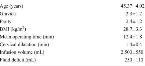
Eighty-four patients out of 153 (age 45.37±4.02) entered the study. The clinical study protocol consisted of modified TCER, followed by the assessment of endometrial cavity by office diagnostic hysteroscopy 3, 12, 24, and 60 months after
the surgery. Table1shows clinical and demographic details of
the population evaluated in the study. Endometrial resection was successfully performed in all patients enrolled. No one of the patients had early or late complications. In 24 out of 84 patients, endometrial resection was associated with the
simul-taneous removal of small polyps (n=15) or myomas (n=9)
(data not shown). In any case, the time required for endome-trial resection with associated polypectomy (13.31±6.09 min) or myomectomy (14.7±8.2 min) did not differ when com-pared to the endometrial resection alone (11.91±4.15 min;
P>0.05), as well as the time spent for cervical dilatation and
the infusion volume needed for uterine distension (data not shown). In 11 cases (13 %), adenomyosis was evidenced by histopathologic examination.
Follow-up outcomes: clinical findings
Eleven women dropped out (13.1 %) and did not complete the first year of follow-up, while 73 (86.9 %) patients completed
the follow-up for at least 60 months (Table2) and 61 (83.6 %)
of these reached a 84-month follow-up. During the observa-tional time interval, bleeding patterns were observed in e u m e n o r r h e a i n 5 0 o u t o f 7 3 w o m e n ( 6 8 . 5 % ) , hypomenorrhea in four patients (5.5 %), and amenorrhea in
10 subjects (13.7 %) (Table2). None of them reported
spot-ting, neither dysmenorrhea onset or worsening, nor medium-/
Table 1 Anthropometric and surgical data related to the population prospectively evaluated
Age (years) 45.37±4.02
Gravida 2.3±1.2
Parity 2.4±1.2
BMI (kg/m2) 28.7±3.3
Mean operating time (min) 12.4±1.8 Cervical dilatation (min) 1.4±0.4
Infusion volume (mL) 2,500±550
Fluid deficit (mL) 250±110
long-term complications such as pregnancy or complications putatively related to the development of intra-cavitary adhe-sions, such as hematometra and/or cornual hematometra, as observed at diagnostic office hysteroscopy.
Seven (9.6 %) patients continued to have menorrhagia, and among them, two of the 11 cases were with adenomyosis (18.2 %). Three patients underwent laparoscopic
hysterecto-my (n=3) after an average interval of 6 months (range, 1–11
months), two patients underwent hormonal treatment by means of levonorgestrel-based intrauterine device, and the remaining two women underwent 4-month GnRH analog
administration (n=2) (data not shown). In these patients, a
second modified TCER was not performed because patients refused such a type of treatment.
In addition, AUB recurred in two women (2.7 %) after 12
and 60 months after endometrial resection (Table 2). They
underwent a new modified TCER or laparoscopic hysterecto-my (for the women in whom recurrence occurred 60 months after modified TCER) (data not shown).
Follow-up outcome: data from interview
At the end of 3-month follow-up, 63 women (86.3 %) stated they were satisfied by the surgery so much that they would recommend it to women with menorrhagia, whereas the re-maining 10 patients (13.7 %) would not, for reasons mainly
related to the fear of general anesthesia (n=8; 80.0 %) or office
hysteroscopy in the follow-up (n=2; 20 %). None of the
patients had symptoms or conditions invalidating and/or lim-iting their quality of life (data not shown).
Follow-up outcome: hysteroscopic findings
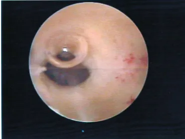
Considering findings recorded at hysteroscopic follow-up, none of the patients was found to have intra-uterine adhesions or cavity contracture after 3 months. It was possible to eval-uate the entire cavity, including the cornual area and the tubal
ostia in all patients (Fig.2). The same findings were obtained
at all time points in the follow-up, even if the uterine cavity at the hysteroscopic evaluation performed after 12, 24, and 60 months from endometrial resection was markedly reduced
to a narrow tube as a result of fibrosis and contracture (Fig.3).
In any case, the entire uterine cavity, including the cornual areas, was found to be open and the tubal ostia were visualized at all time points of follow-up. In addition, histological eval-uation of endometrial biopsy annually performed failed to found cancerous or pre-cancerous endometrial lesions (data not shown).
Discussion
The present study first refers on the clinical efficiency and
patients’perception of a new endometrial resection technique,
by using which menorrhagia was resolved in the majority of patients, without surgical complications, no intrauterine adhe-sions formation in the follow-up, no fluid overload syndrome,
short operative time, and a high degree of patients’satisfaction.
The reasons that led us to devise such a new technique are
related to the fact that theconventionalhysteroscopic TCER is
Table 2 Data related to the clinical findings retrieved at follow-up after modified TCER in women prospectively evaluated
Patients underwent MTCER (n; %) 84 (100 %) Follow-up completed after 3 months (n; %) 84 (100) Follow-up completed after 60 months (n; %) 73 (86.9) Bleeding patterns after TCER
Eumenorrhea (n; %) 50/73 (68.5) Hypomenorrhea (n; %) 4/73 (5.5) Amenorrhea (n; %) 10/73 (13.7) Menorrhagia (n; %) 7/73 (9.6) Recurrence of AUB/DUB (n; %) 2/73 (2.7)
Fig. 2 Hysteroscopic appearance of endometrial cavity at 3-month fol-low-up after MTCER. None of the patients were found to have intrauter-ine adhesions
sometime associated to clinical problems, like surgical (per-foration of the uterus), vascular complications or fluid
over-load syndrome [5–12]. Moreover, like resectoscopic
myomec-tomy, TCER is a surgical procedure suggested only to
expe-rienced surgeons [19]. Consequently, our aim was to simplify
and to accelerate the procedure maintaining the success rate and possibly decreasing the complications in order to make it accessible even to less experienced gynecologists. In addition, we intended to decrease the occurrence of intrauterine
adhe-sions, contractures, and/or hematometra [12,20–22]. These
last side effects are due to the fact that after the endometrium is destroyed or resected, the myometrium is exposed and intra-uterine walls, collapsing on each other, have a natural tenden-cy to grow together. The final result is the intrauterine con-tracture and marked reduction of the endometrial cavity to a narrow tubular structure as a result of fibrosis that often obstructs the cornual area. This mechanism is responsible for the occurrence of synechiae in 40 % of women submitted
to total TCER [2] that may limit the access to the uterine
cavity, hematometra, that usually localizes in the fundus of the uterus, or obstruction of the cornual area in 13 % of cases
[11, 22, 23]. The persistence in these obstructed areas of
islands of endometrial tissue may cause retrograde menstrua-tion or symptomatic cornual hematometra, with an incidence
of even 18 %, causing painful distention of the uterus [24].
The potential of all these complications may limit the clinical efficacy of hysteroscopic TCER and can require hysterecto-my. In our technique, the resection of the endometrium and of the first myometrial layers was limited only on the anterior and posterior and lateral walls, without treating fundal and
cornual areas by using the rollerball, as usually performed [1,
2] The initial hypothesis was that preserving endometrial
mucosa of the fundal and cornual areas could decrease the risk of synechiae or focal hematometra facilitating the
long-term uterine inspection [15, 25]. The possibility that this
approach could result in menorrhagia or bleeding persistence due to the endometrial mucosa not removed was not shown by our study. Moreover, in our case series, we failed to detect intrauterine adhesions at all evaluated time points after 12, 24, and 60 months and it was possible to carry out an endometrial biopsy sampling for histological evaluation despite the pres-ence of some cavity contracture in the long-term follow-up. As possible explanation, one may propose that the integrity of corneal and fundal areas might sustain intrauterine walls.
On this regard, the work of McCausland and McCausland was pioneering, since recommending ablation of only one wall of the uterine cavity and avoiding the cornual areas reduced the incidence of adhesions formation after TCER
[26]. Moreover, other authors have already proposed the
re-section of the entire upper uterine fundus, but sparing the isthmus and the immediate supraisthmic region to prevent hematometra caused by stenosis at the level of the cervical
isthmus [27].
Findings obtained in the long-term follow-up of the present study showed the absence of intrauterine adhe-sions and/or hematometra: no patient reported, both in the hysteroscopic and clinical verification, the appear-ance or worsening of dysmenorrhea, and none of them had long-term complications ascribable to the develop-ment of intrauterine synechiae such as cornual hematometra. These findings lead us to suggest that the modified TCER we are proposing is able to avoid the formation of synechiae or shrinkage of the uterine cavity. The second clinical matter that merits discussion refers to the absence in our study of intra- and peri-operative compli-cations (uterine perforation, hemorrhage, excess fluid absorp-tion, and thermic damages to peri-uterine structures). Cornual myometrium is indeed notoriously thin and thus with low resistance, therefore representing the critical area for any procedure performed at uterine fundus. Deciding not to treat such zone, we simplified the procedure making uterine perfo-rations or thermic damages to peri-uterine structures improb-able or at least less frequent compared to the conventional technique. Nevertheless, in modified TCER, the exposure of a smaller surface of the myometrium and the short operating time needed may together contribute to reduce absorption of hypotonic, electrolyte-free non-conductive distention solu-tion, consequently not allowing the development of the
over-load syndrome [28].
It could be criticized that the residual endometrial tissue not removed in uterine fundus and cornual areas may be the site of putative pre- or markedly malignant lesions. We took care of this criticism, and so, we submitted our patients to an endo-metrial surveillance by endoendo-metrial biopsy under hysteroscop-ic guidance. Our follow-up hysteroscopies consented to visu-alize and collect samples from fundus and corneal areas in every patient. On the contrary, in the conventional TCER technique, the habitual collapse of the uterine walls and the formation of synechiae may hinder endometrial biopsy. Whether such a problem does not seem to affect low-risk population (i.e., patients with pre-ablation biopsy negative for hyperplasia and negative medical history for common risk
factors for uterine neoplasia) [29], modified TCER may
rep-resent a valid therapeutic option for those patients considered at increased risk of developing hyperplasia and endometrial carcinoma. The unquestionable advantage of TCER, as op-posed to new-generation destructive ablation methods, is to provide additional tissue for histological examination of the endometrium so that it is possible to detect any presence of micro foci of neoplasia or a high risk for it in the resected
material previously not diagnosed in pre-surgical biopsy [30].
of meno- or metrorrhagia and also imply a high surgical and anesthesiological risks. The modified TCER we propose can
be even more suitable for those“complex”patients, since it
would have the advantage to be performed more quickly, with less intra- and peri-operative complications that the endome-trial resection used so far.
In conclusion, despite the limitation due to the small sam-ple size, our data on complications and the low (11.3 %) prevalence of bleeding persistence after operative hysterosco-py would suggest that modified TCER is a technique easy to perform, effective in the resolution of long-term menorrhagia and useful in patients with high surgical and anesthesiological risks. In addition, this new approach allows avoiding the formation of synechiae and the shrinkage of the uterine cavity that may be the cause for various long-term complications, such as the delay in the diagnosis of endometrial carcinoma onset.
Informed consent All procedures followed were in accordance with the ethical standards of the responsible committee on human experimen-tation (institutional and national) and with the Helsinki Declaration of 1975, as revised in 2000. Informed consent was obtained from all patients for being included in the study.
Conflict of interest Pietro Litta, Luigi Nappi, Pasquale Florio, Luca Mencaglia, Mario Franchini, and Stefano Angioni declare that they have no conflict of interest.
References
1. Magos AL, Baumann R, Lockwood GM, Turnbull AC (1991) Experience with the first 250 endometrial resection for menorrhagia. Lancet 337:1074–1078
2. Wortman M, Daggett A (1994) Hysteroscopic endomyometrial re-section: a new technique for the treatment of menorrhagia. Obstet Gynecol 83:295–299
3. Farquhar CM, Steiner CA (2002) Hysterectomy rates in the United States 1990–1997. Obstet Gynecol 99:229–234
4. Vilos GA, Harding PG, Ettler HC (2002) Resectoscopic surgery in women with abnormal uterine bleeding and nonatypical endometrial hyperplasia. J Am Assoc Gynecol Laparosc 9:131–137
5. Lethaby A, Penninx J, Hickey M, Garry R, Marjoribanks J (2013) Endometrial resection and ablation techniques for heavy menstrual bleeding. Cochrane Database Syst Rev 8, CD001501
6. Litta P, Merlin F, Pozzan C, Nardelli GB, Capobianco G, Dessole S, Ambrosini A (2006) Transcervical endometrial resection in women with menorrhagia: long-term follow-up. Eur J Obstet Gynecol Reprod Biol 125:99–102
7. MacLean-Fraser E, Penava D, Vilos GA (2002) Perioperative com-plication rates of primary and repeat hysteroscopic endometrial abla-tions. J Am Assoc Gynecol Laparosc 9:175–177
8. Papadopoulos NP, Magos A (2007) First-generation endometrial ablation: roller-ball vs loop vs laser. Best Pract Res Clin Obstet Gynaecol 21(6):915–929
9. Paschopoulos M, Polyzos NP, Lavasidis LG, Vrekoussis T, Dalkalitsis N, Paraskevaidis E (2006) Safety issues of hysteroscopic surgery. Ann N Y Acad Sci 1092:229–234
10. Perino A, Castelli A, Cucinella G, Biondo A, Pane A, Venezia R (2004) A randomized comparison of endometrial laser intrauterine thermotherapy and hysteroscopic endometrial resection. Fertil Steril 82(3):731–734
11. Boujida VH, Philipsen T, Pelle J, Joergensen JC (2002) Five-year follow-up of endometrial ablation: endometrial coagulation versus endometrial resection. Obstet Gynecol 99(6):988–992
12. Propst AM, Liberman RF, Harlow BL, Ginsburg ES (2000) Complications of hysteroscopic surgery: predicting patients at risk. Obstet Gynecol 96(4):517–520
13. Higham JM, O’Brian PMS, Shaw RW (1990) Assessment of men-strual blood loss using a pictorial chart. Br J Obstet Gynaecol 97: 734–739
14. De Angelis C, Carnevale A, Santoro G, Nofroni I, Spinelli M, Guida M, Mencaglia L, Di Spiezio Sardo A (2013) Hysteroscopic findings in women with menorrhagia. J Minim Invasive Gynecol 20(2):209– 214
15. Angioni S, Loddo A, Milano F, Piras B, Minerba L, Melis GB (2008) Detection of benign intracavitary lesions in postmenopausal women with AUB. A prospective study on outpatients hysteroscopy and blind biopsies. J Minim Invasive Gynecol 15(1):87–91
16. Florio P, Puzzutiello R, Filippeschi M, D’Onofrio P, Mereu L, Morelli R, Marianello D, Litta P, Mencaglia L, Petraglia F (2012) Low-dose spinal anesthesia with hyperbaric bupivacaine with intrathecal fenta-nyl for operative hysteroscopy: a case series study. J Minim Invasive Gynecol 19(1):107–112
17. Di Spiezio Sardo A, Bettocchi S, Spinelli M, Guida M, Nappi L, Angioni S, Sosa Fernandez LM, Nappi C (2010) Review of new office-based hysteroscopic procedures 2003–2009. J Minim Invasive Gynecol 17:436–448
18. Daniilidis A, Pantelis A, Dinas K, Tantanasis T, Loufopoulos PD, Angioni S, Carcea F (2012) Indications of diagnostic hysteroscopy, a brief review of the literature. Gynecol Surg 9(1):23–28
19. Litta P, Conte L, De Marchi F, Saccardi C, Angioni S (2014) Pregnancy outcome after hysteroscopic myomectomy. Gynecol Endocrinol 30(2):149–152
20. Overton C, Hargreaves J, Maresh M (1997) A national survey of the complications of endometrial destruction for menstrual disorders: the MISTLETOE study. Minimally Invasive Surgical Techniques–Laser, EndoThermal or Endoresection. Br J Obstet Gynaecol 104(12): 1351–1359
21. Hart R, Magos A (1997) Endometrial ablation. Curr Opin Obstet Gynecol 9(4):226–232
22. Wortman M, Daggett A (2001) Reoperative hysteroscopic surgery in the management of patients who fail endometrial ablation and resection. J Am Assoc Gynecol Laparosc 8(2): 272–277
23. Tapper AM, Heinonen PK (1995) Hysteroscopic endomyometrial resection for the treatment of menorrhagia–follow-up of 86 cases. Eur J Obstet Gynecol Reprod Biol 62(1):75–79
24. McCausland AM, McCausland VM (2002) Frequency of symptom-atic cornual hematometra and postablation tubal sterilization syn-drome after total rollerball endometrial ablation: a 10-year follow-up. Am J Obstet Gynecol 186:1274–1280
25. Litta P, Merlin F, Saccardi C, Pozzan C, Sacco G, Fracas M, Capobianco G, Dessole S (2005) Role of hysteroscopy with endometrial biopsy to rule out endometrial cancer in postmeno-pausal women with abnormal uterine bleeding. Maturitas 50(2): 117–123
26. McCausland AM, McCausland VM (1999) Partial rollerball endo-metrial ablation: a modification of total ablation to treat menorrhagia without causing complications from intrauterine adhesions. Am J Obstet Gynecol 180:1512–1521
28. Witz CA, Silverberg KM, Burns WN, Schenken RS, Olive DL (1993) Complications associated with the absorption of hysteroscop-ic fluid media. Fertil Steril 60(5):745–756
29. Neuwirth RS, Loffer FD, Trenhaile T, Levin B (2004) The incidence of endometrial cancer after endometrial ablation in a
low-risk population. J Am Assoc Gynecol Laparosc 11(4): 492–494