R E V I E W
Open Access
Human motor decoding from neural
signals: a review
Wing-kin Tam
1, Tong Wu
1, Qi Zhao
2, Edward Keefer
3and Zhi Yang
1*Abstract
Many people suffer from movement disability due to amputation or neurological diseases. Fortunately, with modern neurotechnology now it is possible to intercept motor control signals at various points along the neural transduction pathway and use that to drive external devices for communication or control. Here we will review the latest
developments in human motor decoding. We reviewed the various strategies to decode motor intention from human and their respective advantages and challenges. Neural control signals can be intercepted at various points in the neural signal transduction pathway, including the brain (electroencephalography, electrocorticography, intracortical recordings), the nerves (peripheral nerve recordings) and the muscles (electromyography). We systematically discussed the sites of signal acquisition, available neural features, signal processing techniques and decoding algorithms in each of these potential interception points. Examples of applications and the current state-of-the-art performance were also reviewed. Although great strides have been made in human motor decoding, we are still far away from achieving naturalistic and dexterous control like our native limbs. Concerted efforts from material scientists, electrical engineers, and healthcare professionals are needed to further advance the field and make the technology widely available in clinical use.
Keywords: Motor decoding, Brain-machine interfaces, Neuroprosthesis, Neural signal processing
Background
Every year, it is estimated that more than 180,000 peo-ple undergo some form of limb amputation in the United States alone [1]. In 1996, a national survey revealed that there are 1.2 million people living with limb loss [2]. The figure is expected to be more than tripled to 3.6 million by year 2050 [1]. Besides amputations, various neurolog-ical disorders or injuries will also affect one’s movement ability. Examples include spinal cord injury, stroke, amy-otrophic lateral sclerosis, etc. Patients suffering from these conditions lose volitional movement control even though their limbs are still intact. No matter if it is amputation or neurological disorder, affected patients have their every-day life and work significantly disrupted. Some may be forced to give up their original jobs, while some may even lose the ability to take care of themselves entirely.
Fortunately, although part of the signal transduction pathway from higher cortical centers to muscles have
*Correspondence:[email protected]
1Department of Biomedical Engineering, University of Minnesota Twin Cities, 7-105 Hasselmo Hall, 312 Church St. SE, 55455 Minnesota, USA
Full list of author information is available at the end of the article
been severed in those aforementioned conditions, in most of the cases we can still exploit the remaining parts to capture the movement intention of the subject. For ampu-tation, the neurological pathway above the nerve stump is mostly intact. For neurological disorders and injuries, depending on the site of the lesion, usually upper stream structures are still intact and functioning. With mod-ern neural interfacing technology, signal processing and machine learning algorithms, it is now possible to decode those motor intentions and use it to either replace the lost function (e.g. through a prosthesis) or to help rehabilita-tion (e.g. in stroke [3,4]).
The signal for movement control can be intercepted at various points along the neural transduction pathway. Each of these points exhibits different features and poses unique advantages and challenges. Some of the methods are more invasive (e.g. intracortical recording) but also more versatile because they intercept neural signals at the upmost stream, so they are less reliant on the presence of residue functions. However, some others (e.g. surface elec-tromyogram) while are less invasive, rely heavily on the presence of downstream functional structures and thus
any upstream damages undermine their performance. Ultimately, the choice of signal modality to decode from depends on the location, type, and severity of the lesion. In this review, we will discuss the various opportunities available to decode motor intention from human subject at different locations along the motor control pathway. It is our hope that this comprehensive information can help make the most effective clinical decision on how to help the patients.
In this review, we will mainly focus on the decoding of motor intention on human subjects. Although ani-mal studies are an very important and indispensable part of motor decoding research, the application on human subjects is the ultimate goal. Clinical trials on patients may introduce additional and non-negligible challenges to the system and experimental design. For example, in amputees or paralyzed subjects the ground-truth for limb movement is usually unavailable. Special considerations must be incorporated into the experimental design to work around this limitation. Furthermore, although some methods may be working very well on animal studies, their translation into human use may not be straightforward due to safety concerns or surgical difficulties. Therefore, a focus on human studies will allow us to have a more realistic expectation of the current state-of-the-art perfor-mance in the field. This knowledge can then in-turn better inform the decision choosing between risk and benefit of a decoding strategy.
Main text
Neurophysiology of motor control
To decode the motor intention of human subject, it is useful to first understand the natural neurophysiology of motor control, so that we may know where to intercept the control signal and what kind of signal feature that we may encounter.
Motor controls in the human body begins at the frontal and posterior parietal cortex (PPC) [5, 6]. These areas carry out high-level, abstract thinking to determine what actions to take in a given situation [7]. For example, when confronted with a player from the opposing team, a soc-cer player may need to decide whether to dribble, shoot or pass the ball to his teammate. The choice of the best action depends on the location of the player, the opponent and the ball. It also depends on the current joint angles of the knees and ankles in relation to the ball. The PPC receives input from the somatosensory cortex to get information on the current state of the body. It also has extensive inter-connection with the prefrontal cortex, which is responsi-ble for abstract strategic thoughts. The prefrontal cortex may need to consider other factors beside the sensory information about the current environment. For exam-ple, how skillful is the opponent compared to myself? What is the existing team strategy at the current state of
the game, should I play more aggressively or defensively? The combination of sensory information, past experience, and strategic decision in the frontal and posterior parietal cortex determine what sequences of action to take.
The planning of the action sequence is then carried out by the premotor area (PMA) and the supplementary motor area (SMA), both located in Brodmann area 6 of the cortex. Stimulation in area 6 is known to elicit complex action sequence and intracortical recording in the PMA shows that it is activated around 1 second before move-ment and stops shortly after the movemove-ment is initiated [8]. Some neurons in the PMA also appear to be tuned to the direction of movement, with some of them only be acti-vated when the hand move in one direction but not in the other.
After a sequence of action is planned in PMA or SMA, it requires input from the basal ganglia to actually initiate the movement. The basal ganglia contains the direct and indirect pathway [9–11]. The direct pathway helps select a particular action to initiate, while the indirect pathway fil-ters out other inappropriate motor programs. In the direct pathway, the striatum (putamen and caudate) receives input from the cerebral cortex and inhibits the internal globus pallidus (GPi). In the resting state, GPi is sponta-neously activated and inhibits the oral part of the ventral lateral nucleus (VLo) of the thalamus. Thus, inhibition of GPi will enhance the activity of VLo, which in turn excites the SMA. In the indirect pathway, the striatum excites GPi through the subthalamus nucleus (STN), which then sup-presses VLo activity and in turn inhibits SMA. In some neurological disorder like Parkinson’s disease, deficit in the ability to activate the direct pathway will lead to dif-ficulty in initiating a movement (i.e. bradykinesia), while deficit in the indirect pathway will lead to uncontrolled movement in the resting state (i.e. resting tremor).
After the basal ganglia helps filter out unwanted motor programs and focus on the selected programs, the primary motor cortex (M1) will be responsible for their low-level executions [12]. In the layer V of M1, there are popula-tion of large neurons pyramidal in shape that project their axon connections down the spinal cord through the corti-cospinal track. These axons connect with motor neurons in the spinal cord monosynaptically to activate muscles fibers. They also connect with inhibitory interneurons in the spinal cord to inhibit antagonistic muscles. This structure allows one single pyramidal cell to generate coordinated movement in multiple muscle groups.
be rudimentary. Motor neurons in the ventral horn of the spinal cord bundle together to form the ventral root, which exits the spinal cord and joints with the dorsal root to form a mixed spinal nerve. The spinal nerve further branches out to smaller nerve fibers that innervate various muscles of the body. One motor neuron may supply mul-tiple muscle fibers, collectively known as one motor unit. A muscle consists of multiple muscle fibers, grouped into motor units of various sizes, each of which may be sup-plied by different motor neurons. In large muscles such as those in the leg, one motor neuron may supply hundreds of muscle fibers. In smaller muscles, such as those in the fingers, one motor neuron may only supply 2 or 3 muscle fibers, enabling fine movement control.
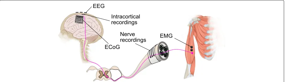
The motor control pathway of the human body goes from the high level associative area of the brain, mediated by the motor cortex, through the spinal cord to the indi-vidual muscle fibers. Each of the stages plays a different role and uses different mechanisms to ensure that a move-ment is carried out in a coordinated and smooth manner. Each of these stages also offers different signal modali-ties and features that can be exploited for motor decoding. We will now discuss these features and strategies to uti-lize them in details below. An overview showing the motor control pathway and various ways to intercept the control signal is shown in (Fig.1).
Cortical decoding of limb movements
All volitional motor controls originate from the brain. The motor cortex of the brain plays an especially impor-tant role in planning and executing motor commands. For some patients, the brain is the only site where motor intention can be captured because they have lost
motor functions in all their extremities (e.g. in tetraplegic patients). Therefore, many efforts have been invested in cortical decoding.
Electroencephalography (EEG)
EEG is the measurement of weak electrical signals from the brain on the surface of the scalp. Its origin is believed to be the summation of postsynaptic potentials of excitable neural tissues in the brain [14]. The skull, dura and cerebrospinal fluid between the brain and the EEG electrodes attenuate the electrical signal significantly, thus the EEG signal is very weak, typically under 150μV. Those structures also act like temporal low-pass filters, limiting the useful bandwidth of the EEG signal to be below 100 Hz [15]. Furthermore, due to the volume conduction effect of current sources in the head, the effect of a single current source spreads to several electrodes. The result is a spatial low-passing of the original signal, leading to a “smearing” of the signal source and reduction in the spatial resolution. Thus most EEG setups for motor decoding only involve 64 or 128 electrodes. Setups with higher than 128 electrodes are uncommon.
EEG signal is traditionally separated into several fre-quency bands (delta: 0 – 4 Hz, theta: 4 – 7.5Hz, alpha: 8 – 13Hz, beta: 13 – 30Hz, gamma: 30 – 100Hz). Of particu-lar importance to motor decoding is the brain oscillation in the alpha band over the motor and somatosensory cor-tex, also known as the μ-rhythm [16, 17]. It has been observed that there is a decrease of the signal power in the 8 – 13 Hz band when a subject is carrying out actual or even imagined movement [18,19]. Similar observations can also be found in the lower beta band (12 – 22Hz). Although some components of the beta band oscillation
Fig. 1Overview of various ways to intercept motor control signals. Motor control signal is relayed from the primary motor cortex of the brain, via the spinal cord and peripheral nerve, to the muscle fibers. The control signal can be intercepted at various points using different techniques.
Electroencephalography (EEG) captures the superimposed electrical fields generated by neural activity on the surface of of the scalp.
may be harmonics of the alpha band signals, the com-mon consensus now is that they are independent signal features due to having different topographic and timing characteristics [18, 20]. The mu-rhythm tends to focus on the bi-lateral sensorimotor area while the beta rhythm concentrates mainly on the vertex. Collectively, the modu-lation of the signal band power over the sensorimotor area is called sensorimotor rhythm (SMR).
This decrease of band power coinciding with an event is called event-related desynchronization (ERD). The oppo-site is called event-related synchronization (ERS), which is the increase of band power coinciding with an event. ERD/ERS is typically calculated with respect to a reference period, usually when the subject is wakefully relaxed and not doing any task [21]:
ERD= R−A
R ×100%
whereRis the band power during the reference period and Ais that during the time period of interest. An example
of ERD topography during motor imagery is shown in (Fig.2).
The ERD topography during movement displays an evolving pattern over time [21]. ERD usually starts around 2 s before actual movement, concentrating on the con-tralateral sensorimotor area, then spreads to the ipsilat-eral side and becomes bilatipsilat-erally symmetrical just before the start of movement. After the movement terminates, there is an increase of beta band power (i.e. ERS) around the contralateral sensorimotor area [21–23], also known as the “beta rebound”. The occurrence of beta rebound coincides with reduction in corticospinal excitability [24], suggesting the rebound may be related to the deactivation of the motor cortex after a movement terminates. Beta rebound occurs in actual as well as in imagined move-ments. An example of the beta rebound can be observed in (Fig2a).
Different kinds of motor imagery (MI) produce differ-ent topograpies of ERD and hence are useful for decoding the motor intention of the subject. For example, imaging
moving one’s hand will elicit ERD near the hand area of the motor cortex, which is in the more lateral position. On the other hand, imaging a foot movement will elicit ERD near the foot area in some of the subjects, which is closer to the sagittal line [25], as can be observed in (Fig.2c). The beta rebound after MI also displays a simi-lar somatotopic pattern [22]. Simultaneous ERD and ERS on different parts of the brain is also evident in some of the subjects. For example, some subjects showed ERD in the hand area and ERS in the foot area during a vol-untary hand movement, and vice versa during a foot movement[22]. ERD may represent an activation of the cortical area controlling the motion while an ERS may represent an inhibition of other unintended movements. As we recall from the neurophysiology of motor control, the indirect pathway of basal ganglia contains mecha-nisms to suppress the thalamic activation to SMA to filter out unintended movements. There are characteristic pat-terns of ERD/ERS during different actual and imagined movements, thus by looking into those patterns we can detect and distinguish the motor intention of different body parts.
The most reactive frequency band at which ERD/ERS occurs may be specific for each subject and even for the type of motor imagery, and its topography may vary slightly across different EEG preparations. Therefore, sig-nal processing and machine learning techniques are usu-ally employed to adapt to the signal features of the subjects automatically.
One of the most important signal processing step in SMR-based motor decoding is the estimation of signal power in the frequency range of choice, typically in the alpha (8–12 Hz) and beta (12–30 Hz) band. There are many methods to achieve this. One of the simplest and most computational efficient method is band-pass filter-ing [3, 26]. The EEG signal is first band-pass filtered in the frequency band of interest, then the sum of the square of the signal is then taken as the power of the signal in the chosen frequency band. Sum-of-the-square is equiva-lent to the variance of the signal, so usually the variance of the signal is used instead. After taking the variance, a log-transform is commonly employed. The log-transform can serve two purposes. First, it transforms skewed data to make them more conforming to the normal distribution [27], which may help improve performance in some clas-sification algorithms. Second, the log-transform empha-sizes the relative change of the signal rather than the absolute difference (e.g.log(110)−log(100)=log(1100)− log(1000)), so it can perform an implicit normalization of the signal and improve the performance of the classifier.
One of the major drawbacks of the simple band-pass fil-tering approach is that it may be difficult to choose the best frequency band to perform the filter, as each patient has their own specific reactive band. To overcome this
limitation, the adaptive auto-regressive (AAR) model is another commonly employed technique [28–31]. It mod-els the signal at current time point as a linear combination of previousppoints:
Yt=a1,tYt−1+a2,tYt−2+ · · · +ap,tYt−p+Xt
whereYt is the signal,Xt is the residue white noise and ap,t the autoregressive coefficients. The core difference with the traditional AR model is that in the AAR model, the coefficientsap,t are dependent on time and are cal-culated for each signal time point using recursive least square [32]. AAR coefficients from multiple electrodes are then concatenated together to form the feature vec-tor used by a classification system. AAR coefficients can be seen as the impulse response of a system and so it con-tains information about the frequency spectrum of the modeled signal. Compared to the traditional band-pass filtering, spectrum estimation using AAR can be more robust against noise. One can also specify the number of spectrum peaks based on domain knowledge (each peak requires two coefficients). Another advantage is that there is no need to choose a subject-specific frequency band beforehand as all model coefficients are used for classification. Another way to choose the subject-specific frequency band automatically is to use a filter bank that consists of multiple band-pass filters in different frequen-cies. After filtering, the most informative frequency band and channels are then selected using some performance metrics, e.g. whether deleting those feature will lead to a reversal of the classification label [33,34].
Due to the volume conduction problem in the human head, a single current source often appears to be “smeared” across several EEG electrodes. Spatial filtering is usually employed to improve the spatial resolution of the EEG signal. Popular spatial filters include the common average reference (CAR) and surface Laplacian [35]. These methods re-reference the signals by subtracting the volt-age at each electrode from the avervolt-age (as in CAR) or from its neighbors (as in surface Laplacian).
VjCAR=Vj− 1 N
k=1
N Vk
VjLAP=Vj− 1 n
k∈Sj Vk
where V is the signal voltage, N is the total number of electrodes, n the number of neighboring electrodes, and S is the set of neighboring electrodes in surface Laplacian (LAP).
the differences in variance between two classes are maximized. A further variation of the method is to add in frequency information by filtering the signal by a set of fil-ter bands and then calculate the CSP for each, and finally select the most informative feature through a mutual information criterion [38].
The performance of EEG-based motor decoding has been improving steadily over the years. While earlier stud-ies can only distinguish between discrete types of motor imagery [39], recent studies have already achieve 2D [40] and 3D control [41–43]. Some of the latest studies even demonstrate that it is possible to decode different move-ments in the same limb [44,45] or even individual finger movements [46].
Besides being used to replace the lost functions, EEG-based motor decoding can also be used a tool for rehabil-itation. For example, it can be used to control a robotic hand to assist in active hand training in post-stroke reha-bilitation [4,47,48]. This application of motor decoding as a tool for training is a very promising area, as it can potentially extend its use to a wider population.
Electrocorticogram (ECoG)
ECoG is the measurement of the electrical signals from the brain on top of the dura, but underneath the skull. ECoG measurement is commonly performed before an epilepsy surgery to delineate the epileptogenic area and identify important cortical regions to avoid during a resec-tion [49]. ECoG signal is not affected by the skull and thus tends to have a higher temporal and spatial resolu-tion than EEG. It also has a larger bandwidth (0 to 500 Hz) [50,51] and higher amplitude (maximum∼500μV [52]). Therefore, generally ECoG has a higher signal-to-noise ratio than EEG although it is also more invasive.
ECoG and EEG likely arise from the same underlying neural mechanisms therefore they share many similarities with each other. Howevers, there are two major signal fea-tures in motor decoding that are unique to ECoG and are specifically exploited. The first is the change of signal band power in the high gamma band (≥75Hz). Many studies have suggested that the high gamma band contains more informative features for motor decoding compared to the alpha and beta band, which are typically used in EEG decoding [53–57]. Interestingly, the high gamma band tends to increase during movement, unlike the alpha and beta band, which typically show desynchronization (i.e. decrease in power). Therefore, high gamma power may be produced by a different neural mechanism than the one that produces the alpha and beta desynchronization.
Another unique feature is the low-frequency amplitude modulation of the raw ECoG signal, coined as the Local Motor Potential (LMP) by Schalk et al. [30, 51]. It was found that the envelop of the raw ECoG shows a strik-ing correlation to the movement trajectory of the human
hand, as measured by a joystick. The amplitude also shows a cosine or sine tuning in relation to the movement direc-tion, similar to what have been observed in intra-cortical recordings. Since this discovery, many group have incor-porated the LMP into ECoG motor decoding in addition to other high frequency features (e.g. [53, 56, 58, 59]). The LMP is a very low frequency component (2-3 Hz) of the raw ECoG signal. It is usually extracted by Guas-sian low-pass filter, running average [30, 53,59], or the Savitzky-Golay filter [58,60,61].
Due to the robustness of the LMP signal, usually a sim-ple linear regression is sufficient to decode the motor intention in many of the previous studies (e.g. [51,62,63]), although a feature selection or regulation step may be needed to first remove the uninformative features. A recent study using deep neural network also show promises [64], however its improvement compared to classical techniques is not always significant.
Because ECoG has a better resolution and higher signal-to-noise ratio, it tends to produce better and finer results than EEG in motor decoding. Beside decoding the move-ment of different body parts as in EEG [65,66], different hand gestures can also be distinguished [56,67]. Using the LMP in addition to frequency features, position and veloc-ity of 2D arm movement can also be decoded from ECoG signals [30,51,58]. Subsequent studies even demonstrate that continuous finger positions can also be decoded [54,59,61,63,64,68]. The correlation coefficient between the predicted and actual finger movement can reach from 0.4 to 0.7 in some of the recent studies [61,64].
The large majority of studies in ECoG motor decod-ing are performed on epilepsy patients without a specific movement disorder or limb injury. However, one of the strongest motivation for motor decoding is that it can compensate the lost motor function of a patient. Given that the brain may re-organize due to disease or injury, it is vitally important that the decoding experiments be repeated on those patient population as well to see if similar decoding performance can be achieved. There are only a few studies to try ECoG motor decoding in stroke patients [57,69] and paralyzed subjects [70], but the results are encouraging.
Intra-cortical recordings
Penetrating electrodes for motor decoding are usually implanted into the primary motor area of the brain. There is a structure in the precentral gyrus resembling a “knob” that houses a majority of the neurons responsible for motor hand function [72]. This “motor hand knob” is typ-ically used as the target for electrode implantation (e.g. in [73–77]). Another potential target for implantation is the posterior parietal cortex (PPC). Although PPC has long been proposed to play an important role in the associative functions, in recent years more and more evidence sug-gests that it also encodes the high-level motor intention of the subject [78]. A recent study suggests that the goal and trajectory of the movement can be decoded from neural activities in human PPC [79].
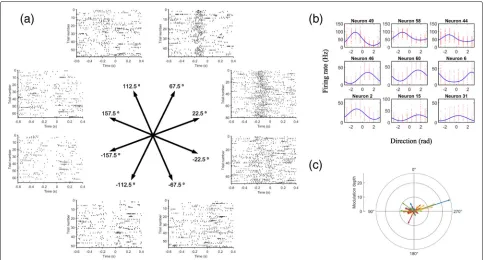
One important property exhibited by the neurons in the M1 is directional tuning. Some of the neurons are broadly tuned to a particular direction. They discharge the strongest when the movement is in their preferred direction, but they will also discharge less vigorously when the movement is in other directions. Their firing rates represent the length of their preferred direction vector. When the vectors of those neurons are summed together, it indicates the final direction of the movement. This pop-ulation encoding of movement is a striking property of
the nervous system. Similar analog of population encod-ing can also be found in the super colliculus representencod-ing the direction of eye movement [80]. An example show-ing the directional tunshow-ing property of M1 in a non-human primate is shown in (Fig.3).
Currently, the only FDA-approved, commercially avail-able microelectrode array for temporary (<30 days) intra-cortical recordings is the Neuroport System (Blackrock Microsystem, Inc, USA). As a result, majority of the work on human intracortical decoding are performed on that platform. Other intracortical electrodes do exist but they are either mainly for acute intraoperative mon-itoring (e.g. Spencer Depth Electrode, Ad-Tech; Neuro-Probes, Alpha Omega Engineering Ltd; microTargeting electrodes, FHC), or for EEG applications (e.g. DIXI Med-ical Microdeep Depth Electrodes).
The activities of the neurons in the implanted site are represented by their action potentials, which manifest as spikes in extracellular recordings. Therefore, detecting the occurrence of a spike is often the first step in intracorti-cal signal processing. There are many methods for spike detection [81,82]. The signal is typically first band-passed filtered in the spike frequency band (e.g. 300-5000Hz), then various methods are used to transform the filtered
signal to improve its signal-to-noise ratio (SNR). A detec-tion threshold is then calculated to distinguish spikes from background noise. One of the most common spike detection methods is to use the root-mean-square of the signal
Thres=C∗
1
N N
n=1 x[n]2
where Thres represents the detection threshold above which a signal time point is considered belonging to a spike. However, the RMS value may be easily contami-nated by artifacts, so another way is to use the median to set the detection threshold [83]
σ=median
|
x| 0.6745
Thres=4∗σ
The non-linear energy operator is also another popular method [83]. It first transforms the signal such that the high frequency component is amplified to improve the SNR.
ψ(x[n])=x[n]2−x[n+1]x[n−1]
Thres=C1 N
N
n=1
ψ[x(n)]
Other more advanced techniques like continuous wavelet transform [84] and EC-PC spike detection [82] can offer a better accuracy but at a higher computational cost. Although there are a lot of ways to detect spike accurately offline, not every one of them are fast enough to be used in real-time. Therefore in online decoding the choices are usually limited to the simpler algorithms. Manually set-ting a threshold by an operator still remains one of the most commonly used method. Another popular method in online decoding is the RMS method due to its high efficiency.
An electrode may record signals from multiple neu-rons nearby. Isolating the activity of a single neuron (i.e. signal-unit activity) from this multi-unit activity usually leads to better results in motor decoding. This process is called spike sorting. There is a large body of literature on spike sorting that cannot be exhausted here. Interested readers are encouraged to consult other excellent reviews [85–87]. In practice, the most popular way to do online, real-time spike sorting is via template matching. A set of spike templates are collected during a period of initial recording, then subsequent spikes are classified by com-paring their similarity with the templates. However, it may
not be really necessary, or may even degrade the decod-ing result, to do online spike sortdecod-ing. The spike clusters obtained from recordings may not be stable across differ-ent sessions of experimdiffer-ents. The total number of single units sorted from recording may change from sessions to sessions [79]. Thus a decoder trained on some sorted spikes may not work well on future sessions. Spike sorting may also introduce additional latency in online decoding, as accurate spike sorting is a computationally expensive process. In fact, many recent decoding studies do not use spike sorting at all, e.g. [79,88–94].
A decoding algorithm reconstructs motor kinematics from neural activity. Since the discovery of the directional tuning property of motor neurons, one of the earliest decoding algorithm for intracortical spike signal is the population vector algorithm[95,96]. In its simplest form, the firing rate of a neuron can be related to its preferred direction by
f =f0+fmaxcos(θ−θp)
wheref is the neural firing rate,f0andfmaxare regression constants andθandθpare the current and preferred direc-tion respectively. However, for cosine funcdirec-tion the width of the modulation is fixed. A more flexible tuning function that allows adjustable width of the modulation is the von Mises tuning function [97]:
f =b+k exp(κcos(θ−μ))
whereb,k,κ,μare the regression constants, andθ is the current movement direction. Whenμ = θ, the function will be at maximum, soμcan also be interpreted as the preferred direction of the neuron. Examples of the von Mises tuning curves are shown in (Fig.3b).
The preferred directions of each of the neurons then can be summed together to predict the target direction [96].
P(M)= N
i=1
wi(M)Ci
whereCiis the preferred direction for thei-th neuron, and wi(M)is the weighting function combining the contribu-tions of each neuron in directionMto the final population vector. However, this method requires a large number of neurons to be accurate and may lead to error if the distri-bution of the preferred direction is not uniform [98]. For example in (Fig.3c), we can see that the preferred direc-tions are not distributed evenly. For this reason, a simple linear regression scheme is usually employed instead in recent studies [73],
u=Rf=R(RTR)−1RTk
motor kinematic values (e.g. joint angles or cursor posi-tions). It has been suggested that this regression scheme can provide more accurate prediction compared to the summation of preferred direction vectors, especially when those vectors are not uniformly distributed [98].
In recent years, the Kalman filter is usually employed instead of the simple linear regression (e.g. in [75–77, 99,100]). The Kalman filter incorporates the information both from an internal process model and actual measure-ment to estimate the states of a system [101]. A Kalman gain variable is used to determine the “mixing weight” of the model and measurements. If the model is more accurate, then it will trust the model more. The same goes for the measurement. Kalman filter is especially useful if the states are not directly observable or if the measurement is very noisy, which are often both true in motor decoding. In motor decoding, the subjects usually lost their limb or ability to move, therefore the internal state (e.g. motor intention) of the system is not directly observable. The observable variables (e.g. neural activity) are also very noisy. A typical Kalman filter for motor decoding assumes no control variable and the system can be formulated as two linear equation [102,103]):
xt = Axt−1+ wt−1
yt = Cxt+ vt
where x is the state of the system one want to decode, e.g. joint kinematics or cursor position.yis the observed variables, e.g. neural firing rate.wtandvtare the process and measurement noises drawn fromwt ∼ N(0,Q)and vt ∼ N(0,R)respectively.A,C,QandRare the Kalman constants that need to be defined according to the decod-ing model. For the internal statex, if it is a cursor position, it can be expressed as
xt=[post,velt, 1]T
With the model defined, the Kalman gainKand the esti-mation error covariancePthen can be updated with the typical two-step update equations:
Predict:
ˆ
x−t = Axˆt−1+But Pt− = APt−1AT+Q
Update:
Kt =
P−tCT CPt−CT+R
ˆ
xt = ˆx−t +Kt(yt−Cxˆ−t ) Pt = (I−KtC)P−t
wherexˆ−andxˆare thea prioranda posteriorstate esti-mates respectively.uis the control variable. Typically it is set to 0 in motor decoding, here we have included it for completeness.
One crucial aspect of performing online motor decod-ing is the traindecod-ing and re-calibration of the decoddecod-ing model. Although the neural features for similar move-ments are relatively stable within a few days [104], the neural tuning curve may start to change when the sub-ject is learning to perform a new task [105]. It is also very difficult to track the same neuron for an extended period of time [106,107], due to the micro-movement of electrodes and fluctuations of other noise sources. Fur-thermore, training data are often acquired in an open-loop fashion, meaning that no feedback is provided by the decoder during training. However, in actual decoding ses-sion, feedback is provided and the subject may attempt to change his motor imagery in order to “learn” the decoder. This may lead to changes in the underlying neural fea-tures [108]. Therefore, re-calibration of the trained model is often necessary and will be ideal if it can be performed online. A successful re-calibration method is the ReFIT-KF algorithm proposed by Gilja et al [109]. ReFIT-KF assumes the subject’s true intention is to move towards the target, so it can generate a pseudo-ground truth from the decoded result automatically even though the prediction of the current model may be wrong. It can then calibrate the model using the estimate ground truth to adapt for the instability of the neural signals. It is able to produce better results than Kalman filter alone [92,93,109].
Due to the more robust signals obtained by intracor-tical recordings, it has been utilized successfully to help tetraplegia patient control the environments in various ways, including 2D cursor control [73,76,94], virtual and real prosthetic hands [77,79,92,110,111] and functional electrical stimulation of the patients’ own paralyzed hands [90,91,93].
Peripheral decoding of limb movements
Signals from the central nervous system (CNS) even-tually arrive at the peripheral nervous system (PNS) and drive the contraction of different muscle fibers. Compared to CNS, signals in the peripheral structures are usually more specific. They contain detailed instructions on the contractions of individual muscle fibers, there-fore potentially can enable dexterous prosthetic control. Surgeries involved in peripheral interface are usually less complicated than those involving the intracortical struc-tures. Therefore, many studies are also devoted to motor decoding in the peripheral structures.
Peripheral nerve recordings
to explore the possibility of decoding efferent periph-eral nerve signals for prosthetic control. Because the peripheral nerves contain low-level information target-ing each muscle, it may be possible to regain high-dexterity and naturalistic control by exploiting this rich information.
One of the major challenges in peripheral nerve record-ings is accessing the axons in the nerves. Axons in spinal nerves are bundled in fascicules and multiple fascicules are grouped together to form a peripheral nerve. Those axons are enclosed in three sheaths of connective tissues – the epineurium that covers the entire nerve, and the perineurium that encloses a fascicle and the endoneurium that holds the neurons and blood vessels together within a fascicle. Due to these multiple layers of lamination around an axon, the amplitude of a peripheral nerve signal is usually very small, can be around 5 – 20μV [112].
There are multiple electrode configurations designed to get a better signal from the peripheral nerves [113]. The cuff electrode [114], as its name suggests, works like a cuff to wrap around a nerve. Its main advantage is that it causes minimal damage to the neural tissues as it does not require any incision on the nerve itself. However, since it only measures the electrical potential at the surface of a nerve, it can only obtain a grand summation of the neural activity in different fascicles. Another variation of the cuff electrode is the flat interface nerve electrode (FINE) [115]. It works like a clip to apply pressure on the nerve and make it flattened into an oval shape, thus increasing its surface area and reducing the distance from the electrode to the fascicles. There are also other types of electrodes that are implanted into the nerves. They offer higher selectivity due to their direct contact with the fascicles. However, they are also more invasive and may cause more damage to the nerve. The longitudinal intrafascicular electrodes (LIFE) are long, thin wires implanted longitudinally into the nerve fascicles [116]. On the other hand, the trans-verse intrafascicular multichannel electrodes (TIME) are implanted transversely into the nerves, accessing multiple fascicles at the same time. There is also the Utah Slanted Electrode Array [117], which consists of an array of elec-trodes with different lengths, such that when the array is inserted into the nerve, the tip of the electrode can get into contact with different fascicles. Recently, there is also development of the regenerative peripheral neural interface (RPNI) [118], which uses a muscle graft to wrap around severed fascicles endings. The nerve endings grow into and innervate with the graft, creating a new interface for acquiring neural signal. Of the different types of elec-trodes introduced, only the cuff electrode is currently used in commercial FDA-approved systems for vagus nerve stimulation (e.g. VNS Therapy, Cyberonics, USA). Most of the others are still in research or undergoing clinical trials [119].
Studies on the human decoding of peripheral signals are still very limited, partly due to the challenge of acquiring nerve signals with sufficient SNR, and may also due to the cross-talk between neural signals and EMG, as the periph-eral nerves are usually located in close proximity with the limb musculature. The majority of existing studies focus on upper limb decoding, as upper-limb amputation tends to have a bigger impact on the everyday life of the patients. Neural recording are performed on the ulnar, medial and/or the radial nerve. Different types of elec-trodes are used, but the more common ones in human decoding are the Utah slate electrode (e.g. in [120,121]) and the LIFE (e.g. [122–124]).
The analysis of peripheral signals commonly involves the detection of action potentials in the nerve. The detec-tion procedures are similar to those used in intracortical studies, but the step of clustering spikes is not usually performed. Due to the low SNR of the peripheral sig-nals, sometimes they need to be first de-noised (e.g. by wavelet [124]) before detection. The firing rate of the action potential can then be fed into a regressor (e.g. in [103, 120–122]) or a classifier (e.g. in [123, 124]) for decoding. The difference in using a regressor or a classi-fier lies in whether a discrete gesture or a continuous joint trajectory is decoded.
Support-vector machine (SVM) is the most commonly used classifier for peripheral decoding (e.g. in [123,124]). For regressor, simple linear regression or a Kalman fil-ter have been used ([103,120–122]). Kalman filter allows the online recursive update of the model in real-time, and is especially helpful when the measurement of the target variable is noisy (as often in the case of motor decoding, since it is not possible to measure the actual movement of the missing limb).
The issue of obtaining ground truth for training the decoder is also very important. While for discrete grasp type classification, it may be sufficient to ask the sub-ject to imagine holding a particular grasp, for position decoding a more precise approach have to be used. One common solution is to show a shadow hand on a screen, and ask the subject to try to follow the movement of the hand, either through a manipulandum controlled by the mirrored movement in the intact hand [121] or through imagined phantom limb movements only.
based on Kalman filter is able to classify 13 different movements offline, but only 2 movements can be decoded online successfully due to the cross-talk between different degree-of-freedoms (DoFs) [121].
The peripheral nerves offer a promising target for motor decoding. It is more downstream in the motor control pathway and contains more specific information about muscle activities. This property can be poten-tially exploited to enable high dexterity control. Access to peripheral nerves is also relatively easier than intracortical structures. However, peripheral recordings are plagued by their low SNRs due to the multiple levels of lamina-tion around an axon. This may be improved by better electrode designs, and ultra-low-noise neural amplifiers that can resolve the small amplitude of the nerve signals (e.g. [126]).
Electromyogram (EMG)
EMG signals are the sum of the electrical activities of the muscle fibers, which are triggered by spike trains, i.e. impulses of activation of the innervating motor neu-rons. EMG signals can be measured in two ways, either on the surface of the skin above a muscle (surface EMG),
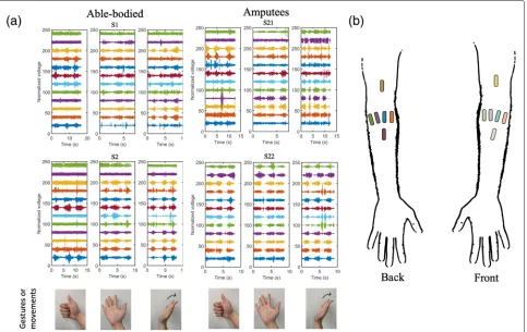
or directly inside a muscle fiber using a needle elec-trode (intramuscular EMG). An example of EMG data in different hand gestures is shown in (Fig.4).
Myoelectric signals have been used as the control source for decades in prostheses, in which muscle sig-nals are recorded and translated into control commands to induce prosthesis motions. Intramuscular EMG sig-nals are believed to be of a higher resolution and less susceptible to cross-talks compared with surface EMG because of its more invasive electrode deployment and direct targeting of specific muscles.
Despite decades of research and development, amputees still do not use state-of-the-art myoelectric prostheses more frequently than the basic, body-powered hooks [127], and an estimate of 40% of upper-limb amputees actually reject using a prosthesis [128]. One primary limitation of clinically available hand-prosthesis is the number of simultaneously and proportionally controllable degrees of freedom (DoFs), which is rarely greater than 2 [129, 130] and has focused mostly on wrist DoFs without the hand [131], although func-tions of hand-movement are more essential for daily living.
Myoelectric control can be categorized into direct con-trol and pattern recognition concon-trol. Direct concon-trol refers to the type of methods that use the amplitude of two surface EMG inputs from an antagonistic muscle pair to control the two directions (ON and OFF) at a prosthetic DoF. Due to the inadequate remaining musculature, signal crosstalk contamination, and attenuation of deep mus-cle signals at the skin level, the number of independent myosites in the residual forearm is typically limited to two, only allowing the control of one DoF at a time. As a result of this constraint, patients need to toggle between modes using quick co-contraction at the myosites to sequentially control multiple DoFs. Pattern recognition control relies on machine learning algorithms to train a separate classifier for each DoF. Multiple classifiers have been proposed and evaluated, including quadratic dis-criminant analysis [132], support vector machine [133], artificial neural network [134], hidden Markov models [135], Gaussian mixture models [136], and more. How-ever, as training of the computational models involves the movement of only 1-DoF, the trained classifiers do not support simultaneous control of multiple DoFs. A more promising approach based on machine learning is adopt-ing a regression-based control scheme (instead of classi-fication) that inherently facilitates continuous control (as opposed to ON and OFF), in which a linear or nonlinear mapping from EMG signal features to the changes of pros-thesis DoFs is learned. Commonly used methods for this purpose include artificial neural networks [137], support vector machine [138], and kernel ridge regression [131]. A major shortcoming of regression-based control is the requirement for large amount of training data that include an exhaustive combination of movements of all prosthesis DoFs, which is impractical to be clinically implemented.
One of the fundamental issues with EMG based pros-thesis control is the scarcity of independent signals with which to control prosthesis DoFs. EMG signals are inher-ently heavily correlated and lacks the resolution and the information capacity needed for simultaneous and pro-portional control of multiple DoFs. A potential solution to this problem is to record motor commands directly from the peripheral nerves, such as ulnar and median nerves that directly innervate all five fingers. However, this comes at the costs of invasive surgical implantation of electrodes and the risks of tissue infection and nerve damage.
There have been works to extract more invariant and independent information from EMG signals without inva-sive recordings. One major group of the efforts focuses on extracting muscle synergy features from EMG record-ings, i.e., the complex muscle activation patterns that are executed by users as high-level control inputs regard-less of any neurological origin [139]. Muscle synergies are believed to be capable of describing complex force and motion patterns in reduced dimensions and can be used
as a robust representation for decoding outputs consis-tent with user’s inconsis-tent. Non-negative matrix factorization (NMF) [140] has been commonly used to extract muscle synergies from multichannel EMG signals for simultaneous and proportional control of multiple DOFs [137,141–143]. Another group of works focuses on directly extracting the neural codes of motor neuron activities that govern the muscle movements through the nerve pathway. This nor-mally requires advanced recording setups such as high-density EMG with a sufficient number of recording sites that are closely spaced. A number of algorithms have been proposed to extract the underlying neural information [144, 145]. Among them, convolution kernel compensa-tion (CKC) has been most extensively used as a type of multichannel blind source separation method [146–149]. Despite the promise of extracting neural contents from high-density EMG signals, the demonstration of utiliz-ing such scheme in online experiments remains difficult. More in-depth investigation and significant efforts are needed to build neural interface and achieve direct neural-based control neural-based on this framework.
Decoding of speech motor activities
Although this review mainly focuses on the decoding of movement in the extremities, recently there are also another line of research in decoding motor speech activ-ities [150,151]. Speech production is a complex process involving multiple areas of the brain and dozens of mus-cles fibers. The muscle activities need to be highly coordi-nated to produce different speech sounds (i.e. phonemes) which concatenate together to form intelligible words and sentences.
Multiple brain regions are associated with language pro-duction [152], but there are two major areas that have received more attentions in speech decoding. The left ventral premotor cortex has been suggested to repre-sent high-level phonemes in speech [153,154], while the ventral sensorimotor cortex contains rich representations of different speech articulators (e.g. lip, tongue, larynx etc.) [155, 156]. Therefore most of the decoding efforts concentrate on these two brain regions.
electrodes in the otherwise healthy eloquent cortex to conduct experiments. Currently, ECoG obtains a greater success in speech decoding due to its high signal quality and less invasive nature. ECoG recordings are also com-monly employed during brain resection to avoid damage to the eloquent cortex, so it is well-integrated into exist-ing surgical procedures. Studies usexist-ing ECoG for speech decoding mainly focus on the high gamma band (70-170Hz), as it has been shown that the high gamma activity correlates strongly with ensemble firing rate [159].
Earlier speech decoding efforts have focused on the direct decoding of simple words or phonemes [150,157, 158,160–162], but their performance is not very satisfac-tory. Decoding from a limited dictionary or phoneme set may produce a higher accuracy (e.g.>80% for 10 words [160] or 9 phonemes [157]), but it can only cover a very narrow range of human spoken expressions. Studies try-ing to decode the full range of English phonemes result in a lower classification accuracy (10-50% [150,155,162]). The low classification accuracy can be partly mitigated by incorporating a pronunciation dictionary and language model (e.g. in [150]), which can limit the output of the decoder to more probable words.
On the other hand, recently attentions have been shifted to focus more on decoding the intermediate representa-tion of speech (e.g. articulator movements) rather than decoding phonemes directly. Part of the shift may be motivated by the growing body of evidence suggesting that the speech motor cortex is able to generate differen-tial activation patterns encoding the kinematics of speech articulators [156, 163–165]. Advances in deep learning has made the prediction of articulator trajectories from acoustic signal (i.e. acoustic-articulatory inversion) accu-rate enough to act as the ground-truth for decoding, as the traditional ways of implanting coils or magnets in the mouth via articulography is invasive and not compatible with neural recordings [166]. In one very recent study [167], a deep neural network is used to decode ECoG fea-tures to articulator trajectories. The trajectories are then decoded by another neural network to acoustic features (e.g. pitch, mel-frequency cepstral coefficients etc.), which are then converted to audible voice using a voice synthe-sizer. Even mimed speech can be decoded, although with a lower accuracy. In another study [168], ECoG features are decoded into mel-scaled spectrograms directly using a neural network, then a neural network vocoder is used to construct the spectrogram into audible waveforms. These recent results show great promises in decoding human speech from ECoG signals. A summary of the different methods of motor decoding is shown in Table1.
Challenges and future direction
Although great strides have been made in decoding human motor intention, there are still some significant
challenges remain to be solved. One of the biggest challenge preventing the adoption of motor decoding out-side the laboratory is the limited longevity of the decoding model. Typically, some calibration session is needed to collect data to train the decoding model, then the model is tested on subsequent sessions on the same or next few days. While it is acceptable in a scientific study due to the limited time and clinical resources available, in actual daily use, the trained model must be able to maintain its performance for an extended period of time.
The limited longevity can be due to several reasons. First is the instability of the electrode interfaces. Micro-movement of the electrodes may cause a shift in the feature space. If the decoder is not robust enough, this shift may result in a deterioration of the decoding per-formance. Another reason is the different environment noises injected into the acquired signals. Neural signals used for decoding usually have a very small amplitude and thus are susceptible to interference by environment noises. A cell-phone, fluorescence lamp or other elec-trical appliances all inject various types of noise in the acquired signal. As the subjects are performing various tasks in daily lives, they may come into the influence of different noise sources not covered in the trained data set and results in performance degradation. The third reason is the slow build up of immune response on the elec-trode interface. Glial scars may encapsulate the elecelec-trode and increase its impedance [174]. Neurodegeneration as a result of immune response will also lead to a weaker sig-nal [175]. The model longevity problem is multifaceted and must be carefully addressed. First, a better electrode design can help secure the electrode onto its anchor-ing structure and reduce their relative movement. An implantable solution will also produce more stable feature than one that requires repeated dismantling and rein-stallation every time (e.g. EEG and EMG). Second, the model should be trained with more robust features and tested in an environment typical of its everyday use. A shielded chamber may help acquire very clean signals that are good for the demonstration of a prototype. However, it is unlikely that the same quality of signals can be acquired in everyday environment. Thus it is also important to con-sider how a decoder is tested rather than just looking at offline numerical metrics. Thirdly, advancement in the electrode materials or special organic coatings can poten-tially reduce its immune response [176]. A flexible instead of rigid electrode may also cause less neuronal damage and inflammation [177,178].
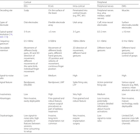
Table 1Comparison of different methods for motor decoding
Cortical Peripheral
EEG ECoG Intra-cortical Peripheral nerves EMG
Decoding site Scalp On the surface of
the brain
Penetrated into cortical tissues (e.g. PPC, M1)
Peripheral nerves (e.g. ulna, median, radial nerves)
Muscles
Types of electrode
Disk electrodes Flexible electrode
array
Utah array Cuff, intra-neural
electrodes
Surface electrodes,needle electrodes
Typical spatial resolution [14,
169–173]
5-9 cm <5 mm 3-5μm 0.5-2 mm >10 mm
Frequency spectrum
0.5-100Hz 0-500Hz 100Hz-20kHz 0.1-10kHz 0.1Hz-10kHz
Decodable intention
Movement of different body parts, 2D and 3D direction of movement, different movements of the same limb, individual finger movement
Movement of different body parts, different hand gestures, 2D position and velocity of movement, continous finger position
2D direction of movement, different hand gestures
Different hand gestures
Different hand gestures, proportional control of grasps
Signal-to-noise ratio
Low Medium High Low High
Signal feature Bandpower,
ERS/ERD
Bandpower, LMP Spike firing rate,
LFP
Action potential firing rate
Various signal features (e.g. RMS, variance, mean absolute value etc.)
Invasiveness Low High Very high Medium Low
Advantages Non-invasive,
easily deployable
Fine-grained and robust feature, mature surgical procedures as part of epilepsy treatment
Fine-grained and robust feature
Less invasive, potentially contains detailed information about muscle activations
Non-invasive, mature
technology, easily deployable
Disadvantages Low
signal-to-noise ratio, high variability of features between sessions, time-consuming to setup
Invasive, long-term implantation not common
Very invasive, require implantation surgery
Low
signal-to-noise ratio
Limited DoF, exessive cross-talk between different channels
provide feedback to the subject based on the decoder outputs. When the decoder output is wrong, the subject may try to correct it deliberately, and that may lead to dis-crepancy in the offline and online performance [179]. One of the solutions is to introduce a small calibration session with feedback at the beginning of the testing session, like in many EEG-based motor decoding studies. The origi-nal model is trained with an open-loop paradigm, then the model is further fine-tuned with feedback in the cal-ibration session. However, this is only possible if a clear ground truth is available. For the case in which the ground truth is not available, e.g. in the case of a tetraplegic patient where it is very difficult to know the true intention
the electrode interface or environmental noises. However, online calibration demands that the model can be updated quickly, which puts an constraint on the complexity of the decoding model. More research is needed to study how to update the decoder efficiently in real-time.
Besides advancement in decoding algorithms, develop-ment of new electrodes and neural amplifiers also play a very important part in advancing motor decoding. Recent trends in electrode development mainly focus on improv-ing four areas of electrode design: density, flexibility, biocompatibility and connectivity. Denser electrode can improve the spatial resolution of neural recordings. High-density electrode has been created from silicon wafer and carbon fiber monofilament [180, 181]. Electrode mate-rial with a flexibility closer to that of brain tissues can reduce neural damage and inflammatory response. Many flexible polymers have been used to make neural elec-trode, including polyimide [182, 183], parylene [184], PDMS [185] etc. Biocompatibility is always an important issue in electrode design because inflammatory response and encapsulation deteriorate signal quality over time and undermine the quality of chronic neural recordings. Strategies to improve biocompatibility including using inert metals like gold or platinum, using flexible materi-als to reduce tissue damage, or coating the electrode with biocompatible materials like conducting polymer [186] and carbon nanotubes [187]. Read-out connection from the electrodes will also quickly become a problem when the density and number of electrode continue to increase. Incorporating transistors into the electrodes directly to enable connection multiplexing is one of the ways to mit-igate this problem [188,189]. Readers interested in neural electrode designs are suggested to consult other more in-depth reviews in this area [119,172,176,177,190].
Development of neural amplifiers also plays a very important role in advancing the science of motor decod-ing, as we first need to acquire a clear neural signal before any processing and decoding can be done. There are mul-tiple lines of research trying to improve the different aspects of the amplifier design. Firstly, the power con-sumption of an amplifier can be reduced by resource shar-ing (e.g. one amplifier sharshar-ing multiple electrodes [191] or multiple amplifiers sharing one analog-to-digital con-vertor [192]), power scheduling (e.g. switching off unused components [193], dynamically adjusting the amplifier parameters [194]), or reduction of supply voltage [195]. Secondly, the channel count can be increased by multi-plexing or integrating amplifiers directly with the elec-trodes [191,196]. Thirdly, the circuit noise can be reduced by trimming [197], chopping [198, 199], auto-zeroing [200] or frequency-shaping [201] etc.. Fourthly, wire-less transmission of power or data can be achieved by an inductive link [193, 202, 203], short-distance power harvest [193, 204] or even ultrasound [205]. Finally, the
functionality of the amplifier can also be expanded by integrating more signal processing on-chip, e.g. spike detection [203], spike sorting [206, 207] and data com-pression [208,209]. Interested readers are encouraged to consult other more focused reviews in this area [210–213].
Conclusions
Every year, a large number of patients suffer from vari-ous degrees of movement disability due to amputation or neurological disorders. Their everyday lives and works are severely affected. With modern neurotechnology, it is now possible to intercept and decode the motor intention at different points along the neuro-muscular control path-way and use that information to drive a prosthetic device to restore movement. In this paper, we have reviewed the various signal features and techniques to decode motor intention in human. Although motor decoding perfor-mance is improving steadily with the advancements in electrode configurations, neural amplifier designs and decoding algorithms, we are still very far away from the goal of achieving naturalistic and dexterous control like our native limbs. The eventual successful clinical appli-cation of motor decoding will depend on the concerted efforts of both healthcare and engineering professionals, and likely also needs to be tailored-made according to the conditions and ability of each patient. We hope our review can provide a useful overview of the current state-of-the-art in motor decoding, so that researchers interested in the field can be aware of the neural features that they can exploit, potential problems they may encounter and the available solutions that they can adopt.
Acknowledgment
Not applicable.
Abbreviations
AAR: adaptive auto-regression; CAR: common average reference; CKC: convolution kernel compensation CSP: common spatial pattern; CNS: central nervous system; DoF: degree of freedom; ECoG: electrocorticogram; EEG: electroencephalography; EMG: electromyogram; ERD: event-related desynchronization; ERS: event-related synchronization; FINE: flat interface nerve electrode; GPi: internal globus pallidus; LIFE: logitudinal intrafascicular electrodes; LMP: local motor potential; M1: primary motor cortex; NMF: non-negative matrix factorization; NHP: non-human primate; PMA: premotor area; PNS: peripheral nervous system; PPC: posterior parietal cortex; RPNI: regenerative peripheral neural interface; SMA: supplementary motor area; SMR: sensorimotor rhythm; SNR: signal-to-noise ratio; STN: subthalamus nucleus; TIME: transverse intrafascicular multichannel electrodes; VLo: oral part of ventral lateral nucleus
Authors’ contributions
WKT is responsible for the majority of the paper. TW writes the section in EMG motor decoding and helps edit the whole paper. QZ provides guidance on machine learning algorithms to decode neural signals. EK provides guidance on physiology. ZY edits the paper and provides overall guidance for this review.
Funding
This work was supported in part by the DARPA under Grants
Availability of data and material
All data analysed during this study are included in their corresponding original articles [214–217].
Ethics approval and consent to participate
This review includes re-analysis of data from other published studies. Each of the cited studies has received ethics approval in their respective institutions.
Consent for publication
Not applicable.
Competing interests
The authors declare that they have no competing interests.
Author details
1Department of Biomedical Engineering, University of Minnesota Twin Cities,
7-105 Hasselmo Hall, 312 Church St. SE, 55455 Minnesota, USA.2Department of Computer Science and Engineering, University of Minnesota Twin Cities, 4-192 Keller Hall, 200 Union Street SE, 55455, Minnesota, USA.3Nerves Incorporated, P. O. Box 141295, Dallas, Texas.
Received: 7 February 2019 Accepted: 21 July 2019
References
1. Ziegler-Graham K, MacKenzie EJ, Ephraim PL, Travison TG, Brookmeyer R. Estimating the Prevalence of Limb Loss in the United States: 2005 to 2050. Arch Phys Med Rehabil. 2008;89(3):422–9.https://doi.org/10.1016/ j.apmr.2007.11.005.http://arxiv.org/abs/arXiv:0803.1592v1.
2. Adams PF, Hendershot GE, Marano MA. Current estimates from the National Health Interview Survey. Vital Health Stat. 1999;10(200):1996.
https://doi.org/10.1037/1099-9809.6.2.168.
3. Wing-Kin T, Kai-yu T, Fei M, Shangkai Gao. A Minimal Set of Electrodes for Motor Imagery BCI to Control an Assistive Device in Chronic Stroke Subjects: A Multi-Session Study. IEEE Tran Neural Syst Rehabil Eng. 2011;19(6):617–27.https://doi.org/10.1109/TNSRE.2011.2168542. 4. Ang KK, Chua KSG, Phua KS, Wang C, Chin ZY, Kuah CWK, Low W,
Guan C. A Randomized Controlled Trial of EEG-Based Motor Imagery Brain-Computer Interface Robotic Rehabilitation for Stroke. Clin EEG Neurosci. 2015;46(4):310–20.https://doi.org/10.1177/
1550059414522229.
5. Bear MF, Connors BW, Paradiso MA. Neuroscience: Exploring the Brain, 4th editio. Philadelphia: Wolters Kluwer Health; 2015.
6. Kandel ER, Schwartz JH, Jessell TM. Principles of Neural Science, 4th editio. New York: McGraw-Hill Medical; 2000.
7. Andersen RA, Buneo CA. Intentional maps in posterior parietal cortex,. Ann Rev Neurosci. 2002;25:189–220.https://doi.org/10.1146/annurev. neuro.25.112701.142922.
8. Weinrich M, Wise SP. The premotor cortex of the monkey,. J Neurosci. 1982;2(9):1329–45.
9. Calabresi P, Picconi B, Tozzi A, Ghiglieri V, Di Filippo M. Direct and indirect pathways of basal ganglia: A critical reappraisal. Nat Neurosci. 2014;17(8):1022–30.https://doi.org/10.1038/nn.3743.
10. DeLong MR. Primate models of movement disorders of basal ganglia origin. Trends Neurosci. 1990;13(7):281–5. https://doi.org/10.1016/0166-2236(90)90110-V.
11. Albin RL, Young AB, Penney JB. The functional anatomy of basal ganglia disorders,. Trends Neurosci. 1989;12(10):366–75.https://doi.org/10.1093/ jhered/esy024.
12. Graziano M. The Organization of Behavioral Repertoire in Motor Cortex. Ann Rev Neurosci. 2006;29(1):105–134.https://doi.org/10.1146/annurev. neuro.29.051605.112924.
13. Splittgerber R. Snell’s Clinical Neuroanatomy, 8th. Philadelphia: Wolters Kluwer; 2019.
14. Buzsáki G, Anastassiou Ca, Koch C. The origin of extracellular fields and currents — EEG, ECoG, LFP and spikes. Nat Rev Neurosci. 2012;13(6):407– 20.https://doi.org/10.1038/nrn3241.http://arxiv.org/abs/NIHMS150003. 15. Webster JG. Medical Instrumentation Application and Design, 4th. New
Jersey: Wiley and Sons; 2009.
16. Chatrian GE, Petersen MC, Lazarte JA. The blocking of the rolandic wicket rhythm and some central changes related to movement.
Electroencephalogr Clin Neurophysiol. 1959;11(3):497–510.https://doi. org/10.1016/0013-4694(59)90048-3.
17. Schomer DL, da Silva FL. Niedermeyer’s Electroencephalography: Basic Principles, Clinical Applications, and Related Fields. Philadelphia: Lippincott Williams & Wilkins; 2011.
18. McFarland DJJ, Miner LAa, Vaughan TMM, Wolpaw JRR. Mu and beta rhythm topographies during motor imagery and actual movements,. Brain Topogr. 2000;12(3):177–86.
19. Han Y, Bin H. Brain-Computer Interfaces Using Sensorimotor Rhythms: Current State and Future Perspectives. IEEE Trans Biomed Eng. 2014;61(5):1425–35.https://doi.org/10.1109/TBME.2014.2312397.http:// arxiv.org/abs/NIHMS150003.
20. Wolpaw JR, Birbaumer N, McFarland DJ, Pfurtscheller G, Vaughan TM. Brain-computer interfaces for communication and control. Clin Neuropsychol. 2002;113(6):767–91.
21. Graimann B, Huggins JE, Levine SP, Pfurtscheller G. Visualization of significant ERD/ERS patterns in multichannel EEG and ECoG data. Clin Neuropsychol. 2002;113(1):43–7. https://doi.org/10.1016/S1388-2457(01)00697-6.
22. Pfurtscheller G, Lopes FH. Event-related EEG / MEG synchronization and desynchronization : basic principles. Clinical Neurophysiology. 1999;110: 1842–57.http://dx.doi.org/10.1016/S1388-2457(99)00141-8.http://arxiv. org/abs/S1388-2457(99)00141-8..
23. Pfurtscheller G, Stancák a, Edlinger G. On the existence of different types of central beta rhythms below 30 Hz,. Electroencephalogr Clin Neurophysiol. 1997;102(4):316–25.
24. Chen R, Yaseen Z, Cohen LG, Hallett M. Time course of corticospinal excitability in reaction time and self-paced movements. Ann Neurol. 1998;44(3):317–25.https://doi.org/10.1002/ana.410440306. Accessed 5 Jan 2019.
25. Pfurtscheller G, Neuper C, Andrew C, Edlinger G. Foot and hand area mu rhythms. Int J Psychophysiol. 1997;26:121–35.
26. Blankertz B, Losch F, Krauledat M, Dornhege G, Curio G, Müller K-R. The Berlin Brain–Computer Interface: accurate performance from first-session in BCI-naïve subjects,. IEEE Trans Biomed Eng. 2008;55(10): 2452–62.https://doi.org/10.1109/TBME.2008.923152.
27. Feng C, Wang H, Lu N, Chen T, He H, Lu Y, Tu XM. Log-transformation and its implications for data analysis,. Shanghai Arch Psychiatry. 2014;26(2):105–9.https://doi.org/10.3969/j.issn.1002-0829.2014.02.009. 28. Schlögl A, Flotzinger D, Pfurtscheller G. Adaptive autoregressive
modeling used for single-trial EEG classification. Biomed Tech. 1997;42(6):162–7.https://doi.org/10.1515/bmte.1997.42.6.162. 29. Pfurtscheller G, Neuper C, Schlögl a, Lugger K. Separability of EEG
signals recorded during right and left motor imagery using adaptive autoregressive parameters. IEEE Trans Rehabil Eng Publ IEEE Eng Med Biol Soc. 1998;6(3):316–25.
30. Schalk G, Kubánek J, Miller KJ, Anderson NR, Leuthardt EC, Ojemann JG, Limbrick D, Gerhardt LA, Moran D, Wolpaw JR. Decoding
two-dimensional movement trajectories using electrocorticographic signals in humans,. J Neural Eng. 2007;4(3):264–75.https://doi.org/10. 1088/1741-2560/4/3/012.
31. McFarland DJ, Wolpaw JR. Sensorimotor rhythm-based brain-computer interface (BCI): Model order selection for autoregressive spectral analysis. J Neural Eng. 2008;5(2):155–62.https://doi.org/10.1088/1741-2560/5/2/ 006.
32. Haykin S. Adaptive filter theory, (5th ed). New Jersey: Prentice-Hall; 2014. 33. Wang T, Deng J, He B. Classifying EEG-based motor imagery tasks by
means of time-frequency synthesized spatial patterns. Clin Neuropsychol. 2004;115(12):2744–53.https://doi.org/10.1016/j.clinph. 2004.06.022.
34. Yamawaki N, Wilke C, Liu Z, He B. An enhanced time-frequency-spatial approach for motor imagery classification,. IEEE Trans Neural Syst Rehabil Eng Publ IEEE Eng Med Biol Soc. 2006;14(2):250–4.https://doi. org/10.1109/TNSRE.2006.875567.http://arxiv.org/abs/NIHMS150003. 35. Mcfarland DJ, Mccane LM, David SV, Wolpaw JR. Spatial filter selection
for EEG-based communication. Electroencephalogr Clin Neurophysiol. 1997;103:386–94.
![Fig. 2 Examples of EEG features in motor decoding. EEG features from one of the subject from the BCI Competition IV 2a dataset [214]](https://thumb-us.123doks.com/thumbv2/123dok_us/9106390.1903301/4.595.56.540.348.675/examples-features-motor-decoding-features-subject-competition-dataset.webp)