Research Paper:
Investigating Morphologic Changes and
Viability of Rats’ Bone Marrow Mesenchymal Stem Cells
in Microgravity
Samira Monfaredi1 , Shahram Darabi1 , Reza Ahadi2 , Farzad Rajaei3*
1. Department of Anatomical Sciences, Faculty of Medicine, Qazvin Branch, Islamic Azad University, Qazvin, Iran. 2. Department of Anatomical Sciences, Rasoul Akram Hospital, Iran University of Medical Sciences, Tehran, Iran. 3. Cellular and Molecular Research Center, Qazvin University of Medical Sciences, Qazvin, Iran.
* Corresponding Author:
Farzad Rajaei, PhD
Address: Cellular and Molecular Research Center, Qazvin University of Medical Sciences, Qazvin, Iran.
Tel: +98 (912) 2817421
E-mail: [email protected]
A B S T R A C T
Keywords:
Cell viability, Mesenchymal
stem cell, Rat, Microgravity
Farzad Rajaei has PhD in Histology & Embryology from Tabriz University of Medical Sciences. He also has Yamaguchi
University Fellowship in Biology of Reproduction, University of Sydney. His research activity is Biology of Reproduction.
Introduction: Mesenchymal Stem Cells (MSCs) are multipotent cells capable of duplication and auto-recovery and distinction from various cells including chondrocytes, adipocytes, chondroblasts, fibroblasts, and osteoblasts. Human stem cells are always subject to local and external mechanical loads. External loads are caused by physical activity in external environment loading to infliction of static and dynamic loads on the body and internal loads are typically caused due to body physiological function. Mechanical factors can affect different parameters such as morphology, proliferation, migration, metabolism and death as well as chemical changes in cells and lead to chemical changes in extracellular matrix and intracellular environment, besides distinction of cells.
Methods: MSCs were isolated from rat’s bone marrow, then cultured in microgravity conditions. Morphologic changes of cells were analyzed by taking pictures at different times.
Results: Results indicated a reduction in cell area and an increase in cell aspect ratio, in microgravity conditions. No significant difference was observed in cell angle of rotation at different time measurements. Also, in measuring viability of these cells using MTT test it was found that microgravity reduces viability of stem cells, considerably.
Conclusion: Microgravity conditions have a considerable impact on morphology of MSCs. Furthermore, viability of MSCs decreased signi ficantly after 48 h, under microgravity conditions.
Citation Monfaredi S, Darabi Sh, Ahadi R, Rajaei F. Investigating Morphologic Changes and Viability of Rats’ Bone Marrow Mesenchymal Stem
Cells in Microgravity. Anatomical Sciences. 2019; 16(1):23-30.
1. Introduction
ore than a century had passed since man’s journey to the moon and it is known that microgravity affects almost all human physiological systems. Physi-ological changes like osteoporosis, and anemia and immune system changes are only some ef-fects of the long-term space journeys on human body [1]. These physiological changes prevent man from inter-planetary journeys. Osteoporosis is among most important physiological changes occurring during space journeys . Space journeys can lead to about 2% bone density loss per month in any astronaut [2]. Some astro-nauts have even lost up to 20% of their bone density dur-ing their mission [3].
Generally, there are 4 types of cells in bones, classi-fied based on morphology, and performance, as follows: MSCs, osteoblasts, osteocytes, and osteoclasts. MSCs are able to differentiate into adipocytes, osteoblasts, and chondroblasts [4]. When mechanical load is increased in bones, osteoblasts create bone and when the mechanical load decreases, osteoclasts decrease bone density [5].
MSCs are multipotent cells that can differentiate into vari-ous cell types [6, 7]. Microgravity exposure results in ex-tensive physiological changes in MSCs [8]. Lack of grav-ity in space can considerably reduce mechanical pressure. Thus, this condition dramatically decrease osteogenesis and increase adipocyte cells growth rate by MSCs [9]. Also, studies indicate that the scale and function of mesenchy-mal cells do not return to former state upon being in nor-mal gravity. Reestablished gravity conditions fail to entirely restore bone texture. Therefore, it is necessary to conduct extensive investigations in this regard to prevent loss in os-sification capacity of bone marrow [10, 11].
Designing laboratories to emulate space-based illumi-nation conditions is very challenging, due to the unique space environment. Environmental conditions change quickly in such state. Therefore, certain equipment are required to simulate these environmental conditions, well [12]. Conducting biologic tests in microgravity conditions on earth is very difficult due to the lack of microgravity conditions on earth. Special equipment are needed to emulate such condition on earth. The present study used Random Positioning Machine (RPM) to em-ulate microgravity conditions [13].
Cells have different behaviors, in respond to various chemical and physical conditions. Morphology and ori-entation of cells, proliferation gene expression, and
dif-ferentiation into other cell types could be affected by environmental conditions. Environmental temperature, stiffness of culture membrane and amount and dura-tion of stress imposed on the cell are among physical environment properties. Culture media type, chemical growth factors and pH of the environment are important chemical conditions of cell culture. Texture engineer-ing has addressed the effect of physiological factors on cell metabolism and biology. Also, problems with tex-ture maintenance and organ transplant has encouraged researchers for stem-cell engineering. Elucidating the mechanobiology and empirical observations in this field can significantly impact the future. This study aimed to quantitatively explore the microgravity effect as an inde-pendent mechanical parameter on morphology, orienta-tion and viability of rats’ bone marrow MCS.
2. Materials and Methods
Animals
Male Sprague Dawley rats (250 to 300 gr) were ob-tained from the Razi Institute (Karaj, Iran) and housed under standard laboratory conditions. They were kept at constant room temperature (21±2℃) under 12:12 h light-dark cycle with free access to food and water.
Cell culture
Rats were initially anesthetized using diethyl ether and their femur and tibia were detached and then bone sur-face textures were cleaned completely. Next, bone ends were cut and bone marrow cells were transferred to the falcon pipe by flushing technique. The falcon pipe was centrifuged for 5 minutes at 1200 rpm and pellet was poured into 1 ml of the DMEM after supernatant re-moval. The surface media was transferred to a flask and supplemented with 10% FBS. Then, it was incubated at 37°C and CO2 concentration of 5%. After 24 hours, the surface media was removed and cells attached to the bot-tom of culture flask were washed via PBS and the new culture media was added. Then, for a period of 14 days, all the 4 culture media were exchanged and when the flask bottom reached confluence, the cells were passaged using 0.25 Trypsin/EDTA
bators at 12, 24, and 48 h. Then, the microscopic photos for morphological changes were obtained.
Investigation of cells morphology
Photos taken of cells were quantitatively investigated for morphological changes considering the parameters of cells area, length-to-width ratio, and cell angle of rotation using ImageJ software. Data were compared, accordingly.
Viability measurement
Metabolic activity of the cells was measured using MTT test. MTT is a yellow water-soluble tetrazolium resuscitated by mitochondrial dehydrogenase enzymes and precipitated in live cells as insoluble formazan crystals. The amount of these produced insoluble purple crystals is proportionate to cell activities. Metabolically active cells perform MTT resuscitation and are considered as live cells. To perform MTT, cells were washed twice with PBS after 21 days in osteogenic media culture and FBS-free culture media was added to them. Then, per 100 microliter of the culture me-dia, 10 µL of MTT solution (5 mg/mL) was added to ev-ery well of the plate and the plates were incubated at 37°C for 4 hours. After this period, the supernatant was removed slowly and the Dimethyl Sulfoxide Solution (DMSO) was added to resulting formazan crystals and light absorption of the resulting solution was read using spectrophotometer at the wavelength of 505 nm.
Statistical analysis
Statistical analysis was performed using ANOVA by SPSS. P≤0.05 was considered as the level of significance.
3. Results
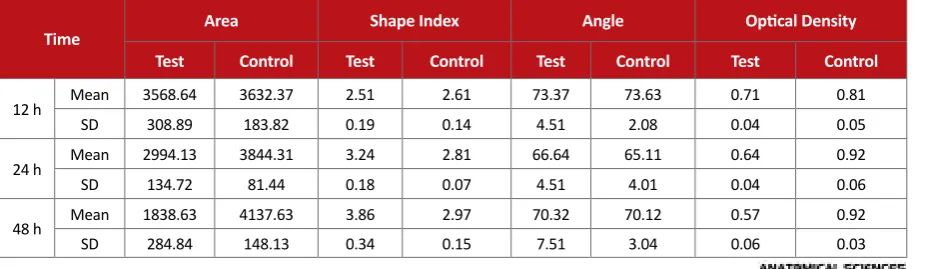
Cell morphology was measured using quantitative parameters and ImageJ software. The obtained data showed that culture of MSCs has a profound impact on
these cells morphology in microgravity conditions
(Fig-ure 1). Cell area, cell aspect ratio, and cell angle of
rota-tion were measured. These parameters were evaluated at 12, 24, and 48 h (Table 1).
One of the morphological parameters on which the ef-fect of microgravity conditions was evaluated was cell areas. Figure 2 shows the extent and quality of cell area changes at 48 h after starting the process. Results show that cell areas have decreased in microgravity. The area occupied by cells under the controlled conditions has increased slightly by 12% over 48 h. However, area of cells under loading had reductions in the first 12 h but the difference was insignificant. The cell area decreased significantly to 59%, after 48 h. It seems that micrograv-ity conditions lead to reduced area of MSCs.
The next morphological parameter evaluated over time was cell aspect ratio. This ratio had 9% increase in the control cells at different times. However, in cells under loading, no significant difference was observed over the first 12 hr from the control group, aspect ratio increased significantly at 24 and 48 h, with the increase after 48 h reaching about 38%. This ratio indicates that cells show a more elongated form under microgravity conditions
(Figure 3). To evaluate the effect of microgravity on cell
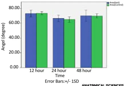
orientation, cell rotation angle was calculated in both groups of test and control using ImageJ software. As shown in Figure 4, lack of significant difference between the 2 groups demonstrates that being exposed to micro-gravity conditions does not have a considerable impact on orientation and alignment of cells.
There were no significant differences between the con-trol and test groups. To evaluate changes in cell mor-phology, cell viability was evaluated under microgravity conditions using MTT test. As illustrated in Figure 5, vi -ability of mesenchymal stem cells decreased significant-ly over time under microgravity conditions ,expressing
Table 1. Results of measuring different parameters related to cell morphology
Time Area Shape Index Angle Optical Density
Test Control Test Control Test Control Test Control
12 h Mean 3568.64 3632.37 2.51 2.61 73.37 73.63 0.71 0.81
SD 308.89 183.82 0.19 0.14 4.51 2.08 0.04 0.05
24 h Mean 2994.13 3844.31 3.24 2.81 66.64 65.11 0.64 0.92
SD 134.72 81.44 0.18 0.07 4.51 4.01 0.04 0.06
48 h Mean 1838.63 4137.63 3.86 2.97 70.32 70.12 0.57 0.92
that microgravity conditions lead to reduced viability of MSCs. Viability of cells exposed to microgravity condi-tions after the start of loading, reduced in a descending fashion, in which after 48 hours, this criterion was about 39% less, compared to cells in static-condition.
4. Discussion
MSCs are capable of long-term proliferation and dif-ferentiation into various types of stromal cells. Various
factors such as microgravity, cellular microenvironment and soluble mixtures affect MSCs. When people get hos-pitalized for a long time or spend a long time under mi-crogravity conditions and different mechanical motives do not affect the cells of this area of body, these cells osteogenesis is weakened [14,15].
Numerous studies evaluated the microgravity effect on bone marrow MSC. Researchers also explored the ef-fect of microgravity conditions on MSCs as well as the
Figure 2. Cell area changes in the test and control groups
**P≤0.01
5,000.00 4,000.00 3,000.00 2,000.00 1,000.00
0.00
Ar
ea (µm
2)
12 hour 24 hour 48 hour Area(test) Area(Control)
Time
Figure 3. Aspect ratio of cells in the control and test groups
**P≤0.01 5.00 4.00 3.00 2.00 1.00 0.00
Shape
Inde
x (µm)
12 hour 24 hour 48 hour
Shape Index(test) Shape Index(Control)
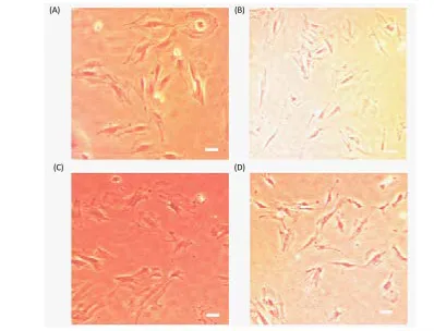
Time Error Bars:+/- SD Figure 1. Morphological changes in MSCs cultured under microgravity conditions
A. Cells cultured under static conditions; B. Cells cultured under microgravity conditions after 12 h; C. After 24 h; and D. After 48 h; Bar represents 20 µm (×40)
(A)
(C)
(B)
mechanism of this effect. In 2004, researchers evaluated the effect of long-term microgravity conditions on mor-phology, meiosis rate and expression of certain cellular markers in cultured human mesenchymal cells. They ob-served the effect of these conditions on cells at different times (1 hour to 10 days). Their obtained results revealed that meiosis rate of these cells reduced considerably un-der microgravity conditions, compared to normal condi-tions. In this experiment, cells cultured under micrograv-ity conditions were more plain, which confirms these mesenchymal cells can sense gravity changes and react to these changes with altered function [16, 13].
Alkaline activity known as an osteoblast differentiation marker decreased by 40% in mesenchymal cells of rats’ bone marrow that had been placed under microgravity conditions, compared to the control group [17,18]. Hav-ing cultured cells for 20 days in zero gravity simulator, meiosis activity of mesenchymal cells decreased; how-ever, number of large flat cells inside the culture media increased [19-21]. Rucci N, et al. reported that decreased gravity leads to suppressed osteogenesis and increased gravity results in further osteogenesis [22].
Findings suggest that the motive of microgravity sup-presses cellular population growth of bone marrow, re-sulting in the lack of differentiation progress to osteo-blasts, which in turn causes physiologic deformities in space explorations [21, 23]. Sheyn et al. clarified that the expression of 882 genes in MSC decrease, compared to the control group and expression of 505 genes increase in microgravity. In this study gene expression related to osteogenesis differentiation was decreased dramatically. However, gene expression was increased in those evolv-ing adipocyte differentiation [24,15].
According to the literature, microgravity leads to wide-spread physiological changes in MSCs. The present study,
quantitatively evaluated the effect of microgravity on mor-phology and viability of MSCs. Three morphologic param-eters including cells area, cell angle of rotation, and cell aspect ratio were evaluated. According to prior studies, mi-crogravity conditions lead to reduced cellular area. Thus, sig-nificant changes in the aspect ratio was observed in which it grew significantly and cells become more elongated under microgravity conditions. Furthermore, angle size of MSCs were compared in microgravity conditions with the control group. Statistical non-significant difference between the 2 groups suggests that cells fail to assume a certain orientation in microgravity conditions. Microgravity conditions have a considerable impact on morphology of MSCs. Furthermore, viability of MSCs decreased significantly after 48 h, under microgravity conditions.
Ethical Considerations
Compliance with ethical guidelines
The research which involved animals (rats) was per-formed in compliance with the principles of the Declara-tion of Helsinki.
Funding
This research did not receive any specific grant from funding agencies in the public, commercial, or not-for-profit sectors.
Authors contributions
The authors contribution is as follows: Samira Mon-faredi: Concept study and design; Reza Ahadi: Acquisi -tion of data; Shahram Darabi: Analysis and interpreta-tion of data; Farzad Rajaei, Reza Ahadi: Drafting the script; Samira Monfaredi: Critical revision of the manu-script for important intellectual content; Shahram Darabi: Figure 4. Cell rotation angle changes in the control and test groups
Area(test) Area(Control)
12 hour 24 hour 48 hour Time
Error Bars:+/- 1SD 80.00 60.00 40.00 20.00 0.00 Ang el (degr ee)
Figure 5. Cell viability changes in the control and test groups measured by MTT test
*P≤0.05; **P≤0.01
Optical density(test) Optical density(Control)
12 hour 24 hour 48 hour Time
Statistical analysis; and Farzad Rajaei: Administrative, technical, and material support, and study supervision.
Conflict of interest
The authors certify that they have no affiliation with or involvement in any organization or entity with any financial interest, or non- financial interest in the subject matter or materials dismissed in this manuscript.
Acknowledgments
The authors wish to thank Rasoul Akram Hospital Clinical Research Development Center (RCRDC) for editorial assist.
References
[1] Brown JH. Physiology of man in space. Cambridge, Massa
-chusetts: Academic Press; 2015.
[2] Stein T. Weight, muscle and bone loss during space flight: An
-other perspective. European Journal of Applied Physiology. 2013; 113(9):2171-81. [DOI:10.1007/s00421-012-2548-9] [PMID] [3] Vico L, Collet P, Guignandon A, Lafage Proust MH, Thomas
T, Rehailia M, et al. Effects of long-term microgravity ex -posure on cancellous and cortical weight-bearing bones
of cosmonauts. The Lancet. 2000; 355(9215):1607-11.
[DOI:10.1016/S0140-6736(00)02217-0]
[4] Calloni R, Viegas GS, Türck P, Bonatto D, Henriques JAP.
Mesenchymal stromal cells from unconventional model
organisms. Cytotherapy. 2014; 16(1):3-16. [DOI:10.1016/j. jcyt.2013.07.010] [PMID]
[5] Manske SL, Boyd SK, Zernicke RF. Muscle and bone fol -low similar temporal patterns of recovery from
muscle-in-duced disuse due to botulinum toxin injection. Bone. 2010; 46(1):24-31. [DOI:10.1016/j.bone.2009.10.016] [PMID] [6] Pittenger MF, Mackay AM, Beck SC, Jaiswal RK, Douglas
R, Mosca JD, et al. Multilineage potential of adult human mesenchymal stem cells. Science. 1999; 284(5411):143-7.
[DOI:10.1126/science.284.5411.143] [PMID]
[7] Khayat G, Rosenzweig DH, Quinn TM. Low frequency me -chanical stimulation inhibits adipogenic differentiation of
C3H10T1/2 mesenchymal stem cells. Differentiation. 2012; 83(4):179-84. [DOI:10.1016/j.diff.2011.12.004] [PMID] [8] Yuge L, Sasaki A, Kawahara Y, Wu S-l, Matsumoto M, Ma
-nabe T, et al. Simulated microgravity maintains the undif -ferentiated state and enhances the neural repair potential of
bone marrow stromal cells. Stem Cells and Development. 2010; 20(5):893-900. [DOI:10.1089/scd.2010.0294] [PMID] [9] Ozcivici E, Luu YK, Adler B, Qin YX, Rubin J, Judex S, et
al. Mechanical signals as anabolic agents in bone. Nature Reviews Rheumatology. 2010; 6(1):50-9. [DOI:10.1038/nr
-rheum.2009.239] [PMID] [PMCID]
[10] Basso N, Jia Y, Bellows CG, Heersche JN. The effect of reload
-ing on bone volume, osteoblast number, and osteoprogeni
-tor characteristics: Studies in hind limb unloaded rats. Bone. 2005; 37(3):370-8. [DOI:10.1016/j.bone.2005.04.033] [PMID] [11] Arfat Y, Xiao WZ, Iftikhar S, Zhao F, Li DJ, Sun YL, et al.
Physiological effects of microgravity on bone cells. Calci
-fied Tissue International. 2014; 94(6):569-79. [DOI:10.1007/ s00223-014-9851-x] [PMID]
[12] Ulbrich C, Wehland M, Pietsch J, Aleshcheva G, Wise P, van Loon J, et al. The impact of simulated and real mi
-crogravity on bone cells and mesenchymal stem cells. BioMed Research International. 2014; 2014:928507.
[DOI:10.1155/2014/928507]
[13] Wuest SL, Richard S, Kopp S, Grimm D, Egli M. Simu -lated microgravity: Critical review on the use of random
positioning machines for mammalian Cell culture. Bi
-oMed Research International. 2015; 2015:971474. [DOI 10.1155/2015/971474]
[14] Özçivici E. Effects of spaceflight on cells of bone marrow origin. Turkish Journal of Hematology. 2013; 30(1):1-7.
[DOI:10.4274/tjh.2012.0127] [PMID] [PMCID]
[15] Kamal KY, Hemmersbach R, Medina FJ, Herranz R. Proper selection of 1 g controls in simulated microgravity
research as illustrated with clinorotated plant cell
suspen-sion cultures. Life Sciences in Space Research. 2015; 5:47-52.
[DOI:10.1016/j.lssr.2015.04.004] [PMID]
[16] Merzlikina N, Buravkova L, Romanov YA. The primary
effects of clinorotation on cultured human mesenchymal
stem cells. Journal of Gravitational Physiology: A Journal of the International Society for Gravitational Physiology. 2004; 11(2):P193-4. [PMID]
[17] Nishikawa M, Ohgushi H, Tamai N, Osuga K, Uemura M, Yoshikawa H, et al. The effect of simulated microgravity by three-dimensional clinostat on bone tissue engineering. Cell Transplantation. 2005; 14(10):829-35. [DOI:10.3727/00 0000005783982477] [PMID]
[18] Gershovich J, Buravkova L. Morphofunctional status and
osteogenic differentiation potential of human mesenchy-mal stromesenchy-mal precursor cells during in vitro modeling of
microgravity effects. Bulletin of Experimental Biology and Medicine. 2007; 144(4):608-13. [DOI:10.1007/s10517-007-0387-1] [PMID]
[19] Wuest SL, Stern P, Casartelli E, Egli M. Fluid dynamics ap -pearing during simulated microgravity using random
po-sitioning machines. PloS One. 2017; 12(1):e0170826.
[20] Brungs S, Egli M, Wuest SL, Christianen PC, Van Loon JJ, Anh TJ, Hemmersbach R. Facilities for simulation of mi
-crogravity in the ESA ground-based facility programme. Microgravity Science and Technology. 2016; 28(3):191-203.
[DOI:10.1007/s12217-015-9471-8]
[21] Meyers VE, Zayzafoon M, Douglas JT, McDonald JM. RhoA and cytoskeletal disruption mediate reduced os -teoblastogenesis and enhanced adipogenesis of human
mesenchymal stem cells in modeled microgravity. Jour
-nal of Bone and Mineral Research. 2005; 20(10):1858-66.
[DOI:10.1359/JBMR.050611] [PMID] [PMCID]
[22] Rucci N, Migliaccio S, Zani BM, Taranta A, Teti A. Char
Wall Vessel bioreactor (RWV). Journal of Cellular Biochem
-istry. 2002; 85(1):167-79. [DOI:10.1002/jcb.10120] [PMID] [23] Chung JH, Ahn CB, Son KH, Yi E, Son HS, Kim HS, Lee SH.
Simulated microgravity effects on nonsmall cell lung
can-cer cell proliferation and migration. Aerospace Medicine and Human Performance. 2017; 88(2):82-9. [DOI:10.3357/ AMHP.4647.2017] [PMID]
[24] Sheyn D, Pelled G, Netanely D, Domany E, Gazit D. The effect
of simulated microgravity on human mesenchymal stem cells cultured in an osteogenic differentiation system: A
bioinfor-matics study. Tissue Engineering Part A. 2010; 16(11):3403-12.