R E S E A R C H
Open Access
A brain proteomic investigation of
rapamycin effects in the
Tsc1
+/
−
mouse
model
Hendrik Wesseling
1, Ype Elgersma
2and Sabine Bahn
1,2*Abstract
Background:Tuberous sclerosis complex (TSC) is a rare monogenic disorder characterized by benign tumors in multiple organs as well as a high prevalence of epilepsy, intellectual disability and autism. TSC is caused by inactivating mutations in theTSC1orTSC2genes. Heterozygocity induces hyperactivation of mTOR which can be inhibited by mTOR inhibitors, such as rapamycin, which have proven efficacy in the treatment of TSC-associated symptoms. The aim of the present study was (1) to identify molecular changes associated with social and cognitive deficits in the brain tissue ofTsc1+/−mice and (2) to investigate the molecular effects of rapamycin treatment, which has been shown to ameliorate genotype-related behavioural deficits.
Methods:Molecular alterations in the frontal cortex and hippocampus ofTsc1+/−and control mice, with or without rapamycin treatment, were investigated. A quantitative mass spectrometry-based shotgun proteomic approach (LC-MSE) was employed as an unbiased method to detect changes in protein levels. Changes identified in the initial profiling stage were validated using selected reaction monitoring (SRM). Protein Set Enrichment Analysis was employed to identify dysregulated pathways.
Results:LC-MSEanalysis ofTsc1+/−mice and controls (n= 30) identified 51 proteins changed in frontal cortex and 108 in the hippocampus. Bioinformatic analysis combined with targeted proteomic validation revealed several dysregulated molecular pathways. Using targeted assays, proteomic alterations in the hippocampus validated the pathways
“myelination”,“dendrite,”and“oxidative stress”, an upregulation of ribosomal proteins and the mTOR kinase. LC-MSE analysis was also employed onTsc1+/−and wildtype mice (n= 34) treated with rapamycin or vehicle. Rapamycin treatment exerted a stronger proteomic effect inTsc1+/−mice with significant changes (mainly decreased expression) in 231 and 106 proteins, respectively. The cellular pathways“oxidative stress”and“apoptosis”were found to be affected inTsc1+/−mice and the cellular compartments“myelin sheet”and“neurofilaments”were affected by rapamycin treatment. Thirty-three proteins which were altered inTsc1+/−mice were normalized following rapamycin treatment, amongst them oxidative stress related proteins, myelin-specific and ribosomal proteins.
Conclusions:Molecular changes in theTsc1+/−mouse brain were more prominent in the hippocampus compared to the frontal cortex. Pathways linked to myelination and oxidative stress response were prominently affected and, at least in part, normalized following rapamycin treatment. The results could aid in the identification of novel drug targets for the treatment of cognitive, social and psychiatric symptoms in autism spectrum disorders. Similar pathways have also been implicated in other psychiatric and neurodegenerative disorders and could imply similar disease processes. Thus, the potential efficacy of mTOR inhibitors warrants further investigation not only for autism spectrum disorders but also for other neuropsychiatric and neurodegenerative diseases.
Keywords:Tuberous sclerosis, Rapamycin, Proteomics, SRM, Animal model
* Correspondence:[email protected]
1Department of Chemical Engineering and Biotechnology, University of
Cambridge, Tennis Court Road, Cambridge CB2 1QT, UK
2Department of Neuroscience, Erasmus Medical Center, Rotterdam 3000, CA,
The Netherlands
Background
Tuberous sclerosis complex (TSC) is a rare multi-system monogenic hamartomatous disorder, which is caused by mutations inactivating the TSC1 (hamartin) or TSC2 (tuberin) genes. TSC is characterized by benign tumors in multiple organs, including the brain, kidneys, heart and eyes [1]. Over 90% of TSC patients develop epilepsy, and around 50% present with neuropsychiatric prob-lems, such as intellectual disability (50%) [2, 3], autism spectrum disorder (ASD) (17–68%), schizophrenia (10– 30%) and anxiety disorders (40%) [4], which account for most of the mortality and morbidity [5].
At the molecular level, both Tsc1 and Tsc2 protein products form hetero-dimers which inhibit the GTP-binding protein RHEB (Ras homolog enriched in the brain). Consequently, mutations within either Tsc1 or Tsc2 lead to increased levels of activated RHEB [6], which causes hyperactivation of mammalian target of rapamycin (mTOR) signaling, a constitutive phosphoryl-ation of eukaryotic translphosphoryl-ation initiphosphoryl-ation factor 4E-binding protein 1 (4E-BP1) and activation of ribosomal protein S6 through S6K1 phosphorylation [7, 8]. The net effect is enhanced protein translation, cell proliferation and growth [9]. Notably, increased mTOR signaling and subsequent changes in global protein synthesis are shared molecular mechanisms of several rare neurodeve-lopmental disorders with an increased prevalence of ASD, such as fragile X syndrome (FXS) [10].
The hyperactivation of mTOR induced by Tsc1 and Tsc2 heterozygosity can be inhibited by mTOR inhibi-tors, such as the macrolide rapamycin. Rapamycin is an immunosuppressant, which is widely prescribed to pre-vent rejection in organ transplantation and exerts anti-tumor properties [11–13]. Rapamycin binds FK-binding protein 12 (FKBP12), and as a complex, rapamycin-FKBP12 directly binds to the mTOR complex 1 (mTORC1), thus reducing phosphorylation of down-stream mTOR targets [14, 15]. Rapamycin and other mTOR inhibitors have been shown to be efficacious in the treatment of several TSC-associated tumors as well as seizures [16–19] and may ameliorate the symptoms of neurodevelopmental disorders in adults [20, 21]. In TSC mouse models, rapamycin limits tumor growth [22, 23], reduces neuropathology and ameliorates epileptic sei-zures as well as learning deficits [24–26]. It was recently reported that rapamycin normalizes social interaction deficits relevant to core disabilities associated with ASD in bothTsc1+/−andTsc2+/−mice [27].
Here, we investigated theTsc1+/− mouse model, which exhibits haploinsufficiency for theTsc1gene, in an attempt to identify molecular changes associated with the neuro-psychiatric phenotype of TSC patients [5]. In this mouse model, the typical human cerebral pathology of spontan-eous seizures, cerebral lesions and giant dysmorphic cells
could not been detected using immuno-cytochemistry and high resolution magnetic resonance imaging, respectively [28]. Furthermore, spine number and dendritic branching are normal [28]. However, theTsc1+/−mouse shows prom-inent behavioural deficits which mimic core symptoms of ASD and other neuropsychiatric disorders [28]. Tsc1+/− mice show hippocampal learning deficits using the Morris water maze test and contextual fear conditioning, as well as social deficits indicated by reduced social interaction and nest building [28]. Consequently, theTsc1+/−mouse is a suitable model to investigate aspects of the molecular pathology associated with neuropsychiatric spectrum dis-orders, especially in relation to ASD and intellectual disability. In this study, we attempted to identify changes in molecular pathways in the frontal cortex and hippocampus of the Tsc1+/− mouse model using a mass spectrometry-based proteomics approach. We also investigated protein changes associated with rapamycin treatment. Findings from this study could aid in the identification of novel drug targets for the treatment of cognitive, social and psychiatric symp-toms in ASD.
Methods
A more detailed description of the materials and methods used in this study can be found in the supple-mentary methods section (Additional file 1).
Animals
Tsc1+/−
in accordance with Institutional Animal Care and Use Committee guidelines.
Rapamycin treatment
Mice were injected intraperitoneally with 5 mg/kg rapamy-cin or vehicle for 5 days and culled 24 h after the last injec-tion [27]. Rapamycin was dissolved in 5% dimethyl sulfoxide diluted with saline to 5 ml/kg. Mice were 5–7 weeks old at the time of injection.
Proteomic sample preparation
Sample preparation was carried out as described previ-ously [30–32]. Based on the lysates, two randomized, blinded, independent sample preparations were prepared for liquid chromatography mass spectrometry (in expres-sion mode; LC-MSE) and selected reaction monitoring mass spectrometry to avoid bias in sample preparations.
Label-free LC-MSEproteomic profiling of brain tissue
Brain tissue analysis and data processing were performed as described previously [31, 33, 34]. The Swiss-Prot hu-man reference proteome (Uniprot release March 2013, 20,252 entries) was used for protein identification searches. Protein abundance changes for the compari-sons between Tsc1+/− and wildtype were determined by the MSstats package [35] based on mixed-effect models using the peptide intensities, following log2
transform-ation and exclusion of intensity values deviating by more than 3 standard deviations from the mean of each group.
Protein set enrichment analysis
Significantly changed proteins were partitioned into three bins, according to their ratio: proteins decreased in abundance (ratio < 1.0), proteins increased in abundance (ratio > 1.0) and a bin to identify general disturbed path-ways which included all proteins with increased and de-creased abundance (ratio > 1 and <1). The R package database org.mouse.eg.db version 2.8.0 was used for gene ontology (GO) term annotation based on entrez gene identifiers and GO-term enrichment analysis was performed usingGOstats.
Label-based SRM mass spectrometry
Abundance alterations of a panel of 43 candidate pro-teins previously implicated in the Tsc1+/− mouse path-ology were measured using a targeted SRM mass spectrometry approach as described previously [32, 36] following the guidelines of Lange et al. [37]. SRMstats was used at default settings [37]. The final transitions, collision energy and retention time windows used for each peptide can be requested.
Results
Label-free LC-MSEproteomic profiling ofTsc1+/−mouse brains
We investigated protein abundance changes in the frontal cortex and hippocampus of the Tsc1+/− mouse. LC-MSE analysis resulted in the identification of 522 proteins (7071 peptides) in the frontal cortex and 463 proteins (5149 peptides) in the hippocampus. Of these, the levels of 51 proteins were altered in the frontal cor-tex (FDR-adjusted *p< 0.05) and 108 in the hippocam-pus (FDR-adjusted *p< 0.05). In the frontal cortex, 17 of the changed proteins were altered by more than 10%, as were 49 of the 108 changed hippocampal proteins (Additional file 2). In the case of the frontal cortex, this included adenylyl cyclase-associated protein 2 (CAP2, ratio = 0.89, FDR-adjusted *p= 0.013), elongation factor 1–α2 (EIF1A2, ratio = 0.97, FDR-adjusted *p = 0.03), eukaryotic translation initiation factor 3 subunit L (eIF3l, ratio = 1.17, FDR-adjusted*p= 0.03) and elongation fac-tor 2 (Eef2, ratio = 0.95, FDR-adjusted*p =0.05), which are all regulators of translation. Copine 6 (ratio = 1.1, FDR-adjusted p = 0.0097) and copine 8 (ratio = 0.8, FDR-adjustedp= 3.9 × 10⁻⁶), which are associated with synaptic plasticity, were changed in the hippocampus. Nine proteins (NCDN⬇⬇, MAP2⬆⬆, SUCB1⬇⬇, MYPR⬆⬇, NDUS7⬇⬇, DPYL2⬇⬇, AT1A2⬇⬆, CRYM⬇⬆, ARP3⬆⬇) were found to be changed in both frontal cortex (first arrow) and hippo-campal tissue (second arrow) (Additional file 2).
Gene set enrichment analysis was employed to investi-gate if the altered 108 and 51 proteins were enriched in biological pathways and cellular compartments. Based on GO enrichment analysis, proteins responsible for the bio-logical pathways “reproductive behaviour” (p = 0.008), “neurological system process” (p = 0.010) and “visual learning”(p= 0.028) were altered in the frontal cortex of theTsc1+/−mouse. In the hippocampus, the proteins were related to the biological pathways“ribonucleotide energy metabolism” (p = 0.0097), “protein polymerisation” (p= 0.005) and “oxidative stress”(p= 0.009). One path-way, “visual learning”, was identified in both the frontal cortex and hippocampus proteomic analyses. Cellular compartment GO association enrichment revealed that the altered proteins were associated with “myelination” and “dendrite” in the frontal cortex and “myelin sheet” and“endoplasmic reticulum-Golgi intermediate compart-ment”in the hippocampus.
Selected reaction monitoring (SRM) validation ofTsc1+/− brain proteomic alterations
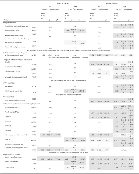
in-house assays. Label-based SRM assays are highly specific and sensitive and outperform immunoblotting analyses for validation [38–41]. This analysis showed that the levels of 5 and 20 of the targeted proteins were significantly changed in the frontal cortex and hippocampus, respectively (p < 0.05, Table 1). Specifically, two proteins (MYPR, PURA) out of five LC-MSEcandidates were validated in the frontal cortex and six (NSF, MYPR, MBP, CPNE6, SODC, MARCS) out of ten LC-MSE-derived protein candidates were validated in the hippocampus (Table 1). Using tar-geted SRM-assays, we confirmed the more prominent proteomic alterations in the hippocampus and further vali-dated GO-terms “myelination”, “dendrite” and “oxidative stress” through confirmation of changes in MYPR, MBP, TSN2 (all myelin specific proteins), MAP2 (dendritic marker) and SODC (oxidative stress marker). We further detected an upregulation in ribosomal proteins and mTOR kinase in theTsc1+/−mice (Table 1).
Label-free LC-MSEproteomic profiling of theTsc1+/− hippocampus following rapamycin treatment
To investigate the effects of rapamycin on the brain proteome, label-free LC-MSE analysis was employed on Tsc1+/−
and wildtype mice treated with rapamycin or ve-hicle (Fig. 1a). Only the hippocampus was studied in this case as this brain region was more affected with regard to significantly changed proteins (Table 1). The hippo-campus plays not only an important role in cognition, but hippocampal dysfunction has also been linked to a wide range of neuropsychiatric symptoms [42, 43]. Defi-cits in consolidating short- and long-term memory and spatial navigation have been shown to be impaired in Tsc1+/−and Tsc2+/−
mice and were reversed by rapamy-cin treatment in Tsc2+/− mice [24]. LC-MSEanalysis led to the identification of 8648 total peptides which trans-lated to 597 proteins, which were detected across all samples. Interestingly, rapamycin treatment exerted a stronger proteomic effect in Tsc1+/− compared to wild-type mice (Fig. 1c (2 and 4)) with significant changes in 231 and 106 proteins, respectively. An overall decrease in protein levels was found in bothTsc1+/−and wildtype mice.
Next, proteins were tested which were affected in all four comparisons. This showed that 9 proteins were changed in common (FRM4A, PEA15, PERQ1, MAP2, BASP, CLD11, ALBU, TCAL3, CLH) and that the levels of 54 proteins were affected by rapamycin treatment in both wildtype and Tsc1+/− mice; of these proteins, 52 corresponded in their fold change direction (37 of the 52 proteins were decreased in abundance and 15 increased, respectively). Pathway analysis linked the 52 overlapping proteins to the biological process of “translation” (p = 0.00082), “macromolecule biosynthetic process” (p = 0.005) and “gene expression” (p = 0.014). Using
KEGG (Kyoto Encyclopedia of Genes and Genomes) an-notation, “ribosome” was the most significant pathway (p= 1.7 × 10⁻⁷) in the enrichment analysis.
We further employed enrichment analysis for the genotype comparisons and the treatment comparisons (Fig. 1c (1–4)). This associated the biological pathways “oxidative stress” and “apoptosis” with the significantly changed proteins identified in the Tsc1+/− vs Wt com-parison (Fig. 1c (1)). Furthermore, cellular compart-ments of myelin sheet and neurofilacompart-ments were affected by rapamycin treatment in both Tsc1+/− and Wt mice (Fig. 1c (2 and 4)). Proteins with decreased levels due to rapamycin treatment mostly related to the biological pathways “translation”, “macromolecular complex as-sembly”and“chromosome organization”(Fig. 1c (2 and 4)). Downregulation of the pathway “chromosome organization” was specifically observed in Tsc1+/− mice following rapamycin treatment (Fig. 1c (4)).
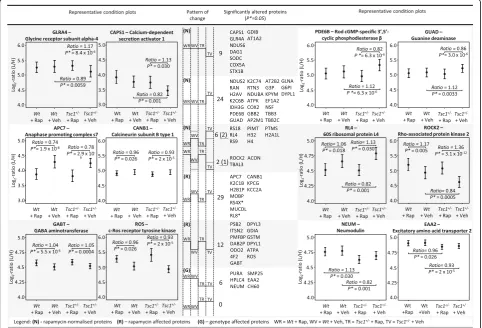
Importantly, 41 proteins which were altered in vehicle-treated Tsc1+/− mice were normalized following rapamy-cin treatment. These proteins include a set of proteins where rapamycin treatment normalizes the genotype-induced protein alterations to wildtype levels (33 pro-teins) and a set of proteins where rapamycin normalizes the genotype-effect below or above baseline levels (8 proteins). The former include the Glycine receptor sub-unit alpha-4 (GLRA4), the Calcium-dependent secretion activator 1 (CAPS1), Rod cGMP-specific 3′,5′ cyclic phosphodiesterase beta (PDE6B) and Guanine deami-nase (GUAD) (Fig. 2 (N)); the latter include Rho-associated protein kinase 2 (ROCK2) and ribosomal pro-teins (RS18, RL4, RS9). All ribosomal propro-teins affected by rapamycin treatment were decreased in their abun-dance levels. Furthermore, proteins were identified that are affected by rapamycin treatment in both wildtype and mutant mice, although there was no difference in their abundance levels between vehicle-treated mutant and wildtype mice (Fig. 2 (R)). This set was comprised of 41 proteins. Amongst them are the anaphase promoting complex s7 (APC7), calcineurin subunit B type 1 (CANB1) and the GABA aminotransferase (GABT). Finally, the levels of six proteins were found to be altered between mutant and wildtype but did not change following rapamycin treatment. Neuromodulin (NEUM), the excitatory amino acid transporter 2 (EAAT2) and SMP25 are examples.
SRM validation of rapamycin treatment effects in theTsc1
+/−hippocampus
Table 1Significantly changed proteins identified by label-based LC-SRM in the frontal cortex and hippocampus ofTsc1+/−mice compared to wildtype mice
The first stage of the analysis consisted of a global profiling approach, followed by validation with a specific and sensitive label-based assay panel.pvalues were determined using SRMstats (linear model with fixed subject effects) and corrected (p*) to control for multiple hypothesis testing (Benjamini-Hochberg) [90]. For reasons of clarity, only ratios and significance levels of significantly changing proteins are shown. For full information, see Additional file3.n.s.not significant,n.d.
LC-MSE discovery study of the Tsc1+/− mouse (SODC, NSF, MAP2, PKCG). The rapamycin-target mTOR was included as a positive control.
This analysis resulted in validation of increased myelin-associated proteins in Tsc1+/− compared to wild-type mice (Fig. 1c (1 and 3)). Furthermore, the decrease
a
b1
b2
c
Fig. 1Label-free LC-MSE and Label-based SRM analysis of Tsc1+/−and wildtype mice under rapamycin and vehicle treatment identifies distinct proteomic changes.a) Flow chart of the experimental design.b) Significant protein changes in the hippocampus of rapamycin-treated WT andTsc1+/−mice identified by label-free LC-MSE.Orange dotsrefer to proteins that are increased in abundance, andgreen dotsrepresent downregulated proteins following rapamycin treatment.c2and4(see below) refer to significantly changed proteins following rapamycin treatment in wildtype (“treatment effect in Wt mice”) or Tsc1
+/−mice (“treatment effect inTsc1+/−mice”). Protein enrichment analysis was performed on the identified proteins.Yellowandblue dotsrepresent the TSC
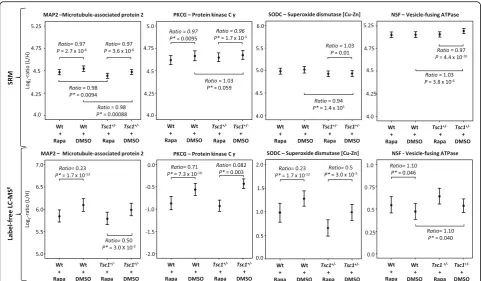
in oxidative stress-related proteins in theTsc1+/− mouse was validated by confirming decreased levels of SODC (Fig. 1c (1)). Additionally, altered levels of NSF and PKCG were also validated (Fig. 1c [targeted] and Fig. 3).
In the case of rapamycin treatment effects, decreased levels of proteins involved in translation could be vali-dated. Specifically, decreased levels of all three tested ribosomal subunits as well as the mTOR-kinase were identified. Moreover, the analysis showed that rapamycin normalized the protein levels of SODC, NSF and PKCG inTsc1+/−mice (Fig. 1c [targeted]).
Discussion
The pathogenesis of psychiatric disorders such as ASD re-mains elusive, and there is accumulating evidence that several neuronal circuits and pathways are affected. This is especially true for the social, cognitive and neuropsychi-atric symptoms associated with these disorders. In an at-tempt to gain further insight into these pathways, this
study combines unbiased and targeted proteomic ap-proaches to investigate the hippocampus and frontal cor-tex of a mouse model of TSC, which is one of the most frequent causes of syndromic ASD [44]. The investigated Tsc1+/−
mouse model exhibits social and cognitive deficits, which are core behavioural symptoms of ASD in humans [45] and other relevant rodent models [46], without any obvious brain pathology (such as tumors or epilepsy). This makes theTsc1+/− mouse an excellent model of pharma-cologically treatable ASD. A previous study has demon-strated the effectiveness of rapamycin to normalize reciprocal social interactions in this model [27]. The aim of the present study was to investigate proteins and path-ways affected by rapamycin treatment, which could sup-port drug discovery efforts and in turn the development of improved treatments for TSC, ASD and possibly other neuropsychiatric disorders.
Proteomic profiling of the frontal cortex and hippo-campus brain tissue in this study identified and
Fig. 2Multivariate analysis of LC-MSEestimates as shown by condition plots illustrating the differences between the WT andTsc1+/−mice with and without rapamycin treatment.X-axis is condition andy-axis is log ratio of endogenous (Llight) over reference (Hheavy) peptides.Dotsrepresent the mean of the log2ratio for each condition, anderror barsindicate the confidence intervals with 0.95 significance. The interval is not related to the model
based analysis. Significant changes as measured by LC-MSEare indicated below each protein. Illustrated are examples of proteins which are affected
quantified a large number of significantly changed pro-teins in the Tsc1+/− mouse model. Differentially expressed proteins and altered molecular pathways were identified and selected candidate proteins were validated using SRM as a highly quantitative method. In a second stage, proteomic analysis of rapamycin treatment effects were investigated to identify down-stream effects of mTOR-pathway inhibition in the hope to gain new in-sights into the molecular underpinnings of social impair-ments in ASD and other psychiatric disorders.
We were able to show that myelin proteins and the translational machinery, specifically several ribosomal subunits, were significantly altered in Tsc1+/− mice treated with rapamycin. Our findings of lower ribosomal subunit abundances are consistent with a rapamycin-induced downregulation of ribosomal biogenesis [47]. Regarding the effects on myelination, previous research has linked the mTOR pathway to oligodendrocyte differ-entiation and axonogenesis [48]. Oligodendrocytes pro-duce myelin, and this is specifically regulated at the late progenitor to immature oligodendrocyte transition stage (as shown by changes in expression of the myelin marker proteins MYPR and MBP). We identified an
increase in myelin proteins in the Tsc1+/− mouse, but not in Wt mice. A recent study has shown that ablation of TSC1 is associated with oligodendrocyte-specific over-activation and subsequent hypomyelination [49]. An increase in myelin proteins in the Tsc1+/− mouse brain may be due to the globally enhanced (Table 1) pro-tein translation and cell proliferation in the context of mTOR hyperactivation. An increase in cell growth and proliferation could interfere with oligodendrocyte matur-ation and thus result in incomplete myelinmatur-ation as seen in some demyelinating diseases, such as multiple scler-osis [50].
MYPR and MBP as well as TSN-2 were amongst the altered myelin proteins. These proteins play an import-ant role in oligodendrocyte differentiation during devel-opment. Furthermore, in vitro studies have shown that MBP mRNA and protein expression are significantly de-creased by mTOR inhibition [48, 51]. Inhibiting mTOR in oligodendrocyte precursor cell/dorsal root ganglion co-cultures potently abrogated oligodendrocyte differen-tiation and reduced numbers of myelin segments. Disor-ganized and structurally compromised axons with poor myelination have already been found in TSC patients,
Fig. 3Multivariate analysis of SRM estimates as shown by condition plots illustrating the differences between Wt andTsc1+/−mice with and without rapamycin treatment.X-axis represents the condition and they-axis the log ratio of endogenous (Llight) over reference (Hheavy) peptides.Dotsrepresent the mean of log2ratio for each condition, anderror barsindicate the confidence intervals with 0.95 significance. The
and this may at least to some extent explain the behav-ioural and cognitive deficits associated with the disorder [52]. Impaired adult myelination has been shown in the prefrontal cortex of socially isolated mice [53]. Import-antly, changes in oligodendrocyte function and myelin-ation abnormalities are amongst the most consistent hallmarks of psychiatric pathology in post-mortem brain studies. Changes were reported for schizophrenia, bipo-lar disorder, depression and ASD [36, 54–58]. In wild-type mice, rapamycin treatment led to a reduction of myelin and myelin protein expression [59, 60]. This is consistent with our findings, where rapamycin affects both wildtype (reduced) and mutant myelin (increased) protein expression.
Interestingly, several proteins that we found altered in the Tsc1+/− mouse model were reversed by rapamycin treatment. One of these proteins, a glycine receptor sub-unit, which abundance was decreased in the mutant and normalized by rapamycin treatment, could be a potential drug target for novel treatments of ASD and schizophrenia-spectrum disorders. The glycine receptor co-localizes with GABAA receptors on hippocampal
neurons [61]. A microdeletion at Xq22.2 implicates GLRA4 to be involved in intellectual disability and be-havioural problems [62]. In a case report, glycine recep-tor antibodies could be detected in a patient with treatment-resistant focal epilepsy, tantrums, clumsiness and impaired speech [63] and in patients with progres-sive encephalomyelitis with rigidity and myoclonus stiff person syndrome [64]. Treatments targeting the glycine transporter are under investigation as novel treatment approaches for schizophrenia [65].
Other altered proteins, which are normalized by rapa-mycin treatment, included the calcium-dependent secre-tion activator 1 (CAPS1) (decreased in Tsc1+/− and normalized by rapamycin), two guanine metabolism as-sociated proteins (guanine deaminase and PDE6B; both increased in Tsc1+/− and normalized by rapamycin) and the vesicle-fusing ATPase NSF (increased in Tsc1+/−, normalized by rapamycin), a molecular component of the exocytosis machinery [66], which is required for membrane fusion [67] and regulates the disassembly of SNARE complexes on early endosomes [68]. The NSF gene has also been linked with cocaine dependence [69] and schizophrenia [36, 70]. Direct interactions with cell surface receptors such as AMPA receptors [71, 72],β 2-adrenergic receptors [73], dopaminergic receptors [74] and the adrenomedulin receptor [75] have been re-ported. Interestingly, a coordinated action of NSF and PKC regulates GABABreceptor signaling efficiency [76].
PKCG was found to be strongly downregulated by rapa-mycin treatment in this study and is known to be in-volved in the regulation of the neuronal receptors GLUR4 and NMDAR1 [77]. It binds and phosphorylates
the GLUR4 glutamate receptor and regulates its function by increasing membrane-associated GRIA4 expression [78]. Several preclinical and clinical trials have investi-gated mGLUR antagonists for the treatment of social deficits in ASD [78, 79] and ASD associated with FXS [80, 81] and PKCG inhibitors could represent a novel treatment strategy to ameliorate cognitive and social def-icits. Notably, Ketamine, which is thought to exert anti-depressant action through modulation of mTOR pathway activity [82], potentiates persistent learning and memory impairment through the PKCG-ERK signaling pathway [83].
Another protein strongly downregulated following rapa-mycin treatment is the anaphase promoting complex S7 (APC7), which is a cell cycle-regulated E3 ubiquitin ligase controlling progression through mitosis and the G1 phase of the cell cycle. The control of APC7 through rapamycin might be a major breakpoint in cell proliferation. Rapamy-cin has already been shown to also downregulate the ex-pression of the APC/C inhibitor Emi1 [84].
Copine 6, which we found upregulated in the hippo-campus of Tsc1+/− compared to wildtype mice, is a calcium-dependent regulator of the actin cytoskeleton in neuronal spines and negatively regulates spine matur-ation during neuronal development [85]. Changes in copine 6 expression may be involved in neurodevelop-mental disorders, as deformed dendritic spines and changes in spine density are hallmarks of many neurode-velopmental conditions, such as Down’s syndrome [86, 87] and FXS [88]. Interestingly, hippocampi from pa-tients suffering from uncontrolled epileptic seizures, typ-ically a problem in tuberous sclerosis patients, exhibit a decrease in spine density [89]. We also found that MAP2, a dendritic spine marker, was increased by Tsc1 heterozygocity and decreased by rapamycin treatment.
Conclusions
Taken together, the results from this comprehensive study represent the first proteomic characterization of the Tsc1+/− mouse model to date. The findings yield novel insights into the molecular Tsc1+/− mouse path-ology as well as the molecular effects of rapamycin treat-ment, which is an effective treatment for several clinical symptoms of the tuberous sclerosis complex. Further-more, the mTOR pathway, which is modulated by rapa-mycin treatment, is a novel drug target for the treatment of ASD, schizophrenia and affective disorders. We hope that the findings from this study will provide evidence and support for future clinical trials in the field of neuropsychiatric disorders.
Additional files
Additional file 1:Supplementary methods. Detailed information of the experimental methods. (DOCX 31 kb)
Additional file 2:Significantly altered proteins identified by label-free LC-MSEanalysis in the frontal cortex and hippocampus of theTsc1+/−
mice compared toWtmice. Overlapping proteins between hippocampus and frontal cortex are bold. (XLSX 23 kb)
Additional file 3:Full information for significantly changed proteins identified by label-based LC-SRM in the frontal cortex and hippocampus ofTsc1+/−mice compared to wildtype mice. (DOCX 31 kb)
Abbreviations
ASD:Autism spectrum disorder; LC: Liquid chromatography; MBP: Myelin basic protein; mTOR: Mammalian target of rapamycin; MYPR: Myelin proteolipid protein; NSF: Vesicle-fusing ATPase; PKCG: Protein kinase C gamma; SODC: Superoxide dismutase C; SRM: Selected reaction monitoring; TSC: Tuberous sclerosis complex; TSC1: Harmartin; TSC2: Tuberin; TSN-2: Tetraspanin 2; Wt: Wildtype
Acknowledgements
We would like to thank Susan Gooden for the generation and treatment of theTsc1+/-mouse model and Jadviga Schreiber for proofreading the manuscript.
Funding
This research was kindly supported by the Stanley Medical Research Institute (SMRI) and the Dutch Fund for Economic Structure Reinforcement (#0908) the NeuroBasic PharmaPhenomics project.
Availability of data and materials
The datasets presented in this study can be made available on reasonable request.
Authors’contributions
HW carried out the label-free LC-MSEexperiments, designed and carried out
the SRM experiments and performed all statistical and bioinformatic data analyses. HW prepared the figures and tables and drafted the manuscript. SB and YE conceived the study and participated in its design and coordination. SB helped to interpret the results and to write the manuscript. All authors read, edited and approved the final manuscript.
Ethics approval and consent to participate
All animal experiments were approved by the Dutch Ethical Committee or in accordance with Institutional Animal Care and Use Committee guidelines. All animal experiments were approved by the Dutch Animal Experiment Committee (Dierexperimenten commissie [DEC]) and in accordance with Dutch animal care and use laws.
Consent for publication
Not applicable.
Competing interests
S.B. is a director of Psynova Neurotech Ltd. and PsyOmics Ltd. The other authors declare no conflict of interest.
Publisher’s Note
Springer Nature remains neutral with regard to jurisdictional claims in published maps and institutional affiliations.
Received: 21 September 2016 Accepted: 14 June 2017
References
1. Islam MP, Roach ES. Tuberous sclerosis complex. Handb Clin Neurol. 2015; 132:97–109.
2. de Vries PJ, Prather PA. The tuberous sclerosis complex. N Engl J Med. 2007; 356(1):92. author reply 93-94
3. Joinson C, O'Callaghan FJ, Osborne JP, Martyn C, Harris T, Bolton PF. Learning disability and epilepsy in an epidemiological sample of individuals with tuberous sclerosis complex. Psychol Med. 2003;33(2):335–44. 4. Roach ES, Smith M, Huttenlocher P, Bhat M, Alcorn D, Hawley L. Diagnostic
criteria: tuberous sclerosis complex. Report of the Diagnostic Criteria Committee of the National Tuberous Sclerosis Association. J Child Neurol. 1992;7(2):221–4.
5. Curatolo P, Moavero R, de Vries PJ. Neurological and neuropsychiatric aspects of tuberous sclerosis complex. Lancet Neurol. 2015;14(7):733–45. 6. Kwiatkowski DJ, Manning BD. Tuberous sclerosis: a GAP at the crossroads of
multiple signaling pathways. Hum Mol Genet. 2005;14 Spec No. 2:R251–8. 7. Ehninger D, de Vries PJ, Silva AJ. From mTOR to cognition: molecular and
cellular mechanisms of cognitive impairments in tuberous sclerosis. J Intellect Disabil Res. 2009;53(10):838–51.
8. Klann E, Dever TE. Biochemical mechanisms for translational regulation in synaptic plasticity. Nat Rev Neurosci. 2004;5(12):931–42.
9. Hay N, Sonenberg N. Upstream and downstream of mTOR. Genes Dev. 2004;18(16):1926–45.
10. Auerbach BD, Osterweil EK, Bear MF. Mutations causing syndromic autism define an axis of synaptic pathophysiology. Nature. 2011;480(7375):63–8. 11. Samuels JA. Treatment of renal angiomyolipoma and other hamartomas in
patients with tuberous sclerosis complex. Clin J Am Soc Nephrol. 2017;12(7): 1196–1202.
12. Hsieh JJ, Purdue MP, Signoretti S, Swanton C, Albiges L, Schmidinger M, Heng DY, Larkin J, Ficarra V. Renal cell carcinoma. Nat Rev Dis Primers. 2017; 3:17009.
13. Phan AT, Dave B. The pivotal role of mammalian target of rapamycin inhibition in the treatment of patients with neuroendocrine tumors. Cancer Med. 2016;5(10):2953–64.
14. Brown EJ, Albers MW, Shin TB, Ichikawa K, Keith CT, Lane WS, Schreiber SL. A mammalian protein targeted by G1-arresting rapamycin-receptor complex. Nature. 1994;369(6483):756–8.
15. Chen J, Zheng XF, Brown EJ, Schreiber SL. Identification of an 11-kDa binding domain within the 289-kDa FKBP12-rapamycin-associated protein and characterization of a critical serine residue. Proc Natl Acad Sci U S A. 1995;92(11):4947–51.
16. Franz DN, Leonard J, Tudor C, Chuck G, Care M, Sethuraman G, Dinopoulos A, Thomas G, Crone KR. Rapamycin causes regression of astrocytomas in tuberous sclerosis complex. Ann Neurol. 2006;59(3):490–8.
17. Bissler JJ, McCormack FX, Young LR, Elwing JM, Chuck G, Leonard JM, Schmithorst VJ, Laor T, Brody AS, Bean J, et al. Sirolimus for angiomyolipoma in tuberous sclerosis complex or
lymphangioleiomyomatosis. N Engl J Med. 2008;358(2):140–51. 18. Davies DM, Johnson SR, Tattersfield AE, Kingswood JC, Cox JA, McCartney
DL, Doyle T, Elmslie F, Saggar A, de Vries PJ, et al. Sirolimus therapy in tuberous sclerosis or sporadic lymphangioleiomyomatosis. N Engl J Med. 2008;358(2):200–3.
20. Ehninger D, Li W, Fox K, Stryker MP, Silva AJ. Reversing neurodevelopmental disorders in adults. Neuron. 2008;60(6):950–60.
21. Citraro R, Leo A, Constanti A, Russo E, De Sarro G. mTOR pathway inhibition as a new therapeutic strategy in epilepsy and epileptogenesis. Pharmacol Res. 2016;107:333–43.
22. Kenerson HL, Aicher LD, True LD, Yeung RS. Activated mammalian target of rapamycin pathway in the pathogenesis of tuberous sclerosis complex renal tumors. Cancer Res. 2002;62(20):5645–50.
23. Kenerson H, Dundon TA, Yeung RS. Effects of rapamycin in the Eker rat model of tuberous sclerosis complex. Pediatr Res. 2005;57(1):67–75. 24. Ehninger D, Han S, Shilyansky C, Zhou Y, Li W, Kwiatkowski DJ, Ramesh V,
Silva AJ. Reversal of learning deficits in a Tsc2+/−mouse model of tuberous sclerosis. Nat Med. 2008;14(8):843–8.
25. Zeng LH, Xu L, Gutmann DH, Wong M. Rapamycin prevents epilepsy in a mouse model of tuberous sclerosis complex. Ann Neurol. 2008;63(4):444–53. 26. Meikle L, Pollizzi K, Egnor A, Kramvis I, Lane H, Sahin M, Kwiatkowski DJ.
Response of a neuronal model of tuberous sclerosis to mammalian target of rapamycin (mTOR) inhibitors: effects on mTORC1 and Akt signaling lead to improved survival and function. J Neurosci. 2008;28(21):5422–32. 27. Sato A, Kasai S, Kobayashi T, Takamatsu Y, Hino O, Ikeda K, Mizuguchi M.
Rapamycin reverses impaired social interaction in mouse models of tuberous sclerosis complex. Nat Commun. 2012;3:1292.
28. Goorden SM, van Woerden GM, van der Weerd L, Cheadle JP, Elgersma Y. Cognitive deficits in Tsc1+/−mice in the absence of cerebral lesions and seizures. Ann Neurol. 2007;62(6):648–55.
29. Wilson C, Idziaszczyk S, Parry L, Guy C, Griffiths DF, Lazda E, Bayne RA, Smith AJ, Sampson JR, Cheadle JP. A mouse model of tuberous sclerosis 1 showing background specific early post-natal mortality and metastatic renal cell carcinoma. Hum Mol Genet. 2005;14(13):1839–50.
30. Wesseling H, Guest PC, Lee CM, Wong EH, Rahmoune H, Bahn S. Integrative proteomic analysis of the NMDA NR1 knockdown mouse model reveals effects on central and peripheral pathways associated with schizophrenia and autism spectrum disorders. Mol Autism. 2014;5:38.
31. Wesseling H, Want EJ, Guest PC, Rahmoune H, Holmes E, Bahn S. Hippocampal proteomic and metabonomic abnormalities in
neurotransmission, oxidative stress, and apoptotic pathways in a chronic phencyclidine rat model. J Proteome Res. 2015;14(8):3174–87. 32. Wesseling H, Rahmoune H, Tricklebank M, Guest PC, Bahn S. A targeted
multiplexed proteomic investigation identifies ketamine-induced changes in immune markers in rat serum and expression changes in protein kinases/ phosphatases in rat brain. J Proteome Res. 2015;14(1):411–21.
33. Wesseling H, Guest PC, Lee CM, Wong EHF, Rahmoune H, Bahn S. Integrative proteomic analysis of the NMDA NR1 knockdown mouse model reveals effects on central and peripheral pathways associated with schizophrenia and autism spectrum disorders. Mol Autism. 2014;5:38. 34. Gottschalk MG, Wesseling H, Guest PC, Bahn S. Proteomic enrichment
analysis of psychotic and affective disorders reveals common signatures in presynaptic glutamatergic signaling and energy metabolism. Int J Neuropsychopharmacol. 2014;18(2).
35. Clough T, Thaminy S, Ragg S, Aebersold R, Vitek O. Statistical protein quantification and significance analysis in label-free LC-MS experiments with complex designs. BMC Bioinformatics. 2012;13 Suppl 16:S6.
36. Wesseling H, Gottschalk MG, Bahn S. Targeted multiplexed selected reaction monitoring analysis evaluates protein expression changes of molecular risk factors for major psychiatric disorders. Int J Neuropsychopharmacol. 2014;18(1). 37. Lange V, Picotti P, Domon B, Aebersold R. Selected reaction monitoring for
quantitative proteomics: a tutorial. Mol Syst Biol. 2008;4:222. 38. Simicevic J, Schmid AW, Gilardoni PA, Zoller B, Raghav SK, Krier I,
Gubelmann C, Lisacek F, Naef F, Moniatte M, et al. Absolute quantification of transcription factors during cellular differentiation using multiplexed targeted proteomics. Nat Methods. 2013;10(6):570–6.
39. Picotti P, Aebersold R. Selected reaction monitoring-based proteomics: workflows, potential, pitfalls and future directions. Nat Methods. 2012;9(6):555–66. 40. Method of the Year 2012. Nat Methods. 2013;10(1):1.
41. Aebersold R, Burlingame AL, Bradshaw RA. Western blots versus selected reaction monitoring assays: time to turn the tables? Mol Cell Proteomics. 2013;12(9):2381–2.
42. Rubin RD, Watson PD, Duff MC, Cohen NJ. The role of the hippocampus in flexible cognition and social behavior. Front Hum Neurosci. 2014;8:742. 43. Beadle JN, Tranel D, Cohen NJ, Duff MC. Empathy in hippocampal amnesia.
Front Psychol. 2013;4:69.
44. Abrahams BS, Geschwind DH. Advances in autism genetics: on the threshold of a new neurobiology. Nat Rev Genet. 2008;9(5):341–55. 45. Happe F, Ronald A. The 'fractionable autism triad': a review of evidence
from behavioural, genetic, cognitive and neural research. Neuropsychol Rev. 2008;18(4):287–304.
46. Silverman JL, Yang M, Lord C, Crawley JN. Behavioural phenotyping assays for mouse models of autism. Nat Rev Neurosci. 2010;11(7):490–502. 47. Iadevaia V, Huo YL, Zhang Z, Foster LJ, Proud CG. Roles of the mammalian
target of rapamycin, mTOR, in controlling ribosome biogenesis and protein synthesis. Biochem Soc Trans. 2012;40:168–72.
48. Tyler WA, Gangoli N, Gokina P, Kim HA, Covey M, Levison SW, Wood TL. Activation of the mammalian target of rapamycin (mTOR) is essential for oligodendrocyte differentiation. J Neurosci. 2009;29(19): 6367–78.
49. Lebrun-Julien F, Bachmann L, Norrmen C, Trotzmuller M, Kofeler H, Ruegg MA, Hall MN, Suter U. Balanced mTORC1 activity in oligodendrocytes is required for accurate CNS myelination. J Neurosci. 2014;34(25):8432–48. 50. Lourenco T, Paes de Faria J, Bippes CA, Maia J, Lopes-da-Silva JA, Relvas JB,
Graos M. Modulation of oligodendrocyte differentiation and maturation by combined biochemical and mechanical cues. Sci Rep. 2016;6:21563. 51. Wood TL, Bercury KK, Cifelli SE, Mursch LE, Min J, Dai J, Macklin WB. mTOR: a
link from the extracellular milieu to transcriptional regulation of oligodendrocyte development. Asn Neuro. 2013;5(1):63–79.
52. Krishnan ML, Commowick O, Jeste SS, Weisenfeld N, Hans A, Gregas MC, Sahin M, Warfield SK. Diffusion features of white matter in tuberous sclerosis with tractography. Pediatr Neurol. 2010;42(2):101–6.
53. Liu J, Dietz K, DeLoyht JM, Pedre X, Kelkar D, Kaur J, Vialou V, Lobo MK, Dietz DM, Nestler EJ, et al. Impaired adult myelination in the prefrontal cortex of socially isolated mice. Nat Neurosci. 2012;15(12):1621-+. 54. Tkachev D, Mimmack ML, Huffaker SJ, Ryan M, Bahn S. Further evidence for
altered myelin biosynthesis and glutamatergic dysfunction in schizophrenia. Int J Neuropsychopharmacol. 2007;10(4):557–63.
55. Tkachev D, Mimmack ML, Ryan MM, Wayland M, Freeman T, Jones PB, Starkey M, Webster MJ, Yolken RH, Bahn S. Oligodendrocyte dysfunction in schizophrenia and bipolar disorder. Lancet. 2003; 362(9386):798–805.
56. Zeidan-Chulia F, de Oliveira BN, Casanova MF, Casanova EL, Noda M, Salmina AB, Verkhratsky A. Up-regulation of oligodendrocyte lineage markers in the cerebellum of autistic patients: evidence from network analysis of gene expression. Mol Neurobiol. 2015.
57. Broek JAC, Lin Z, de Gruiter HM, van 't Spijker H, Haasdijk ED, Cox D, Ozcan S, van Cappellen GWA, Houtsmuller AB, Willemsen R, de Zeeuw CI, Bahn S. Synaptic vesicle dynamic changes in a model of fragile X. Mol Autism. 2016;7:17. 58. Bloemen OJ, Deeley Q, Sundram F, Daly EM, Barker GJ, Jones DK, van
Amelsvoort TA, Schmitz N, Robertson D, Murphy KC, et al. White matter integrity in Asperger syndrome: a preliminary diffusion tensor magnetic resonance imaging study in adults. Autism Res. 2010;3(5):203–13. 59. Flores AI, Narayanan SP, Morse EN, Shick HE, Yin X, Kidd G, Avila RL,
Kirschner DA, Macklin WB. Constitutively active Akt induces enhanced myelination in the CNS. J Neurosci. 2008;28(28):7174–83.
60. Narayanan SP, Flores AI, Wang F, Macklin WB. Akt signals through the mammalian target of rapamycin pathway to regulate CNS myelination. J Neurosci. 2009;29(21):6860–70.
61. Levi S, Logan SM, Tovar KR, Craig AM. Gephyrin is critical for glycine receptor clustering but not for the formation of functional GABAergic synapses in hippocampal neurons. J Neurosci. 2004;24(1):207–17. 62. Labonne JD, Graves TD, Shen Y, Jones JR, Kong IK, Layman LC, Kim HG. A
microdeletion at Xq22.2 implicates a glycine receptor GLRA4 involved in intellectual disability, behavioral problems and craniofacial anomalies. BMC Neurol. 2016;16:132.
63. Wuerfel E, Bien CG, Vincent A, Woodhall M, Brockmann K. Glycine receptor antibodies in a boy with focal epilepsy and episodic behavioral disorder. J Neurol Sci. 2014;343(1–2):180–2.
64. Turner MR, Irani SR, Leite MI, Nithi K, Vincent A, Ansorge O. Progressive encephalomyelitis with rigidity and myoclonus Glycine and NMDA receptor antibodies. Neurology. 2011;77(5):439–43.
65. Harvey RJ, Yee BK. Glycine transporters as novel therapeutic targets in schizophrenia, alcohol dependence and pain. Nat Rev Drug Discov. 2013; 12(11):866–85.
67. Whiteheart SW, Rossnagel K, Buhrow SA, Brunner M, Jaenicke R, Rothman JE. N-ethylmaleimide-sensitive fusion protein: a trimeric ATPase whose hydrolysis of ATP is required for membrane fusion. J Cell Biol. 1994;126(4):945–54.
68. Yan Q, Sun W, McNew JA, Vida TA, Bean AJ. Ca2+ and N-ethylmaleimide-sensitive factor differentially regulate disassembly of SNARE complexes on early endosomes. J Biol Chem. 2004;279(18):18270–6.
69. Fernandez-Castillo N, Cormand B, Roncero C, Sanchez-Mora C, Grau-Lopez L, Gonzalvo B, Miquel L, Corominas R, Ramos-Quiroga JA, Casas M, et al. Candidate pathway association study in cocaine dependence: the control of neurotransmitter release. World J Biol Psychiatry. 2012;13(2):126–34. 70. Imai C, Sugai T, Iritani S, Niizato K, Nakamura R, Makifuchi T, Kakita A,
Takahashi H, Nawa H. A quantitative study on the expression of synapsin II and N-ethylmaleimide-sensitive fusion protein in schizophrenic patients. Neurosci Lett. 2001;305(3):185–8.
71. Nishimune A, Isaac JT, Molnar E, Noel J, Nash SR, Tagaya M, Collingridge GL, Nakanishi S, Henley JM. NSF binding to GluR2 regulates synaptic transmission. Neuron. 1998;21(1):87–97.
72. Osten P, Srivastava S, Inman GJ, Vilim FS, Khatri L, Lee LM, States BA, Einheber S, Milner TA, Hanson PI, et al. The AMPA receptor GluR2 C terminus can mediate a reversible, ATP-dependent interaction with NSF and alpha- and beta-SNAPs. Neuron. 1998;21(1):99–110.
73. Cong M, Perry SJ, Hu LA, Hanson PI, Claing A, Lefkowitz RJ. Binding of the beta2 adrenergic receptor to N-ethylmaleimide-sensitive factor regulates receptor recycling. J Biol Chem. 2001;276(48):45145–52.
74. Heydorn A, Sondergaard BP, Hadrup N, Holst B, Haft CR, Schwartz TW. Distinct in vitro interaction pattern of dopamine receptor subtypes with adaptor proteins involved in post-endocytotic receptor targeting. FEBS Lett. 2004;556(1–3):276–80.
75. Bomberger JM, Parameswaran N, Hall CS, Aiyar N, Spielman WS. Novel function for receptor activity-modifying proteins (RAMPs) in post-endocytic receptor trafficking. J Biol Chem. 2005;280(10):9297–307.
76. Pontier SM, Lahaie N, Ginham R, St-Gelais F, Bonin H, Bell DJ, Flynn H, Trudeau LE, McIlhinney J, White JH, et al. Coordinated action of NSF and PKC regulates GABAB receptor signaling efficacy. EMBO J. 2006;25(12):2698–709.
77. Zheng Z, Keifer J. Protein kinase C-dependent and independent signaling pathways regulate synaptic GluR1 and GluR4 AMPAR subunits during in vitro classical conditioning. Neuroscience. 2008;156(4):872–84.
78. Gomes AR, Correia SS, Esteban JA, Duarte CB, Carvalho AL. PKC anchoring to GluR4 AMPA receptor subunit modulates PKC-driven receptor phosphorylation and surface expression. Traffic. 2007;8(3):259–69. 79. Silverman JL, Smith DG, Rizzo SJ, Karras MN, Turner SM, Tolu SS, Bryce DK,
Smith DL, Fonseca K, Ring RH, et al. Negative allosteric modulation of the mGluR5 receptor reduces repetitive behaviors and rescues social deficits in mouse models of autism. Sci Transl Med. 2012;4(131):131ra151.
80. Berry-Kravis E, Des Portes V, Hagerman R, Jacquemont S, Charles P, Visootsak J, Brinkman M, Rerat K, Koumaras B, Zhu L, et al. Mavoglurant in fragile X syndrome: results of two randomized, double-blind, placebo-controlled trials. Sci Transl Med. 2016;8(321):321ra325.
81. Jacquemont S, Berry-Kravis E, Hagerman R, von Raison F, Gasparini F, Apostol G, Ufer M, Des Portes V, Gomez-Mancilla B. The challenges of clinical trials in fragile X syndrome. Psychopharmacology. 2014;231(6):1237–50.
82. Li NX, Lee B, Liu RJ, Banasr M, Dwyer JM, Iwata M, Li XY, Aghajanian G, Duman RS. mTOR-dependent synapse formation underlies the rapid antidepressant effects of NMDA antagonists. Science. 2010;329(5994):959–64.
83. Huang LN, Liu Y, Jin W, Ji XC, Dong ZM. Ketamine potentiates hippocampal neurodegeneration and persistent learning and memory impairment through the PKC gamma-ERK signaling pathway in the developing brain. Brain Res. 2012;1476:164–71.
84. Shapira M, Kakiashvili E, Rosenberg T, Hershko DD. The mTOR inhibitor rapamycin down-regulates the expression of the ubiquitin ligase subunit Skp2 in breast cancer cells. Breast Cancer Res. 2006;8(4):R46.
85. Reinhard JR, Kriz A, Galic M, Angliker N, Rajalu M, Vogt KE, Ruegg MA. The calcium sensor Copine-6 regulates spine structural plasticity and learning and memory. Nat Commun. 2016;7:11613.
86. Suetsugu M, Mehraein P. Spine distribution along the apical dendrites of the pyramidal neurons in Down's syndrome. A quantitative Golgi study. Acta Neuropathol. 1980;50(3):207–10.
87. Chang KT, Ro H, Wang W, Min KT. Meeting at the crossroads: common mechanisms in Fragile X and Down syndrome. Trends Neurosci. 2013;36(12):685–94.
88. Kaufmann WE, Moser HW. Dendritic anomalies in disorders associated with mental retardation. Cereb Cortex. 2000;10(10):981–91.
89. Swann JW, Al-Noori S, Jiang M, Lee CL. Spine loss and other dendritic abnormalities in epilepsy. Hippocampus. 2000;10(5):617–25. 90. Chang CY, Picotti P, Huttenhain R, Heinzelmann-Schwarz V, Jovanovic M,
Aebersold R, Vitek O. Protein significance analysis in selected reaction monitoring (SRM) measurements. Mol Cell Proteomics. 2011;11(4):M111 014662.
91. Benjamini Y, Hochberg Y. Controlling the false discovery rate—a practical and powerful approach to multiple testing. J Royal Stat Soc Ser B Methodol. 1995;57(1):289–300.
• We accept pre-submission inquiries
• Our selector tool helps you to find the most relevant journal
• We provide round the clock customer support
• Convenient online submission
• Thorough peer review
• Inclusion in PubMed and all major indexing services
• Maximum visibility for your research Submit your manuscript at
www.biomedcentral.com/submit