R E S E A R C H A R T I C L E
Open Access
Combined effect of hyperfiltration and renin
angiotensin system activation on development of
chronic kidney disease in diabetic db/db mice
Stella P Hartono
1,2, Bruce E Knudsen
1, Lilach O Lerman
3, Stephen C Textor
3and Joseph P Grande
1,3*Abstract
Background:Hypertension is a major risk factor for renal disease progression. However, the mechanisms by which hypertension aggravates the effects of diabetes on the kidney are incompletely understood. We tested the hypothesis that renovascular hypertension accelerates angiotensin-II-dependent kidney damage and inflammation in the db/db mouse, a model of type II diabetes.
Methods:Renovascular hypertension was established in db/db and wild-type control mice through unilateral renal artery stenosis (RAS); the non-stenotic contralateral kidneys evaluated 2, 4 and 6 weeks later. Angiotensin-II infusion (1000 ng/kg/min), unilateral nephrectomy, or both were also performed in db/db mice to discern the contributions of hypertension versus hyperfiltration to development of chronic renal injury in db/db mice with RAS. The effect of blood pressure reduction in db/db mice with RAS was assessed using angiotensin-receptor-blocker (ARB) or hydralazine treatment.
Results:Db/db mice with renovascular hypertension developed greater and more prolonged elevation of renin activity than all other groups studied. Stenotic kidneys of db/db mice developed progressive interstitial fibrosis, tubular atrophy, and interstitial inflammation. Contralateral kidneys of wild type mice with RAS showed minimal histopathologic abnormalities, whereas db/db mice with RAS developed severe diffuse mesangial sclerosis, interstitial fibrosis, tubular atrophy, and interstitial inflammation. Db/db mice with Angiotensin II-induced hypertension developed interstitial lesions and albuminuria but not mesangial matrix expansion, while nephrectomized db/db mice exhibited modest mesangial expansion and interstitial fibrosis, but not significant albuminuria. The combination of unilateral nephrectomy and angiotensin II infusion reproduced all the features of the injury albeit in a less severe manner. ARB and hydralazine were equally effective in attenuating the development of mesangial expansion in the contralateral kidneys of db/db mice with RAS. However, only ARB prevented elevation of urinary albumin/ creatinine in db/db mice with RAS.
Conclusion:Renovascular hypertension superimposed on diabetes exacerbates development of chronic renal disease in db/db mice at least in part through interaction with the renin-angiotensin system. Both ARB and hydralazine were equally effective in reducing systolic blood pressure and in preventing renal injury in the contralateral kidney of db/db mice with renal artery stenosis. ARB but not hydralazine prevented elevation of urinary albumin/creatinine in the db/db RAS model.
Keywords:Diabetes, Hypertension, Renal artery stenosis, Renovascular hypertension, Inflammation
* Correspondence:[email protected] 1
Department of Laboratory Medicine & Pathology, Mayo Clinic, 200 First Street SW, Rochester, MN 55905, USA
3
Division of Nephrology & Hypertension, Mayo Clinic, 200 First Street SW, Rochester, MN 55905, USA
Full list of author information is available at the end of the article
Diabetic nephropathy is the leading cause of end-stage renal disease in the United States. In 2008, 44% of new cases of kid-ney failure were attributed to diabetes, and the numbers are expected to increase as the number of Americans with diag-nosed diabetes has reached above 20 million–with another estimated 7 million people with undiagnosed diabetes [1].
Hypertension is a major risk factor for renal disease progression in patients with diabetes. One of the most common causes of secondary hypertension is renal ar-tery stenosis (RAS). Atherosclerosis, the main cause of RAS, shares many similar risk factors with diabetes type II, thereby making it likely for RAS to co-exist in betic type II patients. Indeed, in patients with type II dia-betes and hypertension the incidence of RAS is between 17-44% [2-4] and even subcritical RAS confers a signifi-cant risk for progression to renal failure [3]. However, it is still unclear if this increased risk is due to hyperten-sion alone or contributed by other factors that are in-duced during RAS. It is well recognized that RAS is associated with activation of the renin-angiotensin sys-tem which leads to syssys-temic hypertension. We have pre-viously demonstrated that in our unilateral RAS model, the decrease in blood flow to the stenotic kidney is asso-ciated with an increase in blood flow to the contralateral kidney [5], raising the possibility that the contralateral kidney may be susceptible to hyperfiltration injury. However, few studies have directly addressed potential interactions be-tween hyperfiltration and pathophysiologic activation of renin-angiotensin system in the development of dia-betic renal disease.
We therefore sought to test the hypothesis that activa-tion of the renin-angiotensin system and hyperfiltraactiva-tion interact to produce chronic injury in the contralateral, non-stenotic kidney of db/db mice. We demonstrate that db/db mice with RAS develop diffuse mesangial sclerosis in their contralateral kidney that is not observed in age matched db/db mice or in WT mice with RAS. Unilat-eral nephrectomy, infusion of Angiotensin II, or their combination in age-matched db/db mice failed to repro-duce the glomerular and, in particular, the interstitial lesions observed in db/db mice subjected to RAS. Prophylactic ad-ministration of hydralazine and valsartan yield only modest attenuation of renal damage in the contralateral kidney of db/db mice with RAS, with no difference between the two interventions. We conclude that renovascular hypertension in diabetic db/db mice produced accelerated and progressive renal injury that cannot be explained by increase in blood pressure alone.
Methods Animal models
C57BLKS (WT) (N = 50) and C57BLKS/JLepr (db/db) (N = 166) male mice, 5–6 weeks old, were obtained from
hypertension and RAS was performed using a modified cuff approach as previously described [5,6] at 6–7 weeks of age (N = 10 for each time points). Mice were studied at 2, 4 and 6 weeks post RAS induction. Sham surger-ies (N = 5 for each time points) consisted of a flank in-cision and mobilization of the renal artery without placement of a cuff. To determine the effect of angio-tensin II induced hypertension with or without hyper-filtration, unilateral nephrectomies or sham surgeries [7,8] were performed on db/db mice at 6–7 weeks of age as previously described. Osmotic mini-pump (Alzet model 2004, Durect, Cupertino, CA) loaded with Angio-tensin II (1000 ng/kg/min) or PBS were inserted subcuta-neously on the same day [9-13]. To determine the effect of lowering blood pressure, Hydralazine (10 mg/kg/day) or angiotensin II receptor blocker Valsartan (8 mg/kg/day) was administered in drinking water of db/db mice with RAS on the day of the surgery (N = 10 per each treatment, N = 8 for vehicle treatment).
Blood pressures were measured on conscious acclima-tized mice using tail cuff method (CODA System, Kent Scientific, Torrington, CT) three days prior to surgery and subsequently at two-week intervals. Mice were eu-thanized by exsanguination at 2, 4, and 6 weeks post-surgery. Kidneys and hearts were perfused with sterile PBS, excised, weighed, and either preserved immediately for histology, or shock frozen in liquid nitrogen for Western blotting and PCR analysis. All animal protocols were approved by the Mayo Clinic Institutional Animal Care and Use Committee.
Biochemical analysis
Blood was collected by tail bleed for serial measure-ments and finally by terminal bleed. The plasma fraction was separated by centrifugation upon collection and stored at−80°C until assay. Renin activity in plasma was assessed via production of angiotensin I from angiotensi-nogen using a commercially available GammaCoat Plasma Renin Activity 125I RIA kit (DiaSorin, Stillwater, MN), using porcine angiotensinogen (A2283, Sigma-Aldrich, St. Louis, MO) substrate. Urine albumin and creatinine were measured on spot urine sample using Albuwell and Cre-atinine kit (Exocell, Philadelphia, PA). Commercially avail-able ELISA kits were used for the measurements of serum CCL2 and IL-6 (R&D Systems, Minneapolis, MN).
Histology and immunohistochemistry
Raleigh, NC). H&E slides were used to assess atrophy, glomeruli area and diameter. Atrophy was semi quantita-tively assessed by a renal pathologist by assessing the rela-tive surface area occupied by atrophic tubules compared to the total cortical surface area, as previously described [14]. Mesangial matrix expansion was assessed in PAS sec-tions with a 0–4 scale (0 = normal, 1 = mild mesangial matrix expansion, 2 = moderate matrix expansion with pa-tent capillaries, 3 = severe matrix expansion with seg-mental capillary loop consolidation, 4 = severe matrix expansion with global capillary loop consolidation). Each glomerulus was scored positive or negative for fi-bronectin, and quantified as percent positive glomeruli over total glomeruli per tissue sections. Degree of fi-brosis was quantified in trichrome sections by assess-ment of ratio of surface area of the cortical area (avoiding great vessels and glomeruli) at 200× magnification. Inter-stitial fibronectin deposition and renal microphage infil-tration was similarly quantified in fibronectin and F4/80 sections respectively. All measurements and quantification were performed in a random blinded fashion using an Olympus BX50 microscope (Olympus America, Melville, NY), a Micropublisher 3.3 RTV camera (Q-Imaging, Surrey, BC), and the NIS Elements Imaging Software (Nikon Instruments, Inc., Melville, NY).
Transmission electron microscopy
For transmission electron microscopy, tissue was re-moved from the paraffin block and placed into warm xy-lene for 90 minutes, transferred to warm absolute ethanol for 30 minutes, then transferred to decreasing concentra-tions of ethanol to 60% then placed into Trump’s fixative (1% glutaraldehyde and 4% formaldehyde in 0.1 M phos-phate buffer, pH 7.2) for overnight fixation. Tissue was then rinsed in 0.1 M phosphate buffer, pH 7.2, post-fixed in 1% osmium tetroxide for one hour, rinsed in distilled water, dehydrated, embedded in Spurs resin, and sectioned at 90 nm. Micrographs were taken on a Philips Technai 12 (FEI, Hilsboro, OR) operating at 80KV. Glomerular basement membrane measurement was performed by Mayo Clinic Electron Microscopy Core Facility in a ran-dom blinded fashion.
mRNA analysis
Total RNA was extracted with RNeasy Mini Plus kit (Qiagen, Valencia, CA) and reversed transcribed using iScript cDNA synthesis kit (Bio-Rad, Hercules, CA). Gene expression analysis was determined by quantitative real-time PCR using CFX96 (Bio-Rad, Hercules, CA) and normalized to 18 s. The following primers were
used: Ren1 forward 5’ –GAG GTA GCG ACC CGC
AGC ATT AT- 3’; Ren1 reverse 5’ – GCG CTG CCT CCC AGG TCA AA- 3’; Ccl2 forward 5’ – AGC ACC AGC ACC AGC CAA CTC–3’;Ccl2reverse 5’ –TGG
ATG CTC CAG CCG GCA ACT–3’; Il-6forward 5’ –
TGG TGA CAA CCA CGG CCT TCC – 3’; 1 l-6
re-verse 5’ – TAA GCC TCC GAC TTG TGA AGT
GGT – 3’; 18 s forward 5’ – CTC AAC ACG GGA
AAC CTC AC– 3’;18 s reverse 5’ – CGC TCC ACC
AAC TAA GAA CG–3’.
Statistical analysis
Data are presented as mean ± SE. Comparisons between two groups were done using student t-test for paramet-ric data and Mann–Whitney test for non-parametric data or data without normal distribution. To assess in-teractions between time points and multiple groups, two-way ANOVA followed by a Tukey adjustment for post-hoc comparison across different time points and treatment groups was used. For comparison across mul-tiple groups, one-way ANOVA followed by a Tukey ad-justment was used for post-hoc comparison of the measurements. P values <0.05 were considered significant. Statistical analyses were performed with Graphpad Prism 6 (GraphPad Software, La Jolla, CA).
Results
Wild type (WT) and db/db mice with RAS develop similar degree of hypertension
To determine the effect of renovascular hypertension on the development of diabetic nephropathy in the diabetic db/db mouse, we subjected db/db and wild type mice to unilateral RAS surgery (db RAS and WT RAS) or to sham surgery (db sham and WT sham). WT and db/db mice had similar baseline systolic blood pressure prior to RAS surgery. Both db RAS and WT RAS experienced a similar increase in systolic blood pressure 2 weeks post-surgery that peaks at 4 weeks and remains elevated at 6 weeks (Figure 1A). WT RAS and db RAS mice had similar increases in plasma renin activity at 2 weeks (Figure 1B). However, while plasma renin in WT RAS mice returned to baseline levels after 4 weeks, plasma renin in db RAS mice was further increased at 4 weeks be-fore going back to baseline levels at 6 weeks (Figure 1B).
renalRen1 mRNA levels approached baseline levels in both WT RAS and db RAS (Figure 1C). As expected, Ren1 expression in the contralateral kidney of WT RAS and db RAS was similarly down regulated at 4 weeks (Figure 1D). Although Ren1 expression in the WT RAS mice returned to baseline level by 6 weeks, Ren1 expression in the contralateral db RAS kidney
remained down regulated (p < 0.001 vs. baseline, p < 0.001 vs. WT sham, WT RAS, or db sham).
The hearts of both WT RAS and db RAS underwent hypertrophy, as evidenced by a 15% increase in heart weight to tibial length ratio at 2 weeks following surgery (Figure 1E). However, the hearts were larger in db RAS mice compared to the WT RAS mice at 4 and 6 weeks.
0 2 4 6 8 10 12
WT sham WT RAS db sham db RAS
Heart w
e
ig
h
t
/
tibial
leng
th r
a
tio (mg
/mm)
Baseline 2 weeks 4 weeks 6 weeks
0.5 1.0 1.5 2.0 2.5 3.0
Ren1
fo
ld changes
relat
iv
e
t
o
baseline
Contralateral kidney 0
10 20 30 40 50 60
0 2 4 6
Angiotensin
I
Pr
oduction
(ng
/mL/hr
)
Time post surgery (weeks) 0
50 100 150 200
0 2 4 6
S
y
s
tolic BP
(mmHg
)
Time post surgery (weeks)
WT-Sham WT-RAS db-sham db-RAS
A
B
C
D
50 100 150 200 250 300
Ren1
fo
ld changes
relativ
e
to baseline
Stenotic kidney
E
*** *** ***
***
***
*
§§
** **
*
* **
*** **
§§
§§§ §
** ** ******
†††
††
Figure 1Db RAS and WT RAS mice developed similar degree of hypertension. A. Systolic blood pressure (BP) of db and WT mice subjected
to RAS or sham surgery. Both WT RAS and db RAS experienced similar elevation of systolic BP (WT RAS vs. db RAS, p = ns) compared to their sham control.B. Plasma renin activity in the db and WT mice subjected to RAS or sham surgery as measured by Angiotensin I production.
C-D. RT-PCR analysis of Ren1 mRNA as normalized to 18 s in stenotic(C)and contralateral kidneys(D)of db and WT mice subjected to RAS or sham surgery, relative to baseline.E. Ratio of heart weight to tibial length of db and WT mice subjected to RAS or sham surgery. Data are presented as means ± SE. * p < 0.05, ** p < 0.01, *** p < 0.001 vs. WT sham. §§ p < 0.01, §§§ p < 0.001 vs. WT RAS.††p < 0.01,
Therefore, development of RAS in both WT and db/db mice was associated with renovascular hypertension, in-creased plasma renin content, inin-creased renal Ren1 ex-pression, and cardiac hypertrophy. After 4 weeks, the increase in plasma renin activity, renalRen1 expression, and cardiac hypertrophy were greater in db/db mice than in WT mice subjected to RAS.
The contralateral kidney of db-RAS mice develops accelerated and progressive renal injury
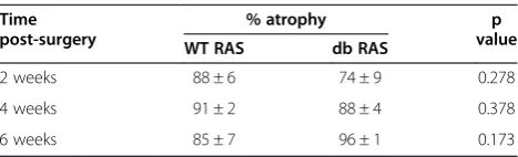
Although the stenotic kidney of db/db mice developed severe atrophy, the glomeruli appeared to be protected from development of diffuse mesangial sclerosis – an early manifestation of diabetic nephropathy –in accord-ance with previous reports on the stenotic kidney of dia-betic patients (data not shown) [15]. Instead, the stenotic kidney of db/db mice developed tubular atrophy to an ex-tent similar to that observed in the stenotic kidney of WT mice at all time-points (Table 1).
As we previously described, the contralateral kidney in WT mice showed mild glomerular enlargement, with no significant interstitial fibrosis, tubular atrophy, or intersti-tial inflammation [5,16]. In striking contrast, the contralat-eral kidney of db RAS mice developed glomerular mesangial matrix expansion that was significantly greater than the contralateral kidney of WT RAS or db sham, as assessed in PAS-stained sections (Figure 2A, 2B) and de novo glomerular fibronectin deposition (Figure 2A, 2C). These histopathologic alterations were observed by 2 weeks following RAS surgery (Figure 2B, 2C) mostly at the juxtamedullary glomeruli. At all time-points be-yond baseline, the severity of diffuse mesangial scler-osis in the contralateral kidney of db RAS mice was significantly greater than that observed in the contra-lateral kidneys of db sham mice or in WT RAS mice (Figure 2B).
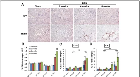
In addition to the glomerular lesions, the contralateral kidney of db RAS mice developed progressive interstitial fibrosis (Figure 2A, 2D, 2E) significantly greater than that of db sham mice, WT RAS, or WT sham mice at the same time point (Figure 2D). Similar patterns were observed in sections stained for the extracellular matrix proteins fibronectin (Figure 2E). The extent of inflam-mation in the contralateral kidney as measured by F4/80+
area was also greater in the db RAS mice compared to both WT RAS and db sham mice (Figure 3A, 3B). We then performed RT-PCR to measure the level of chemo-kine (C-C motif ) ligand 2 (Ccl2) and interleukin 6 (Il-6) mRNA in the contralateral kidney (Figure 3C, 3D). Both were elevated in the contralateral kidney of the db RAS mice in comparison to both WT RAS and db sham mice, indicating presence of inflammation that was not apparent in either the WT RAS or the db sham.
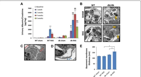
WT RAS mice, but not WT sham mice, developed transient albuminuria that persisted up to 4 weeks post-surgery before returning to baseline (Figure 4A). Db RAS mice, however, developed marked albuminuria (at least 6-fold higher than db sham mice) that persisted throughout the observation period (Figure 4A). To de-termine if basement membrane thickening or podocyte loss (which is some of the characteristic features of diabetic nephropathy) contribute to this transient albuminuria, we performed electron microscopy on the contralateral kid-neys of db/db and WT mice at 6 weeks of hypertension. Mean glomerular basement membrane thickness in the contralateral db RAS kidney was increased by 30% after 6 weeks compared to db sham mice (Figure 4B, 4C, 4E), and their glomeruli also showed extensive podocyte foot process effacement (Figure 4D), which was not observed in the contralateral kidney of db sham, WT sham, or WT RAS mice.
Angiotensin-II induced hypertension does not reproduce the renal injury induced by renovascular hypertension in db mice
A critical role for angiotensin II in the development of chronic renal disease due to etiologies such as diabetes and hypertension has long been recognized [9-13]. We therefore infused db/db mice with angiotensin II (db Ang-II) or PBS for 4 weeks to test the hypothesis that the severe chronic renal damage observed in the contra-lateral kidney of db RAS mice is primarily due to ele-vated angiotensin II levels. Db Ang-II mice developed hypertension comparable to that observed in db RAS mice (Figure 5A) despite lower plasma renin content (Figure 5B). Unlike the db RAS mice, the db Ang-II mice showed a minimal increase in mesangial matrix with no evidence of glomerular fibronectin deposition (Figure 5C). The mean glomerular PAS mesangial matrix score in db Ang-II mice was similar to that of db sham mice, whereas that of db RAS mice was over 4-fold higher (Figure 5D). Both db RAS and db Ang-II developed simi-lar degree of tubusimi-lar atrophy, focal interstitial inflamma-tion and interstitial fibrosis (Figure 5C, 5E), though the db Ang-II mice showed slightly less interstitial fibronectin de-position (Figure 5F).
Despite the lack of mesangial matrix expansion, db Ang-II mice developed significant albuminuria, similar Table 1 Percent atrophy of the stenotic kidney of wild
type (WT) and diabetic (db) mice with RAS at 2, 4, and 6 weeks post-surgery
Time post-surgery
% atrophy p
value
WT RAS db RAS
2 weeks 88 ± 6 74 ± 9 0.278
4 weeks 91 ± 2 88 ± 4 0.378
0.0 0.5 1.0 1.5 2.0 2.5 3.0
WT sham WT RAS db sham db RAS
% A
rea of Fibr
osis
/
200x
HPF
0.0 0.5 1.0 1.5 2.0 2.5 3.0 3.5 4.0
WT sham WT RAS db sham db RAS
P
A
S Sc
or
es (0-4)
Baseline 2 weeks 4 weeks 6 weeks
0 2 4 6 8 10 12 14 16 18
%
FN+ glomeruli /
sections
B
C
D
E
0.0 0.5 1.0 1.5 2.0 2.5 3.0 3.5
WT sham WT RAS db sham db RAS
% FN+
Ar
ea
/ 200x
HPF
WT SHAM WT RAS DB SHAM DB RAS
TRICHR
OME
FN
PA
S
A
4 weeks
6 weeks
***
§§§
* ***
* ***
*** ***
*** ***
*** §§§
§§§
***
**
***
* * ***
*** §§§
†††
**
*** *** †††
††† †††
Figure 2Contralateral kidney of db RAS but not WT RAS or db sham mice developed chronic renal injury. A. Representative
to levels observed in the db RAS mice (Figure 5G). Thus, increased interstitial fibrosis and albuminuria, but not mesangial matrix expansion, can be attributed to angiotensin-II induced hypertension in db/db mice.
Development of renal injury is accelerated in db RAS than in db/db nephrectomized mice (db-UNX)
Given that angiotensin II infusion in db/db mice failed to produce the lesions observed in db RAS mice, we sought to determine whether increased blood flow to the remaining kidney in mice with unilateral nephrectomy (db UNX) was responsible for the development of mesangial sclerosis, interstitial fibrosis, and tubular atro-phy. Unlike db RAS mice, db UNX mice did not develop significant hypertension (Figure 5A), and plasma renin content was lower than that observed in db RAS or db sham (Figure 5B). After 4 weeks, db UNX developed mesangial matrix expansion that was significantly greater than that observed in db sham or db Ang-II mice, but less than in the contralateral db RAS kidney (Figure 5C, 5D). As with db Ang-II, db UNX developed more mod-est interstitial fibrosis compared to db RAS (Figure 5C, 5E) and showed no increased interstitial fibronectin de-position in comparison to db sham (Figure 5C, 5F). Db
UNX developed modest albuminuria, but significantly less than that observed in db RAS mice (Figure 5G).
The severity of injury in the contralateral db RAS kidney exceeds that induced by a combination of UNx and Angiotensin-II induced hypertension
As angiotensin-II induced hypertension and unilateral nephrectomy replicate only some aspects of injury seen in the contralateral kidney of the db RAS mice, we then sought to determine if the combination would produce the severe injury observed in db RAS mice. We thus in-fused angiotensin-II into db/db mice subjected to unilat-eral nephrectomy (db UNX + Ang-II). As with the angiotensin-II infusion alone, db UNX + Ang-II mice de-veloped similar level of hypertension (Figure 5A) with low plasma renin content (Figure 5B). After 4 weeks, we saw a modest increase in the development of mesangial matrix expansion in db uNX + Ang-II mice compared to the db UNX, but lower than the extent of the injury seen in db RAS mice (Figure 5C, 5D). Similarly, we observed an increase in interstitial fibrosis and fibronectin depos-ition in the db UNX + Ang-II mice compared to the db UNX (Figure 5C, 5E, 5F), but similar to those observed in the AngII group. However, the db UNX + Ang-II mice 0
1 2 3 4 5 6 7 8 9 10
Ccl2
Fo
ld
Ch
a
n
g
e
s
***
0.0 0.2 0.4 0.6 0.8 1.0 1.2 1.4
%
F4/80+
Area per HPF
Baseline 2 weeks 4 weeks 6 weeks db/db
WT
Sham 2 weeks 4 weeks 6 weeks
RAS
B
A
C
D
0 2 4 6 8 10 12 14
IL
-6
Fol
d
ch
a
n
g
e
s
††
*** †
***
Ccl2 IL-6
**
§§§
** * ***
§§§
Figure 3The contralateral kidneys of db RAS but not WT RAS mice develop progressive inflammation. A. Representative immunostaining
still developed significantly less fibrosis in comparison to db RAS, indicating other factors that might be con-tributing to the development of this injury. Interest-ingly, db UNX + Ang-II mice developed a similar degree of albuminuria as seen in the db RAS mice at 2 weeks (Figure 5F), but returned to baseline levels at 4 weeks.
Db RAS mice developed greater renal inflammation We and other investigators have shown that the stenotic kidney can become a source of inflammatory cytokines and chemokines [14,17-19] that can cause remote injur-ies. Therefore, we sought to determine if the db RAS mice experienced higher degree of inflammation in com-parison to the control groups. Histological examination showed a significantly higher infiltration of F4/80 renal macrophages in the contralateral kidney of the db RAS mice compared to the other models (Figure 6A, 6B). RT-PCR of Ccl2 and Il-6 as marker of inflammation in the contralateral or remaining kidneys of the mice showed significantly higher elevation of both Ccl2 and Il-6 mRNA in the db RAS compared to the other models (Figure 6C, 6D). In contrast, both db RAS and db
UNX + Ang-II showed similar elevation of serum CCL2 and IL-6.
Reduction of blood pressure ameliorates chronic injury to the contralateral kidney of db RAS mice
To further determine the role of angiotensin II in this process, we sought to determine whether lowering blood pressure by angiotensin-II receptor blocker (ARB) or by hydralazine, which induces vasodilation without direct effects on the renin angiotensin system, would amelior-ate renal damage observed in the contralamelior-ateral kidney of db RAS mice. Treatment of db RAS mice with either ARB or hydralazine was similarly effective in reducing blood pressure to baseline levels (Figure 7A). Both ARB and hydralazine treated mice had no significant eleva-tion of plasma renin content at 4 weeks (Figure 7B). ARB and hydralazine were effective in reducing but not abolishing glomerular mesangial matrix expansion (Figure 8A, 8B), glomerular de novo fibronectin expres-sion, interstitial fibrosis (Figure 8C), and reduced influx of macrophages into the contralateral kidney (Figure 8D). However, only ARB reduced urine albumin excretion in
Figure 4Db RAS mice developed albuminuria to a greater extent that the WT RAS mice. A. db RAS experienced persistent and progressive
albuminuria compared to the transient albuminuria experienced by WT RAS. Albuminuria was measured in spot urine sample and normalized to urine creatinine. Data are presented as means ± SE. ** p <0.01, *** p < 0.001 vs. WT sham with the same time point. §§ p < 0.01, §§§ p < 0.001 vs. WT RAS or db sham with the same time point. Statistical significance was determined by 2-way ANOVA followed by Tukey adjusted post-hoc comparison.B-D. Representative electron microscope (EM) images of contralateral kidneys at 6 weeks post-surgery. db RAS mice showed significant area of mesangial matrix expansion (yellow arrows)(B)in comparison to WT RAS or db sham, along with thickened basement membrane (red arrows)
0 50 100 150 200
0 2 4
S
y
s
tolic BP
(mmHg)
Time post-surgery (weeks)
Sham RAS Ang-II UNX
UNX+Ang-II 0
10 20 30 40 50 60
Sham RAS Ang-II UNX UNX + Ang-II
Angiot
ensin I
(ng
/mL/hr)
B
A
SHAM RAS Ang-II
TRICHR
O
ME
FN
P
AS
UNX UNX + Ang-II
C
0 1000 2000 3000 4000 5000 6000 7000 8000
Sham RAS AII UNX UNX+AII
Urinary
albumin/
c
re
a
ti
n
in
e
(µg /
mg) Baseline
2 weeks 4 weeks
0.0 0.5 1.0 1.5 2.0 2.5 3.0
% Fn
+ Ar
ea
0.5 1.0 1.5 2.0 2.5 3.0
% Ar
ea of Fibr
osis
0 1 2 3 4
P
A
S Scor
e (0-4)
D
E
F
G
** **
***
***
***
***
*
**
§§§
** ***
*** ***
*** *
*** ***
*** *
*
§§
***
*
Figure 5Angiotensin-II induced hypertension (Ang-II), unilateral nephrectomy (UNX), and combination of both angiotensin-II and
db-RAS mice to levels observed in WT-RAS mice (Figure 8E).
Discussion
A role for hypertension in the development of renal le-sions in db/db mice has not been clearly established [20-25]. We found that db sham mice did not develop spontaneous hypertension, while db RAS mice develop hypertension to an extent that is similar to that observed in WT RAS mice, yet associated with transient but more prolonged increases in plasma renin activity and greater renalRen1expression. This persistent increase in plasma renin activity in db RAS mice may reflect interactions
between hemodynamic forces associated with renovascu-lar hypertension and the diabetic mileau. Despite simirenovascu-lar level of systolic blood pressure, the contralateral kidney of db RAS mice developed chronic renal injury charac-terized by development of mesangial matrix expansion, interstitial fibrosis, tubular atrophy, and interstitial in-flammation, as opposed to the contralateral kidneys in a number of other strains of non-diabetic mice subjected to RAS [5,16,26]. Glomerular histopathologic alterations in the contralateral kidney of db/db mice were striking, and reminiscent of those observed in progressive human diabetic nephropathy, with severe and diffuse mesangial matrix expansion, evident as early as 2 weeks following
2 4 6 8 10 12
Ccl2
F
old incr
ease
0.0 0.5 1.0 1.5 2.0 2.5
% Ar
ea F4/80+
B
A
C
F4/80
1 2 3 4 5 6
Il
-6
F
o
ld incr
ease
D
db/db
RAS Ang-II UNX UNX + Ang-II
E
F
0 50 100 150 200
Serum CCL2
(pg
/mL)
0 5 10 15 20
Serum IL-6
(pg
/mL)
*
**
*
*
*
* *
§ Ccl2 IL-6
Figure 6Db RAS mice experienced higher level of inflammation compared to db mice with Angiotensin-II induced hypertension
induction of hypertension. Mesangial matrix expansion consistently was far more extensive than in age-matched db sham mice, and was associated with de novo glom-erular fibronectin expression. Older db/db mice develop glomerular basement membrane thickening, but quanti-tative studies in this model have not yet been reported [27-29]. We found an increase of glomerular basement membrane thickness in the contralateral db RAS kidney by six weeks post-surgery, as assessed by morphometric analysis of electron microscopic images, a well-recognized feature of evolving diabetic nephropathy. Glomeruli in these kidneys (but not in WT RAS) showed extensive ef-facement of visceral epithelial cell foot processes, a mor-phologic correlate of the progressive albuminuria observed in these mice. At all time-points, urine albumin excretion was significantly greater in db RAS than db sham mice. Based on these observations, we conclude that renovascu-lar hypertension markedly accelerates renal disease pro-gression in db/db mice as characterized by glomerular mesangial matrix expansion, progressive interstitial fibrosis and inflammation, and breakdown of the filtration barrier. This is in accordance with clinical observations indicating that progression of diabetic nephropathy is accelerated in patients with hypertension.
We infused db/db mice with angiotensin II for 4 weeks to address a potential role of angiotensin II induced hypertension on renal architecture in db/db mice. These mice developed hypertension to levels similar to those attained in db RAS mice, yet we observed a minimal in-crease in mesangial matrix deposition and no evidence of de novo glomerular fibronectin deposition. Neverthe-less, db Ang-II developed albuminuria similar to that ob-served in db RAS mice and to that reported following angiotensin II infusion to non-diabetic mice [30,31]. Taken together, these observations suggest that the pro-gressive and bilateral renal injury in db RAS mice is not
mechanistically related to elevated angiotensin II levels alone, although angiotensin II plays a major role in de-velopment of albuminuria in this model [7,8]. This find-ing underscores a critical role for activation of the renin-angiotensin system in the development of albuminuria and provides a therapeutic rationale for the widespread use of renin-angiotensin inhibitors in treatment of chronic kidney disease.
We then sought to determine whether hyperfiltration associated with unilateral nephrectomy may underlie the progressive renal damage observed in the contralateral db RAS kidney. Unlike db RAS or db Ang-II mice, db UNX did not develop significant hypertension. Db UNX also did not develop increased urine albumin excretion that was observed in the db RAS or db Ang-II. However, as shown before, db/db mice with unilateral nephrec-tomy did develop greater glomerular mesangial matrix expansion than age-matched db/db mice with two kid-neys [8,32], although its extent was less than that of db RAS mice. Although few investigators have directly re-ported the extent of interstitial fibrosis in this model, db/db mice evaluated at 14–18 weeks post UNX exhib-ited a modest increase in interstitial inflammation, inter-stitial volume, and number of tubules showing dilation or atrophy [8,32,33]. In the current study, we find that db UNX mice, in striking contrast to db RAS mice, do not develop significant interstitial fibrosis or tubular at-rophy at 4 weeks post UNX. Therefore, glomerular mesangial matrix expansion in db/db mice can be attrib-uted at least in part to hemodynamic factors associated with hyperfiltration, whereas elevation of blood pressure appears to play a major role in development of albumin-uria in db/db mice.
As Angiotensin-II induced hypertension and UNX alone only recapitulate some features of renal injury seen in the contralateral kidney of db RAS mice, we combined both 0
50 100 150 200
0 2 4
S
y
s
to
lic BP (mmHg)
Time Post Surgery (weeks)
Sham RAS RAS + ARB RAS + Hyd
A
0 5 10 15 20 25 30 35 40 45 50
Angiot
ensin I
(ng
/mL/hr)
B
*** *
*
Figure 7Both Angiotensin Receptor Blocker (ARB) and hydralazine lower systolic blood pressure in db RAS. A. Systolic blood pressure
0.5 1.0 1.5 2.0 2.5 3.0
% Ar
ea of Fibr
o
sis
RAS+ Vehicle
TRICHR
OME
FN
P
A
S
RAS+ Hydralazine
RAS+ ARB
F4/80
0.0 0.5 1.0 1.5 2.0
% Ar
e
a
F4/80
+
0.0 0.5 1.0 1.5 2.0 2.5 3.0
P
A
S Score
(0-4)
B
C
E
0 500 1000 1500
A
lbu
min/
Creati
nine
(ug/mg)
D
A
* *
**
** **
Figure 8Both Angiotensin Receptor Blocker (ARB) and hydralazine afford similar attenuation of injury in the contralateral kidney of
in db/db mice. Remaining kidneys of db UNX + Ang-II mice developed all the features seen in the db RAS mice, namely mesangial expansion, interstitial fibrosis, tubular atrophy, and albuminuria, but the severity of injury ob-served in the contralateral kidney of db RAS mice was greater than that of db UNX + Ang-II mice.
To examine if hypertension was necessary for the de-velopment of progressive renal fibrosis in the contralat-eral kidneys of db/db mice, we treated them with ARB or the vasodilator hydralazine, which lowered blood pressure to levels similar to those observed in db sham mice without significant changes in plasma renin activ-ity. Reduction of blood pressure was effective in redu-cing mesangial matrix expansion, fibronectin expression, interstitial fibrosis, and tubular atrophy in the contralat-eral kidney of db RAS mice. However, urine albumin excretion was significantly reduced by ARB only. There-fore, we conclude that hypertension plays an essential role for the development of chronic renal lesions in the contralateral kidney of db/db mice subjected to RAS, while increase level of angiotensin II plays a role in the development of albuminuria. Interestingly, while both drug treatments attenuate the development of renal in-jury, both do not abolish it. Given the less severe injury observed in the db UNX + Ang-II, these results point to some other factor independent of blood pressure elevation and hyperfiltration process that is mediated by the sten-otic kidney, possibly by the activated RAAS.
We and other investigators have shown that the sten-otic kidney experienced considerable oxidative stress [14,17] and produced substantial level of inflammatory cytokines [18,19]. Indeed, in comparison to the other models, contralateral kidney of db RAS exhibited signifi-cantly higher expression of the inflammatory chemokine CCL2 and the inflammatory cytokine IL-6, both of which represent prognostic of development of renal jury [34-36]. Nonetheless, db RAS showed similar in-creased in serum CCL2 and IL-6 to db UNX + Ang-II. However, although serum levels of CCL2 might be ele-vated in diabetic patients, they are not associated to the development of albuminuria, renal macrophage influx, or interstitial fibrosis [37-39]. Instead, both urine CCL2 and IL-6 excretion – reflecting production of these in-flammatory molecules within the kidney itself – have been shown to correlate significantly with progression of renal injury [37,38,40]. Furthermore, increased albumin-uria may itself aggravate tubular injury and accelerate development of renal injury by increasing tubular CCL2 and IL-6 production [41,42].
Conclusion
In summary, renovascular hypertension accelerates de-velopment of diabetic renal injury in db/db mice that re-capitulates many of the features of chronic renal disease
in subjects with diabetes and hypertension and markedly accelerated the progression of chronic renal disease. As hypertension induced by angiotensin II infusion was not sufficient to reproduce these lesions, we believe that inter-actions between the diabetic milieu and hemodynamic forces associated with hyperfiltration were necessary to produce progressive renal disease in db/db mice. Although combination of Angiotensin-II infusion and unilateral nephrectomy are able to replicate many features of injury observed in the db RAS, the db RAS model is likely more physiologically relevant to the development of diabetic ne-phropathy in patients with both diabetes and RAS, and will allow the development of mechanistic studies to identify critical pathways related to inflammation, fibrosis, oxidative stress, and cell cycle regulation that are responsible for the development and progression of diabetic renal disease.
Competing interests
The authors declare that they have no competing interests.
Authors’contributions
SPH and JPG conceived the project. SPH, BEK and JPG designed the methods and the experiments. BEK performed all the animal surgeries. SPH carried out the laboratory experiments, analyzed the data, and interpreted the results. SPH and JPG wrote the manuscript. LOL and SCT gave technical support, conceptual advice, and critical review of the manuscript. All authors read and approved the final manuscript.
Acknowledgments
We thank Kim Butters for assistance with primer design, Hui Tang, PhD for assistance with plasma renin content assay, Andrea McConico for assistance with ELISA assay, Mayo Clinic Electron Microscopy Core for assistance with EM imaging and morphometric analysis, Karen Lien for beautifully stained slides and Anuradha Krishnan, PhD for critical reading of this manuscript. These studies were supported by NIH/NIAID R01 AI100911-01, NIH/NHLBI P01 HL85307 and UL1 RR024150. S.P.H. is supported by NIH/NIGMS T32 GM 6584. This paper’s contents are solely the responsibility of the authors and do not necessarily represent the official views of the NIH.
Grant support
NIH R01-AI100911-01; P01-HL85307; T32 GM6584; UL1-RR024150.
Author details
1Department of Laboratory Medicine & Pathology, Mayo Clinic, 200 First
Street SW, Rochester, MN 55905, USA.2Medical Scientist Training Program, Mayo Clinic, 200 First Street SW, Rochester, MN 55905, USA.3Division of
Nephrology & Hypertension, Mayo Clinic, 200 First Street SW, Rochester, MN 55905, USA.
Received: 16 October 2013 Accepted: 31 March 2014 Published: 4 April 2014
References
1. Centers for Disease Control and Prevention. National diabetes fact sheet: national estimates and general information on diabetes and prediabetes in the United States, 2011.Atlanta, GA: U.S. Department of Health and Human Services, Centers for Disease Control and Prevention; 2011. 2. Valabhji J, Robinson S, Poulter C, Robinson AC, Kong C, Henzen C, Gedroyc WM,
Feher MD, Elkeles RS:Prevalence of renal artery stenosis in subjects with type 2 diabetes and coexistent hypertension.Diab Care2000,23(4):539–543. 3. Myers DI, Poole LJ, Imam K, Scheel PJ, Eustace JA:Renal artery stenosis by
three-dimensional magnetic resonance angiography in type 2 diabetics with uncontrolled hypertension and chronic renal insufficiency: prevalence and effect on renal function.Am J Kidney Dis2003,41(2):351–359. 4. Hajsadeghi S, Fereshtehnejad SM, Pourshirmohammadi-Sabzevari M,
angiography study.Arch Iran Med2009,12(3):250–255.
5. Warner GM, Cheng J, Knudsen BE, Gray CE, Deibel A, Juskewitch JE, Lerman LO, Textor SC, Nath KA, Grande JP:Genetic deficiency of Smad3 protects the kidneys from atrophy and interstitial fibrosis in 2K1C hypertension. Am J Physiol Renal Physiol2012,302(11):F1455–F1464.
6. Lorenz JN, Lasko VM, Nieman ML, Damhoff T, Prasad V, Beierwaltes WH, Lingrel JB:Renovascular hypertension using a modified two-kidney, one-clip approach in mice is not dependent on theα1 orα2 Na-K-ATPase ouabain-binding site.Am J Physiol Renal Physiol2011,301(3):F615–F621. 7. Levine DZ, Iacovitti M, Robertson SJ:Modulation of single-nephron GFR in
the db/db mouse model of type 2 diabetes mellitus. II. Effects of renal mass reduction.Am J Physiol Regul Integr Comp Physiol2008,294(6):R1840–R1846. 8. Ninichuk V, Khandoga AG, Segerer S, Loetscher P, Schlapbach A, Revesz L,
Feifel R, Khandoga A, Krombach F, Nelson PJ, Schlondorff D, Anders HJ:The role of interstitial macrophages in nephropathy of type 2 diabetic db/db mice.Am J Pathol2007,170(4):1267–1276.
9. Fraune C, Lange S, Krebs C, Hölzel A, Baucke J, Divac N, Schwedhelm E, Streichert T, Velden J, Garrelds IM, Danser AH, Frenay AR, van Goor H, Jankowski V, Stahl R, Nguyen G, Wenzel UO:AT1 antagonism and renin inhibition in mice: pivotal role of targeting angiotensin II in chronic kidney disease.Am J Physiol Renal Physiol2012,303(7):F1037–F1048. 10. Textor SC, Lerman L:Renovascular hypertension and ischemic
nephropathy.Am J Hypertens2010,23(11):1159–1169.
11. Kobori H, Nangaku M, Navar LG, Nishiyama A:The intrarenal renin-angiotensin system: from physiology to the pathobiology of hypertension and kidney disease.Pharmacol Rev2007,59(3):251–287.
12. Brewster UC, Setaro JF, Perazella MA:The renin-angiotensin-aldosterone system: cardiorenal effects and implications for renal and cardiovascular disease states.Am J Med Sci2003,326(1):15–24.
13. Langham RG, Kelly DJ, Gow RM, Zhang Y, Cordonnier DJ, Pinel N, Zaoui P, Gilbert RE:Transforming growth factor-beta in human diabetic nephropathy: effects of ACE inhibition.Diab Care2006,29(12):2670–2675.
14. Hartono SP, Knudsen BE, Zubair AS, Nath KA, Textor SJ, Lerman LO, Grande JP: Redox signaling is an early event in the pathogenesis of renovascular hypertension.Int J Mol Sci2013,14(9):18640–18656.
15. Berkman J, Rifkin H:Unilateral nodular diabetic glomerulosclerosis (Kimmelstiel-Wilson): report of a case.Metabol Clin Exp1973,22(5):715–722. 16. Cheng J, Zhou W, Warner GM, Knudsen BE, Garovic VD, Gray CE, Lerman LO,
Platt JL, Romero JC, Textor SC, Nath KA, Grande JP:Temporal analysis of signaling pathways activated in a murine model of two-kidney, one-clip hypertension.Am J Physiol Ren Physiol2009,297(4):F1055–F1068. 17. Urbieta-Caceres VH, Lavi R, Zhu XY, Crane JA, Textor SC, Lerman A, Lerman
LO:Early atherosclerosis aggravates the effect of renal artery stenosis on the swine kidney.Am J Physiol Ren Physiol2010,299(1):F135–F140. 18. Eirin A, Gloviczki ML, Tang H, Gossl M, Jordan KL, Woollard JR, Lerman A,
Grande JP, Textor SC, Lerman LO:Inflammatory and injury signals released from the post-stenotic human kidney.Eur Heart J2013,34(7):540–548. 19. Eirin A, Zhu XY, Woollard JR, Herrmann SM, Gloviczki ML, Saad A, Juncos LA,
Calhoun DA, Rule AD, Lerman A, Textor SC, Lerman LO:Increased Circulating Inflammatory Endothelial Cells in Blacks With Essential Hypertension.Hypertension2013,62(3):585–591.
20. Park S, Bivona BJ, Feng Y, Lazartigues E, Harrison-Bernard LM:Intact renal afferent arteriolar autoregulatory responsiveness in db/db mice.Am J Physiol Ren Physiol2008,295(5):F1504–F1511.
21. Brezniceanu ML, Liu F, Wei CC, Chenier I, Godin N, Zhang SL, Filep JG, Ingelfinger JR, Chan JS:Attenuation of interstitial fibrosis and tubular apoptosis in db/db transgenic mice overexpressing catalase in renal proximal tubular cells.Diabetes2008,57(2):451–459.
22. Koya D, Haneda M, Nakagawa H, Isshiki K, Sato H, Maeda S, Sugimoto T, Yasuda H, Kashiwagi A, Ways DK, King GL, Kikkawa R:Amelioration of accelerated diabetic mesangial expansion by treatment with a PKC beta inhibitor in diabetic db/db mice, a rodent model for type 2 diabetes. FASEB J2000,14(3):439–447.
23. Guo C, Martinez-Vasquez D, Mendez GP, Toniolo MF, Yao TM, Oestreicher EM, Kikuchi T, Lapointe N, Pojoga L, Williams GH, Ricchiuti V, Adler GK: Mineralocorticoid receptor antagonist reduces renal injury in rodent models of types 1 and 2 diabetes mellitus.Endocrinology2006,147(11):5363–5373. 24. Moriyama T, Oka K, Ueda H, Imai E:Nilvadipine attenuates mesangial
expansion and glomerular hypertrophy in diabetic db/db mice, a model for type 2 diabetes.Clin Exp Nephrol2004,8(3):230–236.
disrupted blood pressure circadian rhythm in type 2 diabetic db/db mice.Am J Physiol Heart Circ Physiol2008,295(4):H1634–H1641.
26. Wang D, Warner GM, Yin P, Knudsen BE, Cheng J, Butters KA, Lien KR, Gray CE, Garovic VD, Lerman LO, Textor SC, Nath KA, Simari RD, Grande JP:Inhibition of p38 MAPK attenuates renal atrophy and fibrosis in a murine renal artery stenosis model.Am J Physiol Heart Circ Physiol2013,304(7):F938–F947. 27. Hong SW, Isono M, Chen S, Iglesias-De La Cruz MC, Han DC, Ziyadeh FN:
Increased glomerular and tubular expression of transforming growth factor-beta1, its type II receptor, and activation of the Smad signaling pathway in the db/db mouse.Am J Pathol2001,158(5):1653–1663. 28. Like AA, Lavine RL, Poffenbarger PL, Chick WL:Studies in the diabetic mutant mouse. VI. Evolution of glomerular lesions and associated proteinuria.Am J Pathol1972,66(2):193–224.
29. Lubec B, Rokitansky A, Hayde M, Aufricht C, Wagner U, Mallinger WR, Hoger H, Lubec G:Thiaproline reduces glomerular basement membrane thickness and collagen accumulation in the db/db mouse.Nephron1994,66(3):333–336. 30. Mitani H, Ishizaka N, Aizawa T, Ohno M, Usui S, Suzuki T, Amaki T, Mori I,
Nakamura Y, Sato M, Nangaku M, Hirata Y, Nagai R:In vivo klotho gene transfer ameliorates angiotensin II-induced renal damage.Hypertension 2002,39(4):838–843.
31. Jennings BL, Anderson LJ, Estes AM, Yaghini FA, Fang XR, Porter J, Gonzalez FJ, Campbell WB, Malik KU:Cytochrome P450 1B1 contributes to renal dysfunction and damage caused by angiotensin II in mice.Hypertension 2012,59(2):348–354.
32. Ninichuk V, Clauss S, Kulkarni O, Schmid H, Segerer S, Radomska E, Eulberg D, Buchner K, Selve N, Klussmann S, Anders HJ:Late onset of Ccl2 blockade with the Spiegelmer mNOX-E36-3’PEG prevents glomerulosclerosis and improves glomerular filtration rate in db/db mice.Am J Pathol2008,172(3):628–637. 33. Ninichuk V, Kulkarni O, Clauss S, Anders H:Tubular atrophy, interstitial
fibrosis, and inflammation in type 2 diabetic db/db mice. An accelerated model of advanced diabetic nephropathy.Eur J Med Res2007,12(8):351–355. 34. Chow FY, Nikolic-Paterson DJ, Ma FY, Ozols E, Rollins BJ, Tesch GH:Monocyte
chemoattractant protein-1-induced tissue inflammation is critical for the development of renal injury but not type 2 diabetes in obese db/db mice. Diabetologia2007,50(2):471–480.
35. Manhiani MM, Quigley JE, Socha MJ, Motamed K, Imig JD:IL6 suppression provides renal protection independent of blood pressure in a murine model of salt-sensitive hypertension.Kidney Blood Press Res2007,30(4):195–202. 36. Kanamori H, Matsubara T, Mima A, Sumi E, Nagai K, Takahashi T, Abe H,
Iehara N, Fukatsu A, Okamoto H, Kita T, Doi T, Arai H:Inhibition of MCP-1/ CCR2 pathway ameliorates the development of diabetic nephropathy. Biochem Biophys Res Commun2007,360(4):772–777.
37. Banba N, Nakamura T, Matsumura M, Kuroda H, Hattori Y, Kasai K: Possible relationship of monocyte chemoattractant protein-1 with diabetic nephropathy.Kidney Int2000,58(2):684–690.
38. Wada T, Furuichi K, Sakai N, Iwata Y, Yoshimoto K, Shimizu M, Takeda SI, Takasawa K, Yoshimura M, Kida H, Kobayashi KI, Mukaida N, Naito T, Matsushima K, Yokoyama H:Up-regulation of monocyte chemoattractant protein-1 in tubulointerstitial lesions of human diabetic nephropathy. Kidney Int2000,58(4):1492–1499.
39. Kiyici S, Erturk E, Budak F, Ersoy C, Tuncel E, Duran C, Oral B, Sigirci D, Imamoglu S:Serum monocyte chemoattractant protein-1 and monocyte adhesion molecules in type 1 diabetic patients with nephropathy.Arch Med Res2006,37(8):998–1003.
40. Wada T, Yokoyama H, Su SB, Mukaida N, Iwano M, Dohi K, Takahashi Y, Sasaki T, Furuichi K, Segawa C, Hisada Y, Ohta S, Takasawa K, Kobayashi K, Matsushima K:Monitoring urinary levels of monocyte chemotactic and activating factor reflects disease activity of lupus nephritis.Kidney Int 1996,49(3):761–767.
41. Eddy AA, Giachelli CM:Renal expression of genes that promote interstitial inflammation and fibrosis in rats with protein-overload proteinuria. Kidney Int1995,47(6):1546–1557.
42. Takaya K, Koya D, Isono M, Sugimoto T, Sugaya T, Kashiwagi A, Haneda M: Involvement of ERK pathway in albumin-induced MCP-1 expression in mouse proximal tubular cells.Am J Physiol Renal Physiol2003,284(5):F1037–F1045.
doi:10.1186/1471-2369-15-58