C A S E R E P O R T
Open Access
Single-staged uniportal VATS in the supine
position for simultaneous bilateral primary
spontaneous pneumothorax
Kyung Soo Kim
Abstract
Background:Simultaneous bilateral primary spontaneous pneumothorax (SBPSP) is rare, but requires surgery on both sides, in patients with definite bilateral bullae to prevent life-threatening conditions. Recently, uniportal video-assisted thoracoscopic surgery (VATS) has been widely accepted as a less invasive technique for the treatment of pneumothorax. Thus, we introduced single-staged uniportal VATS technique in the supine position, for the management of two cases of SBPSP.
Case presentation:A 17-year-old boy presented with bilateral spontaneous pneumothorax and he underwent single-staged uniportal VATS in the supine position. Single wide draping in consecutive bilateral approaches removes the needs of changing patients’position. Whole thoracoscopic procedure for wedge resection of bullae lesions was conducted without difficulty. The total operation time took 65 min and the patient discharged 3 days after the operation. The patient was followed for 24 months without recurrence of both sides. Another 18-year-old boy was admitted with bilateral spontaneous pneumothorax and single-staged uniportal VATS was also performed in the supine position. The total operation time took 79 min and the patient discharged on postoperative day 4. He was followed for 19 months without recurrence of both sides.
Conclusions:Single-staged uniportal VATS approach yielded satisfactory results from simplicity that not requires position change compared to conventional multi-ports VATS in the lateral position, and with better cosmetics. This technique is thought to be a feasible procedure in selective patients with SBPSP or with contralateral bullae for preventive role.
Keywords:Uniportal, Single port, Pneumothorax, Thoracoscopic surgery, Video Assisted Thoracic Surgery (VATS), Supine position
Background
Bilateral primary spontaneous pneumothorax, either non-simultaneous or simultaneous, has a reported inci-dence of 7.8 to 20% among pneumothorax patients [1]. Simultaneous bilateral primary spontaneous pneumo-thorax (SBPSP) is a rare condition with an incidence of 1%, but single-staged surgery is required on both sides of patients with definite bilateral bullae to prevent the life-threatening condition [2]. Recently, single incisional video-assisted thoracoscopic surgery (VATS) has been accepted for the treatment of primary spontaneous
pneumothorax [3–5]. Treatment of bilateral pulmonary lesions has been reported to be effective using single-staged approach in the lateral position on position change [6]. Bilateral single-port sympathicotomy has also been introduced as a feasible technique in the supine pos-ition [7]. Thus, we introduced a single-staged uniportal VATS technique in the supine position for treatment of two cases of SBPSP, and evaluated technical feasibility with surgical outcomes.
Case presentation
Case 1
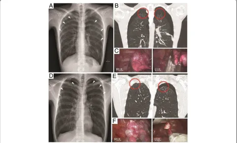
A 17-year-old boy presented with chest discomfort for 1 week. A chest X-ray and preoperative high-resolution computed tomography (HRCT) revealed images of SBPSP
Correspondence:[email protected]
Department of Thoracic and Cardiovascular Surgery, Seoul St. Mary’s Hospital, College of Medicine, The Catholic University of Korea, Banpo-daero 222, Seocho-gu, Seoul 06591, Republic of Korea
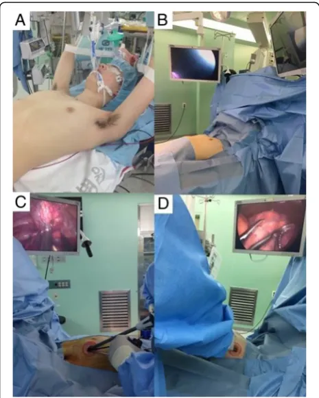
with multiple apical bullae on both lung apices (Fig. 1a-b). Under general anesthesia with double-lumen endotracheal intubation for sequential, selective lung ventilation, the patient was positioned supine with arms hanging to the overhead strut of the ether screen using the arm slings. Then, cushion pads were packed below the midline of the back for chest elevation to avoid collision of thoracoscopic instruments and the operating table (Fig. 2a). After transverse single draping to expose the bilateral chest wall simultaneously, the operative side was slightly ele-vated by table tilting to the opposite side. Two discrete high definition monitors were set up for sequential oper-ation without changing of monitor position (Fig. 2b). Then, a minimal skin incision (less than 2.5 cm in length) was made in the fourth intercostal space at the anterior axillary line. First, full thoracoscopic inspection was con-ducted from the apex to the base using a 5-mm, 30° thora-coscope and thoracoscopic assistant stands on the operator’s sides of the same view-point. Exploration of en-tire lung was possible by lung retractions using endo-grasper under rotated thoracoscopic view (Fig. 2c). An identified apical bullae lesion of the upper lobe was held with grasping forceps and resected with endosta-plers (Echelon 60 Endopath stapler; Ethicon Endosurgery Corp., Cincinnati, OH, USA) through the uniport (Fig. 1c).
Pleural abrasion was not performed, and a saline merging test by temporary lung expansion after infusion of warm physiological saline in the Trendelenburg position con-firmed no air leakage. An absorbable polyglycolic acid sheet (Neoveil; Gunze, Kyoto, Japan) was covered with fibrin glue (Beriplast-P combiset; CSL Behring GmbH, Marbug, Germany). Then, a 16 Fr. chest tube was placed at the anterior part of the uniport. After confirmation of no wound bleeding into direct thoracoscopic view, inter-costal nerve block was conducted. Then, the wound was closed layer by layer using an absorbable suture in a running fashion with 2-0 and 3-0 Braided synthetic absorbable suture (PolysorbTM, Covidien, Mansfield, MA) under both lung ventilation, and the skin was approxi-mated with 4-0 barbed absorbable suture material (V-Loc™ Absorbable Wound Closure Device product line, Covidien, Mansfield, MA). The same procedures were conducted on the contralateral side via a 2.5 cm single incision in the fourth intercostal space at the anterior axillary line (Fig. 2d). Under the selective one lung ventilation by experienced an-esthesiologists, the table was slightly tilted to the opposite direction without changing position into lateral decubitus. During the single-staged operation on the contralateral side, function of the chest tube on opposite side is well detectable, confirming neither air leakage nor tube
compression of the chest wall. The total operation time took 65 min without any intra-operative events. Intraven-ous patient controlled analgesia was applied for relief from postoperative pain. Bilateral chest tubes were removed on postoperative days 1 and 2, serially. The patient recovered uneventfully and discharged 3 days after the operation. The patient was followed at the out-patient clinic for 24 months without a recurrence of pneumothorax on both sides.
Case 2
An 18-year-old boy was conservatively managed for a bi-lateral minimal amount of pneumothorax during follow-ing up of 6 months at the out-patient clinic. He visited the hospital again complaining of chest discomfort. A chest X-ray revealed bilateral spontaneous pneumothorax, and an HRCT revealed multiple, variable-sized apical bullae on both lung apices (Fig. 1d-e). An operation was performed to control recurrent pneumothorax with identified bilateral definite bullae. The operative procedures
were similar to the Case 1 except division procedure of the adhesion band on the apex of the right upper lobe (Fig. 1f, Additional file 1: Video S1). The total operation time was 79 min without any intra-operative events. Bilateral chest tubes were removed on postoperative days 1 and 3. The patient discharged on postoperative day 4 without compli-cations. He was followed for 19 months without recurrence of both sides.
Discussion
Simultaneous bilateral primary spontaneous pneumo-thorax (SBPSP) is a rare clinical event, with a reported incidence as low as 1% [2]. SBPSP may develop in an emergency situation, which requires either urgent chest tube insertion or surgery in cases with definite bilateral bullae to prevent life-threatening conditions. SBPSP has been treated using trans-sternal, bilateral thoracotomy, mini-thoracotomy, or sequential VATS approach [8, 9]. Several reports discussed on a bilateral VATS approach to spontaneous pneumothorax in the lateral position, also multi-ports VATS for SBPSP in the supine position demonstrated its efficacy and low morbidity [10, 11]. Re-cently, uniportal VATS have gained feasibility using min-imal skin incision as well as its lower morbidities due to the least involvement in intercostal space compared to multi-ports VATS [12, 13]. Thus, we transferred the less invasive VATS approach in the supine position into the treatment of SBPSP.
Several advantages were demonstrated by our tech-nique comparing to conventional multi-ports VATS in lateral decubitus position. In supine position, single wide draping in consecutive bilateral approaches after slight tilting of the operation table in the opposite direction removes the needs of changing patients’ position. Thus, the whole procedure can be performed simpler without time-consuming re-draping, position change or possible chest tube compression, which may be required in a lateral position. The positioning of a patient is simple in that both arms are hung overhead, and a cushion is packed below the back to avoid collision of thoracoscopic instruments with the operating table. In addition, this procedure also helps to widen the narrow intercostal spaces [14]. An alter-nating inflation technique using pneumatic cuffs was intro-duced and we thought this may be useful in our technique for treatment of SBPSP. However, excessive chest wall ele-vation was not required because the cushion pad of the back was enough for full exposure to the surgical field without disturbance of instrumental manipulation [15].
Bertolaccini et al. demonstrate the geometric and ergonomic advantages of uniportal VATS approach com-pared to the standard three-port approach in a lateral position which resulting in greater fatigue [16]. In su-pine position, thoracoscopic surgical instruments includ-ing endostaplers can also be introduced into parallel Fig. 2aPatient positioning for bilateral, staged-operation. In a
approaches from the same level of uniport. Thus, articulation technique using endostapler is less needed in supine position compared to the conventional VATS in a lateral decubitus position that surgeon may feel more comfortable to manipulate the instruments as we experienced. Total operation time may also be short-ened by saving time for reconfirming the double-lumen tube position after position change and time for mul-tiple wound closure. The saline submerging test can also be performed without technical difficulties by using Trendelenburg position with 15–30° using less amount of saline infusion.
The trans-mediastinal thoracoscopic approach using multi-ports already has been demonstrated for simul-taneous resection for bilateral bullae lesions with sat-isfactory results [17–19]. In a single port surgery, it is thought to be difficult to manipulate the thoracoscopic instruments for mediastinal dissection through the minimal single incision.. However, the single-stage, uniportal trans-mediastinal approach might also be challengeable because it may be more easily accessible to the supine position due to the ergonomic advantages, as mentioned above.
In our cases, chest tubes were not inserted prior to the operations because both patients had no underlying pul-monary disease and showed stable vital signs without dyspnea. We considered the possible situations of desat-uration during selective one lung ventilation. We thought that air tapping or chest tube insertion on the opposite side can be more easily accessible during a one-side VATS by wide draping in the supine position, compared to the conventional VATS that require pos-ition change in the lateral pospos-ition. We also considered that more collapsed lung is acceptable to the first oper-ation but it is surgeons’preference according to the pa-tient’s condition. If the patient had the chest tube on any side with lung expansion, contralateral lung should be the first choice of approach. In our cases, we could perform the whole sequential procedure without place-ment of additional ports or conversion to thoracotomy be-cause patients were tolerable without desaturation under multi-monitoring by experienced anesthesiologists.
The possibility of using VATS for preventive opera-tions on the contralateral lung lesions for pneumothorax is still controversial. However, more than 50% of ipsilat-eral pneumothorax patients show contralatipsilat-eral blebs or bullae on CT scan [20]. Furthermore, several reports have suggested its cost-effectiveness for saving subse-quent hospitalization or operation, and beneficial effect on preventing contralateral occurrence (0–7%) due to its high recurrence rates (18–50%) [21, 22]. Thus, our tech-nique is thought to be a useful option even for the manage-ment of ipsilateral pneumothorax cases of definite bilateral bullae on CT scans.
Conclusions
Single-staged, uniportal VATS technique in the supine position is thought to be a feasible procedure in selective patients with SBPSP or with contralateral bullae for pre-ventive role.
Additional file
Additional file 1: Video S1.Operative procedures of single staged uniportal VATS technique for SBPSP. (MP4 11779 kb)
Abbreviations
HRCT:High-resolution computed tomography; SBPSP: Simultaneous bilateral primary spontaneous pneumothorax; VATS: Video-assisted thoracoscopic surgery
Acknowledgements
We thank E-world editing for English editing.
Funding
None.
Availability of data and materials
The dataset supporting the conclusions of this article is included within the article and its Additional file 1.
Authors’contributions
KS Kim drafted the manuscript, managed the figures, and performed literature search. All authors read and approved the final manuscript.
Competing interests
The author declares that he/she has no competing interest.
Consent for publication
Written informed consent was obtained from the patient for publication of this Case report and any accompanying images. A copy of the written consent is available for review by the Editor-in-Chief of this journal.
Ethics approval and consent to participate
This study was reviewed and approved by Seoul St. Mary’s Hospital Institutional Review Board (IRB No. KC15ZISE0728).
Publisher’s Note
Springer Nature remains neutral with regard to jurisdictional claims in published maps and institutional affiliations.
Received: 16 November 2016 Accepted: 10 May 2017
References
1. Lee SC, Cheng YL, Huang CW, Tzao C, Hsu HH, Chang H. Simultaneous bilateral primary spontaneous pneumothorax. Respirology. 2008;13:145–8. 2. Graf-Deuel E, Knoblauch A. Simultaneous bilateral spontaneous
pneumothorax. Chest. 1994;105:1142–6.
3. Yang HC, Cho S, Jheon S. Single-incision thoracoscopic surgery for primary spontaneous pneumothorax using the SILS port compared with conventional three-port surgery. Surg Endosc. 2013;27:139–45. 4. Rocco G, Martucci N, La Manna C, Jones DR, De Luca G, La Rocca A, et al.
Ten-year experience on 644 patients undergoing single-port (uniportal) video-assisted thoracoscopic surgery. Ann Thorac Surg. 2013;96:434–8. 5. Ng CS, Rocco G, Wong RH, Lau RW, Yu SC, Yim AP. Uniportal and
single-incision video-assisted thoracic surgery: the state of the art. Interact Cardiovasc Thorac Surg. 2014;19:661–6.
7. Kuijpers M, Klinkenberg TJ, Bouma W, DeJongste MJ, Mariani MA. Single-port one-stage bilateral thoracoscopic sympathicotomy for severe hyperhidrosis: prospective analysis of a standardized approach. J Cardiothorac Surg. 2013;8:216. 8. Ikeda M, Uno A, Yamane Y, Hagiwara N. Median sternotomy with bilateral
bullous resection for unilateral spontaneous pneumothorax, with special reference to operative indications. J Thorac Cardiovasc Surg. 1988;96:615–20. 9. Lang-Lazdunski L, de Kerangal X, Pons F, Jancovici R. Primary spontaneous
pneumothorax: one-stage treatment by bilateral videothoracoscopy. Ann Thorac Surg. 2000;70:412–7.
10. Watanabe S, Sakasegawa K, Kariatsumari K, Suehiro S, Kudama T, Shimokawa S, et al. Bilateral video-assisted thoracoscopic surgery in the supine position for primary spontaneous pneumothorax. Thorac Cardiovasc Surg. 2004;52:42–4. 11. Ayed AK. Bilateral video-assisted thoracoscopic surgery for bilateral
spontaneous pneumothorax. Chest. 2002;122:2234–7.
12. Salati M, Brunelli A, Xiume F, Refai M, Sciarra V, Soccetti A, et al. Uniportal video-assisted thoracic surgery for primary spontaneous pneumothorax: clinical and economic analysis in comparison to the traditional approach. Interact Cardiovasc Thorac Surg. 2008;7:63–6.
13. Jutley RS, Khalil MW, Rocco G. Uniportal vs standard three-port VATS technique for spontaneous pneumothorax: comparison of post-operative pain and residual paraesthesia. Eur J Cardiothorac Surg. 2005;28:43–6. 14. Maruyama R, Anai H. Video-assisted thoracoscopic surgery for bilateral
spontaneous pneumothorax in supine position: the use of a pillow beneath the back for intercostal space widening. Thorac Cardiovasc Surg. 2000;48:370–1.
15. Huang PM, Chang YL, Hsu HH, Chen JS, Lee JM, Lee YC. Supine position with alternating inflation pneumatic cuffs in video-assisted thoracoscopic surgery for bilateral pneumothorax. J Thorac Cardiovasc Surg. 2005;129:437–9. 16. Bertolaccini L, Viti A, Terzi A, Rocco G. Geometric and ergonomic
characteristics of the uniportal video-assisted thoracoscopic surgery (VATS) approach. Ann Cardiothorac Surg. 2016;5:118–22.
17. Wu YC, Chu Y, Liu YH, Yeh CH, Chen TP, Liu HP. Thoracoscopic ipsilateral approach to contralateral bullous lesion in patients with bilateral spontaneous pneumothorax. Ann Thorac Surg. 2003;76:1665–7. discussion 1667. 18. Cho DG, Cho KD, Kang CU, Jo MS, Kim YH. Thoracoscopic simultaneous
bilateral bullectomy through apicoposterior transmediastinal access for bilateral spontaneous pneumothorax: a challenging approach. World J Surg. 2011;35:2016–21.
19. Song N, Jiang G, Xie D, Zhang P, Liu M, He W. Bilateral bullectomy through uniportal video-assisted thoracoscopic surgery combined with contralateral access to the anterior mediastinum. J Bras Pneumol. 2013;39:32–8. 20. Sihoe AD, Yim AP, Lee TW, Wan S, Yuen EH, Wan IY, et al. Can CT scanning
be used to select patients with unilateral primary spontaneous pneumothorax for bilateral surgery? Chest. 2000;118:380–3.
21. Chou SH, Li HP, Lee JY, Chang SJ, Lee YL, Chang YT, et al. Is prophylactic treatment of contralateral blebs in patients with primary spontaneous pneumothorax indicated? J Thorac Cardiovasc Surg. 2010;139:1241–5. 22. Huang TW, Lee SC, Cheng YL, Tzao C, Hsu HH, Chang H, et al. Contralateral
recurrence of primary spontaneous pneumothorax. Chest. 2007;132:1146–50.
• We accept pre-submission inquiries
• Our selector tool helps you to find the most relevant journal • We provide round the clock customer support
• Convenient online submission • Thorough peer review
• Inclusion in PubMed and all major indexing services • Maximum visibility for your research
Submit your manuscript at www.biomedcentral.com/submit