J Nanostruct 9(2): 326-330, Spring 2019
RESEARCH PAPER
Studies on Sol-gel Dip-coated Nanostructured ZnO Thin Films
Anil Ramdas Bari*,1, Prapti Anil Bari1 and Ramesh Harishchandra Bari21Department of Physics, Arts, Commerce and Science College, Bodwad, 425 310, Maharashtra, India
2Nanomaterials Research Lab., Department of Physics, G.D.M. Arts, K.R.N. Commerce and M.D. Science College, Jamner, 424 206, Maharashtra, India
* Corresponding Author Email: [email protected] ARTICLE INFO
Article History:
Received 25 December 2018 Accepted 18 February 2019 Published 01 April 2019
Keywords: Grain Size
Nanostructured ZnO Sol-Gel Dip Coating Technique Thin Films
ABSTRACT
How to cite this article
Anil R. Bari, Prapti A.Bari, Ramesh H. Bari. Studies on Sol-gel Dip-coated Nanostructured ZnO Thin Films. J Nanostruct, 2019; 9(2): 326-330. DOI: 10.22052/JNS.2019.02.014
Nanostructured ZnO thin films were prepared by sol-gel dip coating technique. Zinc acetate and ammonium hydroxide were used as precursors and ethanol was as solvent. Ammonium hydroxide (NH4OH) solution was added drop-wise under vigorous stirring to obtain the sol-gel of different pH (varying from 6.9 to 7.2). ZnO thin films were obtained by dipping the glass substrates for few seconds and then dried in air at room temperature. This process was repeated for different number of coats for the typical sol. Different numbers of coating cycle was employed to obtain the films with varying thicknesses. These films were annealed at 5000C and were characterized by x-ray powder diffraction (XRD), scanning electron microscopy (SEM) and energy dispersive analysis of x-rays (EDAX). ZnO thin films obtained from sol-gel dip-coating technique were observed to nanostructured. Average particle size was observed to be smaller than 50 nm. The most of the particles were observed to be spherical in shape. ZnO films were observed to be nonstoichiometric (Zinc deficient) in nature. The results were discussed and interpreted.
INTRODUCTION
Nowadays, research in the field of metal oxide nanoparticles is proceeding vigorously [1]. ZnO has attracted intensive research effort for its unique properties and versatile applications in ultraviolet (UV) light emitters, short-wavelength nano-lasers, and piezoelectric devices, ultrasensitive, spin electronics, field-effect transistors, and field emitters. ZnO is a typical n-type semiconductor, in which the density of holes in the valence band is exceeded by the density of electrons in the conduction band; the major charge carrier in ZnO semiconductors is electrons in the conduction band. Zinc oxide nanoparticles are used in various applications of catalyst [2], photocatalyst [3], U-V absorption and antibacterial treatment [4]. Zinc oxide is a wide band gap semiconductor with a
band gap of 3.37 eV. Pure nonstoichiometric ZnO is n-type semiconductor. Its optical and electrical properties are not very stable at high temperature [5]. It is used for high power devices, ferroelectric memories, transparent conductive films used in displays, solar cells, various optoelectronic devices [6-8] and gas sensors [9- 16]. ZnO based material have been widely used as dielectric, ceramics, pigments, catalyst and sensing materials [17]. ZnO thin films have been grown using several deposition techniques, such as: spray pyrolysis [18], magnetron sputtering [19, 20], pulsed laser deposition [21, 22], chemical vapor deposition [23, 24] and sol-gel techniques [25].
Currently, there is a great interest in the methods of creating nanostructures on the surfaces for the next generation high performance
This work is licensed under the Creative Commons Attribution 4.0 International License.
327 J Nanostruct 9(2): 326-330, Spring 2019
nano-devices and for the number of molecular electronics applications. Therefore, synthesis and characterization of the nanostructured ZnO thin films and nanocrystalline powder have been an active area of research for nearly half a century and is still an active area of high priority research in nanoscale research [26-28]. ZnO exists in variety of nanostructures. Therefore, it is expected that it will be the next most important nanomaterial after the carbon tubes. In the present work, nanostructured ZnO thin films were prepared by sol- gel dip-coating technique and characterized by various analytical techniques.
MATERIALS AND METHODS
Zinc acetate [(CH3COO)2 Zn.2H2O GR grade] and ethanol was used as precursor materials. 0.2 M zinc acetate was dissolved in 50 ml ethanol under vigorous stirring at 800C. This solution was refluxed for 3 hours and then cooled to a room temperature as explain elsewhere [18]. Ammonium hydroxide (NH4OH) was drop-wise added to obtain the sol-gel of different pH (varying from 6.9 to 7.2). ZnO thin films were obtained by dipping the glass substrates and then dried in air at room temperature. This process was repeated for different number of coats (dipping cycles) for the typical sol. ETCL –01 dip coater was used for coating the films. The films, so obtained were annealed at 5000C. The sol-gel was washed and dried at 5000C to obtain ZnO powder.
The possible chemical reactions responsible to
form ZnO could be represented as follows:
Zn(CH3COO)2+2NH4OH→2CH3COONH4+Zn(OH)2 (1a)
Zn(OH)2 → ZnO + H2O (1b)
Addition of ammonium hydroxide into zinc acetate would result into ammonium acetate and zinc hydroxide (Eq.1 (a)). Heat treatment would convert zinc hydroxide into ZnO (Eq.1 (b)).
The overall chemical reaction can be written as:
Zn(CH3COO)2 + 2NH4OH → ZnO + 2CH3COO NH4 + H2O (2)
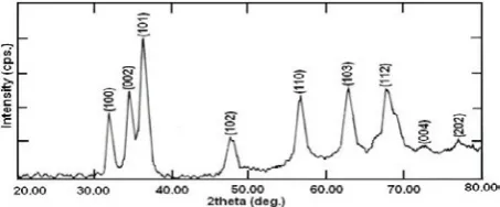
ZnO powder and films could therefore be obtained successfully prepared by sol-gel dip-coating technique. As prepared ZnO powder was examined by Philips X-ray diffractometer (Model PW 1730). The surface morphologies of the films were studied by JEOL 6300(LA). The quantitative elemental analysis of the films was carried out by using JEOL-energy dispersive spectrometer (Model JED-2300). The transmission electron microscopy of the film was studied by Tecnai 20 G2 (FEI make). RESULTS AND DISCUSSION Structural analysis Fig. 1. shows the x-ray diffractogram of the powder after drying the gel. The diffraction peaks from various planes and d values, as represented in Table 1, are matching well with reported JCPDS data for ZnO which confirmed the powder to be Fig.1. X-ray diffractogram of nanostructured ZnO. Peak d values Intensity Planes No Reported Observed Reported Observed 1 2.816 2.811 71 50 100
2 2.602 2.604 56 67 002
3 2.476 2.479 100 100 101
4 1.911 1.916 29 37 102
5 1.626 1.624 40 63 110
6 1.477 1.478 35 67 103
7 1.379 1.384 28 68 112
328
Anil R. Bari et al. / Dip-coated Nanostructured ZnO Thin Films
J Nanostruct 9(2): 326-330, Spring 2019
of ZnO. The confirmation of the ZnO powder, in turn, would confirm that the material deposited on substrate to be of ZnO.
Scherrer’s formula for grain size calculation
t = 0.9 λ/β cos θ (3)
Where, λ = Wavelength of X-ray, β = FWHM of peak, cos θ = Corresponding angle of the peak.
The average grain size calculated from Scherrer’s formula was about 27 nm.
Elemental analysis by EDAX
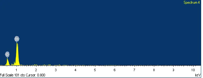
Fig. 2 shows the energy dispersive spectra of the sample prepared with 5 coats and pH = 7.1. The mass% of Zn and O in stoichiometric ZnO (at%
of each of Zn and O is 50) are expected to be 80.3 and 19.7 respectively. The observed values of mass % of Zn and O are represented in Table 2.
The elemental composition from Table 2, clearly indicates that the films are nonstoichiometric in nature. The films are observed to be zinc deficient.
Surface morphology using SEM
Effect of coating on particle size
Fig. 3(a), (b) and (c) are the SEM images of the thin films obtained after 5, 6 and 7 coats respectively from pH 7.1. The average particle sizes were observed from SEM image is presented in Table 3. The particle size distribution was observed to be reasonably narrow.
It is clear, from Table 3 that as the particle sizes increases with increase in number of coats.
Fig.2. EDS spectra of sample prepared with 5 coats and pH = 7.1.
Sample No. of mass % at. % No. coats Zn O Zn O 1 5 50.97 49.03 39.90 60.10 2 6 53.30 46.70 31.42 68.58 3 7 52.36 47.64 34.33 65.67
No. of coats 5 6 7 Particle Size (nm) 30.6 34.6 37.0
Table 2: Elemental compositions of typical ZnO thin films
329 J Nanostruct 9(2): 326-330, Spring 2019
Anil R. Bari et al. / Dip-coated Nanostructured ZnO Thin Films
Effect of pH on particle size and morphology
Fig. 4. (a), (b) and (c) show the SEM images of the film samples 1, 2 and 3 obtained from solutions with pH 6.9, 7.1 and 7.2 respectively. The average particle sizes of the films with pH 6.9, 7.1 and 7.2 were observed to be: 29.8 nm, 30.6 nm and 30.0 nm respectively. The particle size distribution seems to be reasonably narrow. The SEM images clearly indicate the elliptical or spherical shaped nanoparticles.
Microstructure using TEM
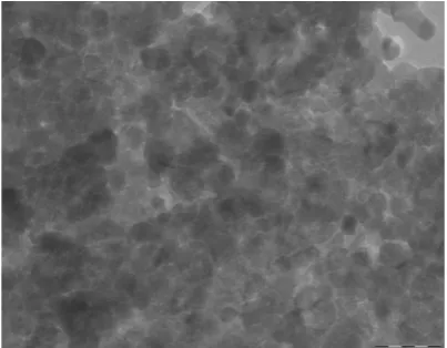
Fig. 5 shows the transmission electron microscopy image of the sample prepared with 5 coats and pH = 7.1. It is clear from TEM image that there are uniformly distributed spherical-shaped grains with the average grain size of 25 nm.
CONCLUSION
ZnO films obtained from sol-gel dip-coating technique were observed to nanostructured. The average grain size calculated from Scherrer’s formula was about 27 nm. Average particle size
Fig.4. SEM images obtained from solutions with pH: (a) 6.9, (b) 7.1 and (c) 7.2
Fig.5. TEM image of sample prepared with 5 coats and pH = 7.1
was observed from SEM is to be smaller than 40 nm. The average grain size observed from TEM was about 25nm. The most of the particles were observed to be spherical in shape. ZnO films were observed to be nonstoichiometric (Zinc deficient) in nature. Number of coats used to synthesize the films was increase with increase in size of nanostructured ZnO.
ACKNOWLEDGMENT
The authors are thankful to the Head, Department of Physics and Principal, Arts, Commerce & Science College, Bodwad.
CONFLICT OF INTEREST
The authors declare that there are no conflicts of interest regarding the publication of this manuscript.
REFERENCES
330
Anil R. Bari et al. / Dip-coated Nanostructured ZnO Thin Films
J Nanostruct 9(2): 326-330, Spring 2019 2. Huang W-J, Fang G-C, Wang C-C. A nanometer-ZnO catalyst to
enhance the ozonation of 2,4,6-trichlorophenol in water. Colloids and Surfaces A: Physicochemical and Engineering Aspects. 2005;260(1-3):45-51.
3. Annapoorani R, Dhananjeyan MR, Renganathan R. An investigation on ZnO photocatalysed oxidation of uracil. Journal of Photochemistry and Photobiology A: Chemistry. 1997;111(1-3):215-21.
4. Sánchez L, peral J, Domènech X. Degradation of 2,4-dichlorophenoxyacetic acid by in situ photogenerated fenton reagent. Electrochimica Acta. 1996;41(13):1981-5. 5. Bari A R, Sawadekar N P, Bari P A, Bari R H, Suryawanshi D
N, Pathan I G, Patil L A. Synthesis and characterization of zinc oxide nanowires, Int. Jn. Chem.and Phy. Sci., 2018; 7: 371-374.
6. Shionoya S, Yen W M, Phosphor Handbook (CRC Press, Boca Raton, 1999), p. 255.
7. O’Regan B, Grätzel M. A low-cost, high-efficiency solar cell based on dye-sensitized colloidal TiO2 films. Nature. 1991;353(6346):737-40.
8. Tang Z K, Wang G K, Yu L P, Kawasaki M, Ohtomo A, Koinuma H, Appl. Phys. Leet., 1997; 70: 2230.
9. Zang Q, Xie C, Zange S, Wang A, Zhu B, Wang L, Yang Z. Sens. Actuators B., 2005; 110: 370-376.
10. Lin H M, Tzeng S J, Hsiau P J, Tsia W L. Nano Struct. Mater. 2005; 10: 370.
11. Patil LA, Bari AR, Shinde MD, Deo V, Kaushik MP. Detection of dimethyl methyl phosphonate – a simulant of sarin: The highly toxic chemical warfare – using platinum activated nanocrystalline ZnO thick films. Sensors and Actuators B: Chemical. 2012;161(1):372-80.
12. Bari AR, Patil LA. LPG sensing performance of nanostructured zinc oxide thin films. AIP; 2013.
13. Bari AR, Patil LA, Pathan IG, Surawanshi DN, Rane DS. Characterizations of Ultrasonically Prepared Nanostructured ZnO powder and NH 3 Sensing Performance of its Thick Film Sensor. Procedia Materials Science. 2014;6:1798-804.
14. Patil LA, Bari AR, Shinde MD, Deo V. Ultrasonically prepared nanocrystalline ZnO thin films for highly sensitive LPG sensing. Sensors and Actuators B: Chemical. 2010;149(1):79-86.
15. Patil LA, Bari AR, Shinde, Deo V. Ultrasonically synthesized nanocrystalline ZnO powder-based thick film sensor for ammonia sensing. Sensor Review. 2010;30(4):290-6. 16. Patil D, Patil L. Cr2O3-modified ZnO thick film resistors as
LPG sensors. Talanta. 2009;77(4):1409-14.
17. Wagh MS, Jain GH, Patil DR, Patil SA, Patil LA. Modified zinc oxide thick film resistors as NH3 gas sensor. Sensors and Actuators B: Chemical. 2006;115(1):128-33.
18. Goyal D, Solanki P, Marathe B, Takwale M, Bhide V. Deposition of Aluminum-Doped Zinc Oxide Thin Films by Spray Pyrolysis. Japanese Journal of Applied Physics. 1992;31(Part 1, No. 2A):361-4.
19. Nakada T, Ohkubo Y, Kunioka A. Effect of Water Vapor on the Growth of Textured ZnO-Based Films for Solar Cells by DC-Magnetron Sputtering. Japanese Journal of Applied Physics. 1991;30(Part 1, No. 12A):3344-8.
20. Damiani LR, Mansano RD. Zinc oxide thin films deposited by magnetron sputtering with various oxygen/argon concentrations. Journal of Physics: Conference Series. 2012;370:012019.
21. Hiramatsu M, Imaeda K, Horio N, Nawata M. Transparent conducting ZnO thin films prepared by XeCl excimer laser ablation. Journal of Vacuum Science & Technology A: Vacuum, Surfaces, and Films. 1998;16(2):669-73. 22. Sing A V, Mehera R M, Yoshida A, Wakahara A, J. Appl. Phys.,
2001; 90: 5661.
23. Hu J, Gordon RG. Textured aluminum-doped zinc oxide thin films from atmospheric pressure chemical-vapor deposition. Journal of Applied Physics. 1992;71(2):880-90. 24. Minami T, Sato H, Takata S, Ogawa N, Mouri T. Large-Area Milky Transparent Conducting Al-Doped ZnO Films Prepared by Magnetron Sputtering. Japanese Journal of Applied Physics. 1992;31(Part 2, No. 8A):L1106-L9. 25. Bari A R, Shinde M D, Deo V, Patil L A, Effect of solvents
on the particle morphology of nanostructured ZnO, Ind. J. Pure & Appl. Phys., 2009; 47: 24-27.
26. Patil LA, Bari AR, Shinde MD, Deo V, Kaushik MP. Effect of precursor concentrations on structural, microstructural and optical properties of nanocrystalline ZnO powder synthesized by an ultrasonic atomization technique. Physica Scripta. 2010;82(3):035601.
27. Patil LA, Bari AR, Shinde MD, Deo V. Effect of pyrolysis temperature on structural, microstructural and optical properties of nanocrystalline ZnO powders synthesised by ultrasonic spray pyrolysis technique. Journal of Experimental Nanoscience. 2011;6(3):311-23.
28. Patil LA, Bari AR, Shinde MD, Deo V, Kaushik MP. Effect of aerosol carriers on ultrasonically prepared nanocrystalline
ZnO powders. Advanced Powder Technology.