Standardized protocol proposed for clinical
use of L-PRF and the use of L-PRF Wound
Box
®
Alessandro Crisci1, Domenico Lombardi2, Edda Serra3, Guido Lombardi4, Federica Cardillo5, Michela Crisci6
1Unit of Dermosurgery Cutaneous Transplantations and Hard-to-Heal Wound, “Villa Fiorita” Private Hospital, 81031 Aversa CE, Italy. 2School of Medicine, University of Salerno Italy, 84084 Fisciano SA, Italy.
3Laboratory Clinical Patology and Microbiology, Institute for the Study and Treatment of Diabetes, 81022 Casagiove CE, Italy. 4General Surgery, Hospital S. Maria della Pietà, 80026 Nola NA, Italy.
5Veterinarian freelancer, 67039 Sulmona AQ, Italy.
6Faculty of Medicine and Surgery, Vasile Goldis Western University of Arad, 310025 Arad, Romania.
Correspondence to: Dr. Alessandro Crisci, Unit of Dermosurgery Cutaneous Transplantations and Hard-to-Heal Wound, “Villa Fiorita” Private Hospital, 81031 Aversa CE, Italy. E-mail: [email protected]
How to cite this article: Crisci A, Lombardi D, Serra E, Lombardi G, Cardillo F, Crisci M. Standardized protocol proposed for clinical use of L-PRF and the use of L-PRF Wound Box®. J Unexplored Med Data 2017;2:77-87.
Aim: The hypothesis is that the production of leukocyte and platelet-rich fibrin (L-PRF) is
easy in horses without modifying human protocol, thus allowing better standardization of human protocol. In this study, the aim is to standardize the production of L-PRF in horses to direct it to human production. Methods: The authors took 9 mL of blood from the jugular vein of 6 horses to produce L-PRF membranes at a temperature > 21 °C by measuring the size of
the membranes (height, length, thickness, weight, surface) and clot. Therefore we analyzed
their microbiological characteristics. Results: The production of leukocyte and platelet-rich
fibrin (L-PRF) is easy in horses without modifying human protocol, thus allowing better
standardization of human protocol. The parameters found in the horse are clearly similar to the parameters found in humans. In optical microscopy, most of the cellular bodies were found and concentrated in the proximal portion of each membrane, with the last 1/4 observed in the center; the distal part only had residual traces of cellular bodies. The L-PRF is composed constantly when the production process below described is strictly respected. The success
of L-PRF depends entirely on blood collection, on the quick transfer to a centrifuge (within 1 min) and on centrifugation and squeezing temperatures (between 21 and 30 °C) of the clot.
Conclusion: The experiments on horses for the standardization of L-PRF production will improve our understanding about wound healing, particularly in the regenerative therapy of chronic skin lesions in humans. The data collected show that the best preparation method is the 2 min compression of the clot after 0 min of blood sampling, using the 9 mL vacutainer system and not a 9 mL syringe.
Key words: Autologous, buffy coat, growth factor level,
platelet-rich fibrin,
thrombocyte concentrate
ABSTRACT Article history:
Received: 26 Jun 2017 First Decision: 24 Jul 2017 Revised: 12 Sep 2017 Accepted: 12 Sep 2017 Published: 22 Nov 2017
Quick Response Code:
Original Article
This is an open access article licensed under the terms of Creative Commons Attribution 4.0 International License (https://creativecommons.org/licenses/by/4.0/), which permits unrestricted use, distribution, and reproduction in any medium, as long as the original author is credited and the new creations are licensed under the identical terms.
For reprints contact: [email protected]
Open Access
DOI: 10.20517/2572-8180.2017.17
Journal of
factor, and insulin-like growth factor-1.
Fibrinogen can improve the healing of a wound, increasing both proliferation and cell migration, and it
is assembled with fibronectin into fibrils regardless of
the formation of the fibrin. The fibrin is an important factor in wound healing; in fact, the result of the healing of a wound is influenced by the structure of
fibrin (the thickness of the fibres, the number of branch
points, the porosity, and the permeability of the clot
at the site of injury)[3]. The PRF clots also constitute a
bioactive reservoir. After the first massive release of growth factors, platelets synthesize and secrete new ones for the rest of their lives (7-10 days).
Fibrin gels are crucial as scaffolds in tissue engineering for several reasons. The most important reason is the innate compatibility with the fibrin of the cellular organism which is different according to the many components and processes involved in the manufacture of scaffolds. Although platelet growth factors play an important role in the biology of PRF, the architecture of the fibrin and the content of leukocytes are two key parameters.
A valid method of preparation of PRF® must effectively
separate the platelets from red blood cells and concentrate them without damaging or lyse the platelets themselves. Growth factors contained within
the α-granules are not active during the discharge;
they blend with the platelet-activating membrane. Consequently, if platelets are damaged during the production of PRF, they will not secrete more of the bioactive growth factors. In fact, they are particularly labile and sensitive to any kind of stressful event during the processing step and application; for this reason, the concentration of growth factors can also
be influenced by manipulation during processing of the
blood. Thus, it is crucial to standardize the preparation procedure and the type of centrifugation that is carried out must have certain characteristics, which include: at the beginning starting slowly, a central phase at
high rpm, and a final slow phase before stopping[4],
and should take place at a certain temperature and for
a definite time.
Our hypothesis is that the L-PRF is easy to produce
in horses, without modification to the human protocol,
even allowing better standardization of the human protocol.
It is necessary to establish a standard protocol to obtain L-PRF with these main features: (1) the growth factors present in the platelets must be stored to stimulate the surrounding host cells; (2) platelets must
INTRODUCTION
The platelet-rich fibrin (PRF) of Choukroun et al.[1] is a new step in the therapeutic concept of platelet gel
with a simplified design and little artificial biochemical
changes. Unlike other platelet concentrates, this technique does not require anticoagulants, thrombin, or any other gelling agent, which makes it no longer
than natural blood centrifuged without additives[2].
Although platelets and leukocyte cytokines play an important role in the biology of this biomaterial, the supporting fibrin matrix certainly constitutes the real therapeutic potential of PRFs. The absence of the anticoagulant leads to the formation of the endogenous thrombin which successively activates most of the platelets contained in the sample and at the same time triggers fibrin formation. The PRF protocol is a simple technique developed in France by
Choukroun et al.[1] and can be regarded as a
second-generation platelet concentrate because it is a natural product without any anticoagulant or gelling agents. Platelets and white blood cells are collected with high efficiency using this method and the leucocytes are preserved. However, platelets are activated during this process, which leads to a substantial embedding of platelets, leukocytes, and growth factors in the fibrin matrix.
Fibrinogen is initially concentrated in the upper part of the tube, until the effect of the circulating autologous thrombin transforms it into a network of fibrin. The result is a fibrin clot containing the platelets located in the centre of the tube, just between the lower layer of red blood cells and the plasma acellular on the top. The PRF clot is then placed on the grill
in the PRF Box® and covered with the compressor
cover; it produces a membrane of autologous fibrin
in approximately 1 min. The PRF Box® is designed
to produce a constant thickness of the membrane that remain hydrated for several hours and allows the recovery of the serum exudate expressed by fibrin clots which is rich in proteins, such as vitronectin
and fibronectin[2]. The PRF clot is produced by a
natural polymerization process during centrifugation, and its natural fibrin architecture seems to be responsible for the slow release of growth factors and
glycoproteins from the matrix (≥ 7 days). The adhesive
proteins fibrinogen, fibronectin, vitronectin, and
thrombospondin-1 are abundant on the fibrin structure.
Among the growth factors stored in platelets, and which are essential for the repair of wounds, there are: platelet-derived growth factors, with -AB and -C; they are also present as vascular endothelial growth factor
A, transforming growth factor beta 1, fibroblast growth
be stored in the fibrin structure with minimal damage or activation; (3) the 3-dimensional fibrin lattice should
be used as a scaffold for the surrounding host cells.
METHODS
Preparation
The blood collection must be followed by an immediate centrifugation as a prerequisite in the specification of the PRF output. It is formulated to produce a homogeneously-moisturized thick membrane and an exudate rich in platelets, leukocytes, vitronectin, and
fibronectin expressed by fibrin clots[5].
Overall, the L-PRF is mechanically resistant, able to support loads, has a capacity of two times stretching under tension and retains surgical sutures well enough
as deforms itself significantly before laceration[6].
The production technique of PRF is very simple and requires only a blood sample and a table centrifuge
[Figure 1].
The protocol followed is that of “Choukroun et al.[1]”:
the blood samples are collected in 9 mL tubes, without anticoagulant or gel separator, and are immediately centrifuged according to the following program: 30 s acceleration, 2 min at 2,700 rpm, 4 min at 2,400 rpm, 3 min at 3,000 rpm, and 36 s deceleration and stopping.
After centrifugation, three parts are localized in the
tube: the red blood cells at the bottom, a fibrin clot that
represents the PRF in the middle, and the acellular plasma at the top. We can obtain the PRF extracting the matrix from the tube with forceps and removing
the red clot. The success of this technique depends entirely on the blood collection and the transfer
speed in the centrifuge[7,8]. Equine blood was used by
Textor et al.[9] for the production of platelet concentrate (PRF). Written consent from the owners has been obtained for all of the horses and the blood collection procedure was performed according to the current AVMA guidelines.
We performed a prospective study of equine blood, which has been collected in test tubes without anticoagulant plastic-coated glass or a gel separator (BD vacutainer tubes for serum 9.0 mL), for the production of L-PRF clots and membranes by 6 healthy horses of various ages (average ± SD, 10 ± 4.1 years, ranging from 4 to 17 years), gender and breed.
The 9 mL of blood was harvested quickly with sterile 10 mL syringes as well as with the blood collection system of vacutainer tubes with pre-attached prefabricated Ref. 368652 Becton, Dickinson & C. USA (22 “mean value less than 25” per tube) from the vein and immediately (within 1 min) centrifuged according to the previous description at a temperature of 21-30 °C in a benchtop model 3225 Centrifuge.
The temperatures of the inner and outer surface of the centrifuge were recorded before and during centrifugation with a digital thermometer with an internal probe (Tronic) [Figure 1].
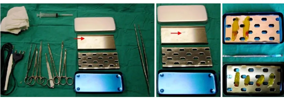
A new device has been tested for the preparation and standardization of L-PRF in clots and membranes: the
L-PRF Wound Box®[Figure 2]. Using the PRF Wound
Box, the compression process of the membrane in the clots is performed through a slow and homogeneous
slight compression, and the final membrane always remains homogeneously wet and soaked in serum.
This method avoids the extraction and the loss of a significant amount of the growth factors. The L-PRF
Wound Box® was made by us using a metal container
17.5 cm × 7.6 cm × 2 cm containing a perforated steel plate of 150 m × 68 m × 1.5 m. There was a second steel plate which acts as a compressor, 150 mm ×
68 mm × 1.5 mm, with a weight of 148 g [Figure 2].
This second shaped plate exerts a pressure of
142.437 Pa/cm2. In this study, the compression has
been exerted on the clot for 2, 5, 10, and 15 min to produce membranes.
Macroscopic analysis
After centrifugation, the L-PRF clot was removed from the test tube using sterile tweezers and a smooth spatula to gently release the red clot from the buffy coat. Each L-PRF clot obtained was placed on a tray to measure the weight and size with a goldsmith digital scale [Figure 1].
The compression of the clot was carried out with
the L-PRF Wound Box®, applying a pressure of
142.437 Pa/cm2 constant for 2, 5, 10, 15 min. This
method allowed us to obtain, from each clot, the L-PRF membranes, which were individually weighed and
measured with a digital gauge [Figure 1]. The surface
area in square centimetre of clots and the membranes were measured with the Calcderm measurement software[10].
Optical microscopy procedure
The membranes were fixed in 10% neutral buffered formalin for 24 h at ambient temperature, then were
added in paraffin. Subsequent sections of 4 mm were
performed along the midpoint of the membranes and were stained with haematoxylin-eosin. Each section
was divided into 3 equal-sized areas: proximal (head), centre (body), and distal (tail). Each area of these sections was observed through an optical microscope and analyzed by counting the visible cell bodies (marked in dark purple) at the centre of each observed
area with a magnification of 25×, 40×, 60×, and 100×
(immersion). The total number of counted cell bodies were used to correlate their distribution between the 3 membrane areas (head, body, and tail).
Smears of blood prepared by residual blood in the tubes were also examined for a morphological assessment after removal of the PRF clot with a spatula (2 for each tube), differentiating the clot at 0 and 60 min by centrifugation and fixing them with
alcohol 90% for a may-grǔnwald-giemsa colouring
stain in order to identify the various corpuscular elements, in particular platelets and neutrophils, to compare them in relation to the examination blood count. A blood sample was also taken from each horse to perform a blood count using K3E 5.4 mg EDTA tubes (VacuMed).
According to the study of Peck et al.[11], three blood
samples were taken from the left jugular vein of each horse through a 14-gauge needle, two for the production of PRF and one for the blood count. After centrifugation, the clot was removed immediately
[group A (n = 6)] and after 60 min [group B (n = 6)].
The release derived from the pressing with the Wound
Box® membrane was differentiated between the two
groups and was preserved in a test tube with K3E 5.4 mg EDTA for blood count analysis. It was compared with the basal one and with the corpuscular elements of the counts performed on smears derived from the red clot as an indirect measurement of the platelet and the leukocyte concentrations of the L-PRF. The two samples taken with the vacutainer system without gels were randomly assigned to two groups (A and B),
using a coin toss.
The supernatant derived from the pressing of L-PRF at 0 and 60 min were analysed with a blood count in standard lodging. Although a direct measurement of platelet concentration of the PRF is not yet possible, we calculated the residual platelet concentration. The examinations were performed with a Cell Dyn 3500 R cell counter (Abbott).
Determination of the parameters of the membranes
Immediately after formation, the L-PRF membranes were stored at 4 °C until delivery to the laboratory in order to avoid damage from storage.
The size of the membranes was measured in height, length, and width (in mm) with a digital gauge, and the
irregularly-shaped areas were measured in cm2 with
Calcderm software[10].
The content of erythrocytes, platelets, WBCs (neutrophils, lymphocytes, monocytes, basophils) present in the membrane was calculated by comparing the basic blood count with one obtained from the supernatant, as performed on blood smears of the red clot.
Weight reduction after compression was also evaluated using blood samples taken with syringes and with vacutainer. The use of vacutainer results in less variability in the characteristics of the blood sample.
Statistical analysis
The statistical significance for the differences between
two groups was calculated using Student t-test and
ANOVA for repeated measures for parametric variables and with the Chi-square for non-parametric variables.
P values < 0.005 were considered statistically
significant. The data were analysed using the discipline
biomedical statistics of Stanton Glantz 2007 software package, version 6.0.
RESULTS
The procedure was well-tolerated in all animals. No significant differences were found in the basic haematological confrontation, which had an average
concentration of RBC 9.8 × 10³/mL [range: (7-13) ×
103/mL], WBC 5.1 × 10³/mL [range: (5-13) × 103/mL] (±
0.37, 95%CI) (P = 0.24) and a platelet count average
of 106.8 × 10³/mL [range: (100-350) × 103/mL] (± 15.3,
95%CI) (P = 0.5) (P is the comparison of the results
between the 6 horses). It was not possible to directly quantify the platelet concentration and WBC trapped inside the L-PRF clot, which was derived indirectly by comparing the mean values of whole blood, the mean values of the supernatant obtained after compression of the clot at 2 min, and the average values obtained counting the smears of the red clot after the removal of the L-PRF clot.
Table 1 compares the characteristics of clots and
membranes of L-PRF obtained in humans (centrifugal
Intraspin) reported by Pinto et al.[12] and Dohan
Ehrenfest et al.[13,14] and those we observed in the
horse model. In this comparison, it was verified that
there are significant differences in the characteristics
of the clot, but these differences are eliminated when the membranes derived from the compression were examined. This observation, in our opinion, would be assigned to a different content of exudate (weight of the exudate is 1.47 ± 0.13 g in humans, 3.05 ± 0.11 g in
horses, P = 0.000).
The fact that they have been used in comparison to two different types of centrifuges, according to our opinion, is irrelevant, as the results are overlapping.
In this study, no relation was found between the size of the membranes and the haemoglobin content or the content of erythrocytes encountered in the blood
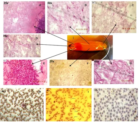
count baseline. In optical microscopy [Figure 3], most
of the cell bodies (stained in dark purple for the nuclei) were concentrated in the proximal part (head) of each membrane, the last 1/4 was observed at the centre, and the distal part had only residual traces of cell bodies. However, optical microscopy has not allowed the observation of the exact state of these cell bodies in greater detail. Most of the cells were concentrated in the proximal area (head), closest to the red clot.
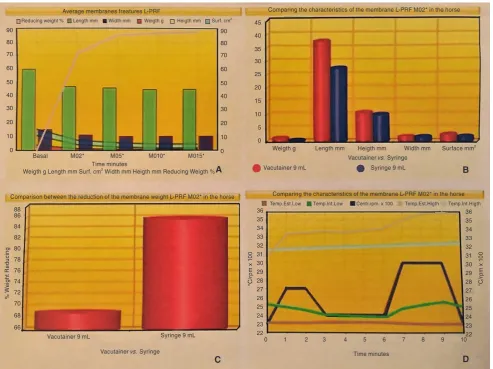
Figure 4A shows the average characteristics of a
membrane obtained with the L-PRF Wound Box after several minutes of compression (2, 5, 10, and 15 min). The slight variations between M05 min, M10 min, M15 min, M02 min are compared to make us reach out towards the use of the membrane after 2 min into the
compression [Table 2].
The working temperature must be between 21 and 30 °C because the clot is not produced if the
temperature is lower than that. Figure 4D shows the
Table 1: Comparison of membranes obtained from human blood and from equine blood (average ± SD)
Variable Human (n = 8) Horse (n = 6) Student t-test (P) Significance
Final temperature test of tube (°C) 27.5 ± 0.66
Weight of the clot (g)* 2.09 ± 0.19 4.23 ± 0.55 0.000 S
Weight of the membrane (g) 0.62 ± 0.15 0.78 ± 0.08 0.036 NS
Exudate weight (g) 1.47 ± 0.13 3.05 ± 0.11 0.000 S
Length of the clot (mm) 35.69 ± 3.43 44.38 ± 3.83 0.000 S
Width of the clot (mm) 12.81 ± 0.75 14.74 ± 1.23 0.003 S
Height of the clot (mm) 7.02 ± 1.09
Surface area of the clot (mm2) 4.10 ± 0.86
Length of the membrane (mm) 34.81 ± 2.95 36.81 ± 3.18# 0.248 NS
Width of the membrane (mm) 12.25 ± 0.71 13.02 ± 1.01# 0.119 NS
Height of the membrane (mm) 3.02 ± 0.51#
Surface area of the membrane (cm2) 3.08 ± 0.5#
Weight ratio clot/blood sample (%) 10 mL 20.94 ± 2.4 32.53 ± 0.54 0.000 S
*: the difference of the weight clot is due to a difference of exudate content; #: average values (± SD) after 2 min compression at 30 °C. The
values are not in relation to the content of Hb and erythrocytes in whole blood; S: significant; NS: not significant
Figure 3: Membrane L-PRF 0 min after compression (hematoxylin-eosin staining). (A) III proximal 25× white blood cell-pattern fibrin; (B) medium-III 60× erythrocytes pattern fibrin; (C) III distal 60× pattern fibrin; (D) III proximal 25× erythrocytes-fibrin; (E) III proximal 60× fibrin on the right, the center lymphocytes, erythrocytes, and granulocytes neutrophils on the left; (F) average III 25× pattern of fibrin; (G) III distal 60× pattern fibrin; (H) smear of red clot 40× presence of monocyte in a carpet of red cells; (I) red clot smear 40× presence of red blood cells,
monocytes and platelets; (J) red clot smear 100× presence of platelets in a carpet of red cells (coloration: may-grǔnwald-giemsa)
A B C
D
E F G
Using 9 mL syringes instead of the vacutainer
sampling system the membranes that are produced are smaller in size weight, length, width and total useful area. After a [Figure 4B], both in terms of
Figure 4: (A) Average characteristics of the L-PRF membrane features obtained in the horse model with the L-PRF Wound Box for several minutes of compression: weight (g), height (mm), length (cm), width (cm), size (cm2), and % weight reduction during compression; (B) average characteristics of an L-PRF membrane obtained in the horse model with the L-PRF Wound Box: weight (g), height (mm), length (mm), width (mm), surface area (cm2), A is the measure in different times and B in M02; (C) % weight reduction during compression; (D) variations of external and internal temperature during centrifugation at 23 °C (B) and at 30 °C (A), picking with vacutainer system and with 9 mL syringe after 2 min of compression
Table 2: Test of significance on the variations of the parameters of the membrane L-PRF to compression with the
L-PRF Wound Box for 2, 5, 10, and 15 min, (P)
Variable Basal and
M02 min Basal and M05 min Basal and M10 min Basal and M15 min M02’-M05’-M10’-M15’ Parameter Student’s
t ANOVA Student’s t ANOVA Student’s t ANOVA Student’s t ANOVA c2
Weight (g) 0.006 0.006 0.002* 0.005* 0.002* 0.005* 0.001* 0.006 1.000
% reduction in weight (g) / / / / / / / / 0.852
% reducing clot (g) 0.000* 0.000* 0.000* 0.000* 0.000* 0.000* 0.000* 0.000* /
Length of the membrane (mm) 0.031 0.004 0.039 0.002* 0.019 0.002* 0.021 0.000* 1.000
Width of the membrane (mm) 0.015 0.021 0.001* 0.005* 0.001* 0.006 0.001* 0.008 1.000
Height of the membrane (mm) 0.018 0.054 0.005* 0.020 0.002* 0.014 0.002* 0.008 1.000
Surface area of the membrane (cm2) 0.506 0.137 0.083 0.079 0.058 0.078 0.038 0.066 1.000
*P < 0.005, significant difference; L-PRF: leukocyte platelet-rich fibrin; /: this value could not be calculated
A B
C D
Average membranes freatures L-PRF Comparing the characteristics of the membrane L-PRF M02* in the horse
90 80 70 60 50 40 30 20 10 0 90 80 70 60 50 40 30 20 10 0 45 40 35 30 25 20 15 10 5 0
Basal M02* M05* M010* M015* Weigth g Length mm Heigth mm Width mm Surface mm
2
Weigth g Length mm Surf. cm2 Width mm Heigth mm Reducing Weigth %
Vacutainer vs. Syringe Vacutainer 9 mL Syringe 9 mL
Vacutainer 9 mL Syringe 9 mL
Vacutainer vs. Syringe Time minutes
0 1 2 3 4 5 6 7 8 9 10 Comparing the characteristics of the membrane L-PRF M02* in the horse Comparison between the reduction of the membrane weight L-PRF M02* in the horse
88 86 84 82 80 78 76 74 72 70 68 66 36 35 34 33 32 31 30 29 28 27 26 25 24 23 22 36 35 34 33 32 31 30 29 28 27 26 25 24 23 22 % W ei gh t R ed uc in g °C /r
pm x 1
00
°C
/r
pm x 1
00
Time minutes
Reducing weight % Length mm Width mm Weigth g Heigth mm Surf. cm2
2-min compression, the reduction of the weight of the membrane compared to the clot is 85% if it’s picked up with a syringe, and 70% if it is taken with the
vacutainer system [Figure 4C].
The results of the blood counts of whole blood and of the supernatant obtained from the clot after 0 min, and after 60 min of compression are compared with the counts of erythrocytes, platelets, and the WBC smear of the red clot after 0 and 60 min, and are shown with
the corresponding statistical tests in Table 3, while the
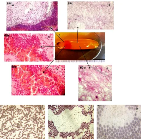
microscopic images after 60 min are shown in Figure 5.
There is also a statistically significant difference between the content of RBC, WBC, and PLT in smears obtained from the red clot at 0 and 60 min, as shown
in Figures 3 and 5A-C at various magnifications.
Tables 3, 4 and 5 also shows the hypothetical contents
of RBC, WBC, and PLT in L-PRF membranes derived from the difference of these corpuscular elements
Figure 5: Membrane L-PRF of 60 min by the compression horsepower (hematoxylin-eosin staining). (A) III proximal 25× White Blood Cell-Erythrocytes-pattern Fibrin; (B) medium-III 25× pattern Fibrin; (C) III proximal 60× pattern of Fibrin on the right, the center lymphocytes, erythrocytes and granulocytes Neutrophils to left; (D) III proximal 60× Fibrin on the right, the center lymphocytes, erythrocytes and
granulocytes neutrophils to left; (E) average III 60× pattern of fibrin with Lymphocyte; (F) smear of red clot 40× presence of platelets in a carpet of red cells; (G) red clot smear 40× presence of erythrocytes and many platelets; (H) red clot smear 100× presence of many platelets
in a carpet of red cells (may-grǔnwald-giemsa staining)
A B
C
D E
between whole blood, the supernatant at 0 and 60 min,
and the smear of the red clot at 0 and 60 min. The t
-test shows significant differences between RBCs at 0
and 60 min, and between PLT at 0 and 60 min in L-PRF membranes.
The content of RBC in the membranes is 0.0028%, that of WBC is 99.24%, and that of PLT is 99.0%, compared to the content in whole blood. The membranes after 60 min of compression have a content of RBC,
WBC, and PLT lower than those at 0 min [Table 5].
DISCUSSION
The success of this technique depends entirely on
the blood collection and the transfer speed in the centrifuge[7,8]. Equine blood was used by Textor et al.[9] for the PRF.
The study performed by McLellan and Plevin[15]
has shown that the equine PRF is similar to that of humans, providing an immediate and constant source of tissue growth factors. Our study has attempted to standardize the preparation of the L-PRF procedure which, while remaining a technique of easy execution and low-cost, does not require specialized equipment, but has a certain constancy in the production of a membrane in terms of L-PRF macroscopic and microscopic features. The autologous platelet
concentrates are promising in the field of regenerative
Table 3: Full blood emocromocytometer examination compared with erythrocytes, plateles and WBC counts on red clot strip at 0” and 60” with significance tests
Type CBC blood Smear C.R. 0 min Smear C.R. 60 min Between Red Clot smear 0-60 min Average ± SD Average ± SD Average ± SD t-test* c2
RBC 7,648,000 ± 11,309.81 7,399,440 ± 27,039.76 7,322,960 ± 81119.29 P = 0.333 NS P = 0.000 S
WBC 5,150 ± 369 8.5 ± 2.12 0.5 ± 0.71 P = 0.037 S P = 0.908 NS
PLT 106,780 ± 153.51 500 ± 707.11 6,000 ± 1414.21 P = 0.039 S P = 0.000 S
Neutrophil 3,046 ± 857 / / / / / /
Basophils 4.2 ± 1.3 / / / / / /
Lynphocites 1,606 ± 668 / / / / / /
Monocytes 490.2 ± 138.06 / / / / / /
Eosinophils 5.4 ± 5.37 / / / / / /
c2: processing performed on two comparisons. P > 0.05 = +0.5% no significant difference; P < 0.01 = -1% significant difference; /: this value could not be calculated
Table 4: Hemocromocytometric examination on supernatant at 0' compared to 60' with significance tests
Type CBC supernatant at 0 min CBC supern.at 60 min supernatant 0-60 minBetween CBC
Average ± SD Average ± SD t-test* c2
RBC 13,428 ± 21,345 73,714 ± 186,233 P = 0.411 NS P = 0.000 S
WBC 30 ± 27.99 10,914 ± 172.95 P = 0.255 NS P = 0.000 S
PLT 479 ± 77.614 3,627 ± 3,401 P = 0.031 S P = 0.000 S
Neutrophil 0.29 ± 0.76 1.29 ± 2.21 P = 0.280 NS P = 0.991 NS
Basophils 2.29 ± 2.14 2 ± 1.73 P = 0.785 NS P = 0.611 NS
Lynphocites 19 ± 23.15 98.43 ± 167.69 P = 0.238 NS P = 0.000 S
Monocytes 4.57 ± 7.68 2.86 ± 5.01 P = 0.631 NS P = 0.928 NS
Eosinophils 4 ± 9.71 4.57 ± 7.96 P = 0.906 NS P = 0.316 NS
c2: processing performed on two comparisons. P > 0.05 = +0.5% no significant difference; P < 0.01 = -1% significant difference
Table 5: Count of erythrocytes, plateles and WBC on L-PRF membranes derived from clots at 0' compared with those derived from clots at 60' with significance tests
Type Membrane 0 min Membrane 60 min Between Membrane L-PRF 0-60 min
No./mL % No./mL % t-test* c2
RBC 21,6012 0.0028% 193,966 0.0025% P = 0.000 S P = 0.266 NS
WBC 5,111.15 99.24% 5,036.86 97.80% P = 0.007 NS P = 0.993 NS
PLT 105,801 99.00% 97153 91.00% P = 0.002 S P = 1.000 NS
medicine due to the abundance of growth factors.
The L-PRF represents a very significant advance in the evolution of platelet concentrates since it is essentially a fibrin membrane with platelets and leukocytes trapped within. These solid membranes possess excellent handling characteristics, and can be firmly sutured in an anatomically-desired location during open surgery. However, the physical and biological properties are relatively unknown. The L-PRF will form when the steps described above are strictly observed.
The PRF Wound Box that exists on the market come in a variety of shapes and availability, through the compression plate, different pressures according to the weight, giving rise to a membrane of varying thickness, width, and length.
One of the most important considerations to generate a good L-PRF membrane is the delay in the time between blood collection and centrifugation, as well as the processing temperature. The success of the L-PRF technique depends entirely on the speed of collection of blood and the immediate transfer into a centrifuge, usually within 1 min, and by a centrifugation temperature and higher squeeze at 21 °C. You cannot generate a clot of well-structured L-PRF (with its
specific cellular content, architecture of the matrix, and profile of the release of growth factors) if the collection
of blood is prolonged and not homogeneous, or if the centrifugation temperature is between 21 and 30 °C; instead, it will form an inconsistent, crumbly mass of
fibrin with unknown contents. The L-PRF works as a
provisional extracellular matrix, which is transformed into functional tissue during healing, and can be subjected to mechanical forces and healing outcomes with success, depending on the structural integrity of the L-PRF so it is important to clarify its physical properties.
The L-PRF looks like dense connective tissue with superior handling characteristics. With an elastic modulus of 0.470 MPa (SD = 0.107) the L-PRF membrane stretches twice its initial length before breakage (of 215% strain). This data is in accordance
with the published literature[6] who reported a low
rigidity (1-10 MPa) and a high voltage (up to 150%) before breaking down.
Based on these results, it is clear that L-PRF is a new biomaterial with unique features: the anticipated preparation of autologous blood, the simplicity of the protocol, the defined architecture, the impressive mechanical properties, and the abundance of derived
growth factors from activated platelets.
The L-PRF membranes produced from the horse by the authors are assimilable to human ones both in terms of their morphological and microbiological aspects. Biochemical behavior was not investigated in this study. In the future our experiments on equine blood will undoubtedly be able to improve our understanding of healing wounds in humans, as well as helping to
promote the field of personalized medicine.
Our experiments on horses for the standardization of L-PRF production will undoubtedly improve our understanding of wound healing, particularly in the regenerative therapy of chronic skin lesions in humans. The data collected show that the best preparation method is the 2 min compression of the clot after 0 min of blood withdrawal, using the 9 mL vacutainer system and not a 9 mL syringe. The limit of this study is the lack of in-depth biochemical characteristics of horse membranes compared with human ones. The problems related to the conservation of these membranes will greatly limit their use. Cryopreservation at 4 °C may be a solution to this problem, but at the moment the data reported in the literature are not encouraging because of the reduced half-life of cryopreserved platelets (<
2 days) compared to fresh (3.5-3.8 days)[16]. The data
concerning the maximum storage time and the ideal temperature for the conservation of PRF are largely lacking. In future studies we would like to test the
hypothesis of Peck et al.[11] comparing the parameters
observed at 0 and 60 min by centrifugation using dry
vacutainer tubes vs. vacutainer tubes with separating
gel, analysing the prepared product with a scanning electron microscope. We would also consider how long the PRF (clot or membrane) is stable after its establishment and if the exudate collected in the L-PRF Wound Box can be used effectively to fix the autologous grafts (L-PRF membrane, or bio-conductive or bio-inductive dermis).
DECLARATIONS
Authors’ contributions
Main frame work of the paper: A. Crisci, E. Serra Manuscript correction: D. Lombardi, F. Cardillo Preparation of the manuscript: M. Crisci, G. Lombardi
Financial support and sponsorship
The first author bore all the expenses. There was
neither sponsorship nor financial support.
Conflicts of interest
Patient consent
A written consent of the owners has been obtained for all of the horses.
Ethics approval
As the owners of the horses have given the consent so we did not go for ethical liberation.
REFERENCES
1. Choukroun J, Adda F, Schoeffler C, Vervelle A. Une opportunite´ en
paro-implantologie: le PRF. Implantodontie 2001;42:55-62. (in French) 2. Toffler M, Toscano N, Holtzclaw D, Del Corso M, Dohan Ehrenfest D.
Introducing Choukroun’s platelet rich fibrin (PRF) to the reconstructive
surgery milieu. J Impl Adv Clin Dent 2009;1:21-32.
3. Del Corso M, Choukroun J, Simonpieri A, Zampetti P, Bucci Sabbatici V. Accelerazione dei processi di cicatrizzazione tissutale con un nuovo
biomateriale: la fibrina ricca di piastrine (PRF). Rivista degli amici di Brugg 2007;4:361-6. (in French)
4. Nurden AT, Nurden P, Sanchez M, Andia I, Anitua E. Platelets and wound healing. Front Biosci 2008;13:3532-48.
5. Hotwani K, Sharma K. Platelet rich fibrin - a novel acumen into regenerative endodontic therapy. Restor Dent Endod 2014;39:1-6. 6. Madurantakam P, Yoganarasimha S, Hasan FK. Characterization
of leukocyte-platelet rich fibrin, a novel biomaterial. J Vis Exp
2015;(103):53221.
7. Crisci A. Le membrane L-PRF® utili in chirurgia. J Plast Dermatol 2015;2:75-90.
8. Crisci A, Placido F, Crisci M, Bosco A. A new instrument aid of plastic
surgeon: membranes L-PRF (platelet-rich-fibrin). J Plast Dermatol 2015;3:162-72.
9. Textor JA, Murphy KC, Leach JK, Tablin F. Ultrastructure and growth factor content of equine platelet-rich fibrin gels. Am J Vet Res 2014;75:392-401.
10. Crisci A, Crisci M, Boccalone E. Final results of an experimental research about a technique of measurement of skin lesions. Esperienze Dermatologiche 2014;16:147-52.
11. Peck MT, Hiss D, Stephen L, Satti A, Majeed A. Platelet-rich fibrin
(PRF) - The effect of storage time on platelet concentration. S Afr Dent J 2015;70:448-51.
12. Pinto N, Pereda A, Jiménez P, Del Corso M, Kang BS, Wang, HL, Quirynen M, M Dohan D. The impact of the centrifuge characteristics and centrifugation protocols on the cells, growth factors and fibrin architecture of a Leukocyte-and Platelet-Rich Fibrin (L-PRF) clot
and membrane. Part 2: macroscopic, photonic microscopy and Scanning Electron Microscopy analysis of 4 kinds of L-PRF clots and membranes. Poseido J 2014;2:141-54.
13. Dohan Ehrenfest DM, Del Corso M, Kang BS, Lanata N, Quirynen M, Wang HL, Pinto NR. The impact of the centrifuge characteristics and centrifugation protocols on the cells, growth factors and fibrin
architecture of a leukocyte- and platelet-rich fibrin (L-PRF) clot and
membrane. Part 3: comparison of the growth factors content and slow
release between the original L-PRF and the modified A-PRF (advanced platelet-rich fibrin) membranes. POSEIDO 2014;2:155-66.
14. Dohan Ehrenfest DM, Pinto NR, Pereda A, Jiménez P, Corso MD, Kang BS, Nally M, Lanata N, Wang HL, Quirynen M. The impact of the centrifuge characteristics and centrifugation protocols on the cells,
growth factors, and fibrin architecture of a leukocyte- and platelet-rich fibrin (L-PRF) clot and membrane. Platelets 2017;24:1-14.
15. McLellan J, Plevin S. Temporal release of growth factors from
platelet-rich fibrin (PRF) and platelet-platelet-rich Plasma (PRP) in the horse: a
comparative in vitro analysis, Intern. J Appl Res Vet Med 2014;1:48-57. 16. Agnoli C. Terapie trasfusionali nel cane parte I. Il sangue intero e i suoi
componenti-raccolta, produzione e stoccaggio. Veterinaria 2015;5:7-15.