*Corresponding author:Anil Kumar Reddy ISSN: 0976-3031
Research Article
THE PROTECTIVE EFFECTS OF FOLIC ACID SUPPLEMENTATION ON TERATOGENECITY
INDUCED BY THE VALPROIC ACID IN DEVELOPING CHICK EMBRYO
Anil Kumar Reddy
1, Joy A Ghoshal
2, Ujwal Gajbe
3and R R Fulzele
41,2
Department of Anatomy, All India Institute of Medical Sciences, Mangalagiri, Andhra Pradesh
3,4Department of Anatomy, Jawaharlal Nehru Medical College, Sawangi, Wardha, Maharashtra
DOI: http://dx.doi.org/10.24327/ijrsr.2019.1010.4114
ARTICLE INFO ABSTRACT
Background: Teratogenicity of the drugs are tested by injecting into the developing chick embryo & is not new to the world. Several studies have been reported on the teratogenicity of AEDs on chick embryo. Sodium valproate, an analog of VPA is found to be effective against seizures. Aim & Objectives: The present experimental study was undertaken to find out dose-related teratogenic effects of VPA on developing chick embryo and to know the impact of folic acid supplementation on teratogenicity induced by VPA in developing chick embryo. Materials & Methods: The current experimental single-blinded study was done in the Department of Anatomy. A Total of 240 Fresh Fertilized White Leghorn Chicken Eggs were utilized for the study. Control group eggs were injected with Distilled water and experimental groups were injected with various doses of Valproic acid (VPA) with or without folic acid. Results: Increased percentage of Mortality rate, Gross malformations, Histopathological changes in Brain, Heart, and Liver were observed in experimental groups. Conclusion: Teratogenicity was found in developing chick embryos with the administration of VPA and the injection of folic acid along with the drug in early embryogenesis period would reduce the effects.
INTRODUCTION
Testing teratogenicity of various drugs by injecting into developing chick embryo is not new to the world. The chick as a model was chosen for the study because of its easy availability compared to rats or mice and organogenesis in chick embryo is comparable to the human embryonic period[1]. Various authors have done experiments on chick embryo to know the teratogenicity of Anti Epileptic Drugs (AEDs). However chick embryo experimental studies always have advantages over the pregnancy registry to know the graded malformations, dose-related effects, and to understand the manifestations of malformations[2, 3].
Sodium valproate or valproate sodium is the sodium salt of valproic acid (VPA). Sodium valproate, an analog of VPA is found to be effective against seizures. VPA and sodium valproate ionize to the valproate ion at body pH[4].
In pregnancy, valproate has the highest risk of birth defects of any of the commonly used AEDs. The drug crosses the placenta and its levels in umbilical cord blood are found to be five times higher than maternal serum, which imposes an increased risk of embryotoxicity[5]. VPA use during early
pregnancy carries a 3-fold increased risk of congenital malformation, with 1-2% increase in NTD (including anencephaly and exencephaly) and that elevates to 5.4% with a higher daily dose of 1000mg/day of VPA[6]. The dominant teratogenic characteristic is lumbosacral meningomyelocele (spina bifida aperta); it also causes cardiovascular, genital and limb abnormalities, hypospadias, developmental delay, autism, and the specific-Fetal Valproate Syndrome. VPA like other AEDs possess the antifolate activity and disrupts gene expression, enhances embryonic oxidative stress and alters protein synthesis[7]. These disturbances may result in NTD, cardiac anomalies, and neurodevelopment delay.
The strongest evidence for drugs causing neural tube defects exists for valproic acid and carbamazepine[3, 8]. Besides these anti-epileptics, drugs that act as folate antagonists are also known to cause teratogenicity in the form of NTDs. The mechanism is the inhibition of folate transporters at the cellular level, which can lead to folate deficiency and, predispose to neural tube defects[9].
Even though Valproic acid (VPA) is known teratogen, the detailed survey of the experimental studies of VPA on developing chick embryo revealed that supplementation of
International Journal of
Recent Scientific
Research
International Journal of Recent Scientific Research
Vol. 10, Issue, 10(E), pp. 35497-35501, October, 2019
Copyright © Anil Kumar Reddy et al, 2019, this is an open-access article distributed under the terms of the Creative Commons Attribution License, which permits unrestricted use, distribution and reproduction in any medium, provided the original work is properly cited.
DOI: 10.24327/IJRSR
CODEN: IJRSFP (USA)
Article History: Received 4th July, 2019 Received in revised form 25th
August, 2019
Accepted 23rd September, 2019 Published online 28th October, 2019
Key Words:
folic acid was not tested to know the impact on teratogenicity. The present experimental study was undertaken to find out dose-related teratogenic effects of VPA on developing chick embryo and to know the impact of folic acid supplementation on teratogenicity induced by VPA in developing chick embryo.
MATERIALS & METHODS
Type of Study: Experimental study, Single-blind study
Place of Study: Present study conducted in the Department of Anatomy, J N Medical College, Sawangi (Meghe), Wardha.
Approval of Ethical Committee: The study was approved by the Institutional Ethical Committee (IEC) and Institutional Animal Ethical Committee (IAEC), which has duly authorized by the Committee for Control and Supervision of Experiments on Animals (CPCSEA).
Sample size: Total 240 Fresh Fertilized White Leghorn Chicken Eggs obtained from government registered poultry farm.
Inclusion criteria: The pathogen-free, fresh fertilized white leghorn chicken eggs are selected for the study.
Exclusion criteria: Unfertile eggs, cracked shell eggs, double yolks, long stored eggs, and decomposed eggs are excluded from the study.
METHODOLOGY
Eggs were divided into six groups i.e. control group A (A1, A2),
experimental groups B, C, D, E, and F. 40 eggs were allotted to each group. In the control group, 20 eggs were kept without any injection (A1), the remaining 20 eggs were injected with
distilled water (A2) equal to the drug volume injected in the
experimental group on the 3rd day of incubation. Experimental group B, C, D were injected with antiepileptic drug doses minimum effective dose, maximum effective dose, higher the effective dose respectively on 3rd day of incubation (Table 1). Experimental group E treated with a drug dose of maximum effective dose along with 0.4µg folic acid (FA) on 3rd day of incubation, whereas group F treated with a drug dose of maximum effective dose on 3rd day of incubation and 0.4µg folic acid administered on 5th day of incubation (Table 1).
All the eggs were first candled in the order to discard the defective ones and to outline the exact location of the air cell cavity with a pencil. The eggs were incubated in an egg incubator at a temperature of 37 ±1 0C, relative humidity inside the chamber maintained at 60-80 percent. The drug solution or distilled water injected to the air cell cavity at the blunt/round
The eggs were removed from the incubator on the 19th day of incubation and embryos were removed by breaking the eggshell. The number of live and dead embryos was noted. Parameters for gross malformation were noted down. All groups of chicks were sacrificed according to CPCSEA guideline and dissected carefully for collection of Brain, Heart and Liver tissue. Samples were taken for morphological and histological examination by light microscopy. The guidelines set up by the CPCSEA and animal care committee was followed concerning the disposal of embryos.
Observations
Mortality rate
Our study showed a considerable increase in the mortality rate of chick embryos of all experimental groups treated with various concentrations of VPA than the control group embryos. The mortality rate was found to be maximum 5% in chick embryos of both the control groups i.e. without injection, DW treated controls. The VPA injected experimental groups showed a maximum of 60% mortality rate, a minimum of 25% mortality rate (Table 2).
Table 2 Percentage of mortality seen in chick embryos in control, experimental groups treated with VPA and Folic acid.
S. No
Control group VPA injected groups VPA & FA injected groups
A1 (n=20) A2
(n=20) B (n=40) C (n=40) D (n=40) E (n=40) F (n=40)
1 Mortality rate 0 5% 25% 40% 60% 20% 30%
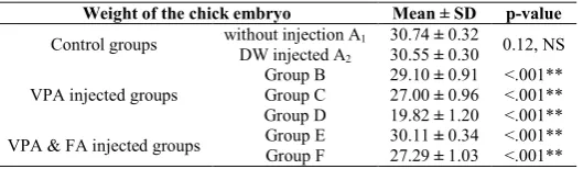
Weight of the chick embryo
Table 3 Showing the effect of VPA injection on the weight of chick embryos in comparison to DW injected control group.
Weight of the chick embryo Mean ± SD p-value
Control groups without injection A1 30.74 ± 0.32 0.12, NS
DW injected A2 30.55 ± 0.30
VPA injected groups
Group B 29.10 ± 0.91 <.001** Group C 27.00 ± 0.96 <.001** Group D 19.82 ± 1.20 <.001** VPA & FA injected groups Group E 30.11 ± 0.34 <.001** Group F 27.29 ± 1.03 <.001** *The mean difference is significant at the 0.05 level, NS: Not Significant
Congenital Anomalies
Table 4 Percentage of gross malformations seen in chick embryos in control and experimental groups treated with VPA.
S.No Deformities
Control group VPA VPA & FA
A1 (n=20) A2
(n=20) B (n=40) C (n=40) D (n=40) E (n=40) F (n=40)
1. Beak deformity 0 0 40% 70.8% 100% 18.7% 42.8%
2. Limb deformities 0 0 43.3% 58.3% 75% 25% 35.7%
3. Thin ventral body wall 0 0 33.3% 45.8% 12.5% 6.2% 21.4%
4. Ectopia Cordis 0 0 33% 62.5% 87.5% 6.2% 21.4%
5. Visceroptosis 0 0 0 37.5% 68.7% 0 0
Histological features
Liver
On histological examination of liver sections of the control group showed normal histological architecture with a central vein, radiating hepatic cords are observed. Hepatic sinusoids are little larger in chick embryo liver, possibly due to the presence of extravascular clusters of hemopoietic cells.
The assessment of the liver sections of VPA injected groups Table 1 Showing concentration of VPA injected in various
groups of chick embryos.
Groups Subgroups Number of eggs ‘n’
Drug (mg) or Distilled water (cc) Folic Acid (µg) Day of Injection Control Group A
A1 20 --- --- No injection
A2 20 0.1ml --- 3rd day
Experimental Group
B 40 0.25mg/egg --- 3rd day
C 40 0.5mg/egg 3rd day
D 40 1mg/egg 3rd day
E 40 0.5mg/egg 0.4µg/egg 3rd day
F 40 0.5mg/egg 0.4µg/egg 3
rd day and 5th
degeneration) with indefinite borders. Sections show that degenerated cells present slight cytoplasmic mass forming a thin peripheral rim. A central vein was dilated with disturbed endothelial lining. Larger areas occupying the anisonucleated hepatocytes with lymphocytic infiltration confirms severe injury at tissue level (Fig. 1.c, 1.d).Severe hepatic damage with gross morphological changes in the liver. There is no evidence of radiating hepatocyte arrangements, occasionally dilated central vein found (Fig. 1.b, 1.d). Most of the area shows cells without a nucleus, vacuoles in the cytoplasm.
Figure 1 Photomicrographs of Liver sections of the chick embryos in Control group (a),
Valproic acid-treated experimental groups (b,c,d,e).Central vein (CV), Dilated Central Vein (DCV), Large Vacuoles (LV), inflammatory cells in the portal triad (black arrow),
Nuclear damage progressed with pyknosis (yellow arrow), Hepatic degeneration (e), Macrovesicular (MV) changes, inflammatory infiltration.
Heart
The light microscopy assessments of the samples were conducted to observe morphological changes of heart highlighted in previous studies. Findings were confirmed excluding artifacts through comparison with control samples. The changes observed in VPA treated chick embryos of heart muscle are frequent separation of cardiac muscle fibers, degeneration of fibers, edema, congestion of cardiac muscle detected under endocardium, hemorrhagic spots in myocardium, infiltration of lymphoid cells, focal necrosis of the myocardium, and loss of nuclei (Fig. 2.b, 2.c, 2.d, 2.e).
Figure 2 Photomicrographs of histological sections of the heart of the chick embryos in
Control group (a), VPA treated experimental groups (b,c,d,e). Separation of fibers (d), Congestion of the myocardium (b), Haemorrhagic spots (white arrow), Degeneration of fibers with vacuolations (e), Infiltration of the lymphoid cells (red arrow).
Cerebrum
The serial sagittal and midsagittal histological sections, no changes were observed in the morphology of neurons and supporting cells in all sections of the cerebral cortex of DW treated control group and without an injection control group (Fig. 3.a&3.b). Experimental groups treated with VPA reveals mild changes in the organization of different layers in the cerebral cortex, the number of neurons is reduced in comparison to control sections. Thickening in the external granular layer was observed with more number of glial cells (Fig.3a, b). Maximum effective dose embryo section shows shrunken somata appeared in most of the pyramidal cells, hypertrophy of several different types of glial cells seen. Large vacuoles seen in hypertrophied glial cells and the nucleus pushed to the periphery. Degenerative changes are demarcated with necrosis at some locations. Lymphocytic infiltrations in the cerebral cortex, enlarged oligodendrocytes with nucleus pushed to one side and increased atrophy of pyramidal neurons were seen (Fig. 3.c). Cerebral sections of higher the effective dose-treated embryos, show more injury than the other groups. The architecture of the cerebral cortex was completely lost, the molecular layer was intensely infiltrated with lymphocytes. Increased inflammation with more amount of neuronal damage is a characteristic feature of all most all the sections in these groups. The Photomicrographs of higher magnification demonstrates that the nucleus being fragmented (Fig. 3.e).
Figure 3 Photomicrographs of histological sections of the cerebral cortex of the chick
embryos in Control group (3.a, 3.b), VPA treated experimental groups (3.c,3.d,3.e) (H&E 100x, 400x). Neuron (N), Neuroglial cells (G), Neuropil (Np), Fragmented nucleus (FN), Degenerative neurons, Perineuronal edema, Haemorrhage, cytoplasmic vacuolations (black arrow), Inflammatory infiltration.
Histomorphometric Parameters
The nuclear diameter of Hepatocytes:Histological sections of
Table 6 Multiple Comparisons of Nuclear diameters of hepatocytes (µm) in VPA treated chick embryos.
Post HOC Test: Tukey HSD - Nuclear diameters of hepatocytes (µm)
Group Mean Difference SEDM Sig.
A1
A2 .20000 .31161 .968
Group B .17500 .26986 .966
Group C .67500* .26986 .101
Group D 1.72500* .26986 .001
A2
Group B .02500 .26986 1.0
Group C .87500* .26986 .015
Group D 1.92500* .26986 .001
Group B Group C .85000
* .22034 .002
Group D 1.90000* .22034 .001
Group C Group D 1.05000* .22034 .001
*. The mean difference is significant at the 0.05 level.
DISCUSSION
VPA at Effective concentrations inhibits histone deacetylase (HDACs), which explains its anticancer properties[5]. VPA teratogenic effects are thought by some to be mediated through HDACs inhibition rather than antiepileptic activity. VPA inhibits HDACs, preferentially class I HDACs. It decreases histone acetylation, and thus changes in the chromatin structure and the activity of transcription factors of the genes may be induced. This action may interrupt the cell cycle, induce growth arrest and apoptosis, and in turn affect cell proliferation, which may explain its teratogenic properties[6, 9]. VPA triggers the active methylation of DNA along with acetylation of H3 Histone in human embryonic kidney cell culture. This is achieved by the direct action of VPA as an HDAC inhibitor, thus increasing demethylase accessibility to DNA, and the demethylation of DNA induces changes at the gene level. Such changes may produce various epigenetic abnormalities[9, 10].
Folic acid is essential for the body to make DNA, RNA, and metabolize amino acids that are required for cell division. Folic acid affects the disposition of anticonvulsants. The beneficial effect of additional folic acid in the periconceptional period to prevent neural tube defects, orofacial clefts, and conotruncal heart defects in the offspring has been shown. Folate shortage results in homocysteine accumulation[11]. Elevated levels of homocysteine have been related to neural tube defects. Folic acid addition increased neuroepithelial cell outgrowth and increased neural crest cell differentiation into nerve and smooth muscle cells[12].FA supplementation in the chick returned Hex and Isletgene expression to normal, and often to higher than control, levels. Although it is known that folic acid deficiency is one of the factors which can cause spina bifida and other NTD but the exact pathophysiology is still not clear[11, 12].
Amy I. Whitsel et. al [10], was studied on Effects of VPA
The observations of their study were increased mortality, growth delay, anomalies in the neural tube, cardiovascular, limb and skeletal. Similar kind of observations is found in the present study. Ercan Tureci et. al [13], was reported on injection of VPA and levetiracetam on chick embryos. The observations were recorded as several abnormalities found in the developing chick embryos.
The mortality rate, gross malformations, histological observations reported in the current study conformed with the reports on teratogenic effects of Dilantin on chick embryo by M Singh et al. [14], Shah GL[15]. Similar kind of results was also been reported by Mandavi Singh[16], Wilson RW [17].
We also observed that the fatality was less in all drug experiments on those embryos supplemented with FA on the 3rd day and 5thday of the incubation period. This probably might be due to selected AEDs disturbs the FA cycle along with several signaling molecules that may affect cell differentiation[4, 18]. Folic acid supplements, free radical scavengers, and antioxidant enzymes were found to counteract drug-induced teratogenic effects[19].
CONCLUSION
The gross morphological anomalies and statistical evaluation of the present study emphasize that the administration of folic acid in the early embryogenesis period would reduce the effects of AEDs, especially on AEDs alters the folic acid metabolism. The data obtained in the present study concludes that a noticeable impact of folic acid administration seen on teratogenic effects induced by Valproic acid. Further experimental studies with biochemical evaluation are required to specify the role of folic acid alteration in developing chick embryo as a cause of teratogenicity.
Conflict of Interest: None to Declare
References
1. Perry MM. A complete culture system for the chick embryo. Nature.1988;331(6151): 70-2.
2. Stern C D. The chick embryo–past, present, and future as a model system in developmental biology. Mech Dev. 2004;121:1011-3.
3. Reddy A. K., Fulzele R R, U L Gajbe. Study on Effects of carbamazepine and Phenytoin on Development of Chick Embryo; International Journal of Research and Development in Pharmacy and LifeSciences. October - November 2016, Vol. 5, No.6, pp 2388-2396.
4. Chateauvieux S, Bastien, Frank Morceau, Marc Diederich M. Molecular and Effective Potential and Table 5 Group-wise Comparison of Nuclear diameter of hepatocytes (µm) in VPA treated chick embryos using One Way
ANOVA
Groups N Mean SD SEM 95% CI df F-stat P-value
LL UL
Control groups A1 20 3.300 0.674 0.213 2.81 3.78
4,155 17.956 <.001**
A2 20 3.100 0.737 0.233 2.57 3.62
VPA
B 40 3.125 0.582 0.130 2.85 3.39
C 40 3.975 0.734 0.164 3.63 4.31
D 40 5.025 0.751 0.168 4.67 5.37
5. Umka J, Mustafa S, ElBeltagy M, Thorpe A, Latif L, Bennett G, et al. Valproic acid reduces spatial working memory and cell proliferation in the hippocampus. Neuroscience.2010;166(1):15-22.
6. Kostrouchová M, Kostrouch Z, Kostrouchová M. Valproic acid, a molecular lead to multiple regulatory pathways. Folia Biol (Praha).2007;53(2):37-49.
7. Detich N, Bovenzi V, Szyf M, Detich N, Bovenzi V, Szyf M. Valproate induces replication-independent active DNA demethylation. Journal of Biological Chemistry. 2003;278(30): 27586-92.
8. Kwan P, Sills GJ, Brodie MJ. The mechanisms of action of commonly used antiepileptic drugs. Pharmacology & Effective. 2001; 90(1):21-34.
9. Güney O, Canbilen A, Konak A, Acar O. The effects of folic acid in the prevention of neural tube development defects caused by phenytoin in early chick embryos. PubMed NCBI. 2003;28(5):4425.
10. Amy I. Whitsel, Casey, Johnson, Cynthia J. An In Ovo Chicken Model to Study the Systemic and Localized Teratogenic Effects of Valproic Acid. Clinical and Molecular Teratology 2002;66 (4): 153-63.
11. Gamal M. Bekhet, Mohamed A. Al-Kahtani, and Ashraf M. Abdel-Moneim. The protective effects of vitamin C and folic acid against methylmercury teratogenicity in chick embryo. Journal of Cell and Animal Biology. 2013;7(7);77-84.
12. Wegner C, Nau H. Alteration of embryonic folate metabolism by valproic acid during organogenesis: implications for mechanism of teratogenesis. Neurology. 1992;42(5):17-24.
13. Ercan Tureci, Ziya Asan, Mediha Eser, Taner Tanriverdi, Faruk Alkan, Pamir Erdincler. The effects of valproic acid and levetiracetam on chicken embryos. J Clin Neurosci. 2011;18(6):816-20.
14. M Singh, GL Shah. “Teratogenic effects of dilantin on thoraco-abdominal organs of developing chick embryos”. Indian J Exp Biol. 2000; 38(10):1026-30. 15. Shah GL, Singh M. The teratogenic effect of dilantin on
cerebellum in developing chick embryos. Journal of the Anatomical Society of India. 1988; 37(1): 1-8.
16. Mandavi Singh, G.L. Shah. Teratogenic Effects of Phenytoin on Chick Embryos. Teratology. 1989; 40:453-458.
17. Wilson Rw, Kalmus Gw And Pennington Sn. Alcohol Dehydrogenase Activity In The Developing Chick Embryo. Comp Biochem Physiol B. 1984;77:191-6. 18. Reddy AK, Fulzele RR, Gajbe U. Teratogenic Effects of
Lamotrigine on Thoraco-abdominal Organs of Developing Chick Embryos. Sch. J. App. Med. Sci., 2017; 5(5C):2003-2007.
19. Kim J, Kondratyev A, Gale K. Antiepileptic drug-induced neuronal cell death in the immature brain: effects of carbamazepine, topiramate, and levetiracetam as monotherapy versus polytherapy. J Pharmacol Exp Ther 2007;32:165–73.
How to cite this article:
Anil Kumar Reddy et al., 2019, The Protective Effects of Folic Acid Supplementation on Teratogenecity Induced By The Valproic Acid In Developing Chick Embryo. Int J Recent Sci Res. 10(10), pp.35497-35501.
DOI: http://dx.doi.org/10.24327/ijrsr.2019.1010.4114