RESEARCH ARTICLE
The evolution of the materials used
in the yun technique for the decoration
of Burmese objects: lacquer, binding media
and pigments
Diego Tamburini
1*, Verena Kotonski
2, Anna Lluveras‑Tenorio
3, Maria Perla Colombini
3and Alexandra Green
4Abstract
A series of Burmese lacquered objects decorated with the yun (incised) technique and belonging to the British Museum’s collection was analysed with the aim to investigate the decorative materials—lacquer, binding media, additives and pigments—used in the production of these objects, which span chronologically from the eighteenth century to the late twentieth century. As the manufacturing process is supposed to have remained very similar over this time period, especially regarding the use of materials, we were interested in scientifically assessing for the first time the nature of these materials and the correspondence to the written sources in the relation to their specific use. Gas chromatography mass spectrometry (GC–MS) and analytical pyrolysis with in situ silylation coupled with GC–MS (Py(HMDS)GC–MS) were used for the identification of the organic components in several samples taken from the coloured areas of the objects. Fibre optic reflectance (FORS) and Raman spectroscopies were used to identify the pig‑ ments after a visual investigation of the samples by digital microscopy. Burmese lacquer was detected in all objects and trends in its degradation were highlighted. Lipids, proteins and saccharide material were found to be mixed with lacquer, and they appeared to be applied with specific pigments, in good agreement with the written records, apart from proteins, which are not mentioned. The use of synthetic pigments, such as phthalocyanines blue and green and chrome yellow, was assessed in the most recent objects, showing an evolution in the use of pigments. Indigo, although expected, was not identified in any of the green samples and Prussian blue appeared to be the main source of blue colour. All this information is of fundamental importance for conservation practices and corrects the general opinion about the production materials of these objects. These results also open the way to future research dedicated to exploring the chemical interaction between Burmese lacquer, proteins, lipids, gums and pigments, with the aim to predict possible differences in degradation pathways.
Keywords: Burmese lacquer, Binding media, Pigments, Yun technique, GC–MS, Analytical pyrolysis, Raman spectroscopy, Conservation
© The Author(s) 2019. This article is distributed under the terms of the Creative Commons Attribution 4.0 International License (http://creat iveco mmons .org/licen ses/by/4.0/), which permits unrestricted use, distribution, and reproduction in any medium, provided you give appropriate credit to the original author(s) and the source, provide a link to the Creative Commons license, and indicate if changes were made. The Creative Commons Public Domain Dedication waiver (http://creat iveco mmons .org/ publi cdoma in/zero/1.0/) applies to the data made available in this article, unless otherwise stated.
Open Access
*Correspondence: [email protected]
1 Department of Scientific Research, The British Museum, Great Russell Street, London WC1B 3DG, UK
Introduction
Burmese1 lacquer (thitsi) has probably been used for more than a millennium, but no archaeological evidence is available before the thirteenth century, possibly due to the limited archaeological work undertaken in Myan-mar in the last 60 years [1]. Damaged objects were also discarded and replaced in Burmese traditional practices, due to the belief that old and defective objects may bring bad luck. As a consequence, many Burmese lacquered objects in European collections date to the nineteenth to twentieth centuries and are mainly vessels that have been used domestically in private homes or in monasteries [2,
3].
Most information about the techniques of lacquer pro-duction is also relatively recent and starts with the Brit-ish colonial period [1]. Burmese lacquer is obtained from the sap of the Melanorrhoea (Gluta) usitata tree, which belongs to the Anacardiaceae family and grows in Laos, Myanmar, Thailand and Cambodia. The sap is harvested from the tree by tapping and has a straw colour, which quickly turns glossy black. Its main function is to water-proof and heatwater-proof the object and has excellent adhesive properties, as well as good stability in hot, acid and alka-line conditions. It is therefore a good binding medium for pigments and it has been applied to a variety of inorganic (metal and ceramic) and organic (wood, cane, palm-leaf, leather) substrates [1]. Nowadays, most containers, such as vessels, bowls and cups, are made of split coiled bam-boo, but other wood substrates and basketry have also been used in the past.
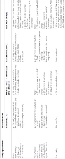
A number of different decorative techniques are known across Myanmar, with the yun (incised) technique tradi-tionally being the most common one. The term yun refers to a form of decoration where a surface is engraved and the grooves filled with colour, but yun is also used as the generic term for lacquer in Myanmar [4]. A small number of first-hand accounts of local craft practices are available describing this technique in some detail [1, 4–6]. A com-parison of these accounts, collected by researchers travel-ling to Myanmar, is summarised in Table 1. This reveals that materials and manufacturing processes remained similar over the period from the late nineteenth to twenty first centuries, which is a testament to a continuous tra-dition of craftsmanship. Any decorative layers require a smooth substrate, which is achieved by applying a paste called thayo to fill any interstices the work piece might show. Thayo paste consists of lacquer mixed with fillers,
varying from coarse particles (e.g. sawdust ash) for the initial layers to fine particles (e.g. cow bone or cow dung ash) for layers closer to the decorative surface. The thayo ground is then covered with a series of 5–20 lacquer coatings without filler. The number of thayo and lac-quer applications, the drying time between each appli-cation and the care taken smoothing the layers before a new layer is applied determine the quality of the finished object. Items earmarked for engraving are often of black or red colour, which will later show as the background colour after engraving. The yun technique relies on the principle that the colour will adhere only to the incisions of the engravings according to the dictates of the design and can be removed from non-engraved areas. Multi-col-oured designs require repeated applications of differently coloured coatings. Different methods of applying the col-our have been reported: the pigment is either mixed with a binder to form a paste and this paste is rubbed into the grooves, or the grooves are filled with binding media and the dry pigment in rubbed into still moist binder [6]. To avoid contamination of previously applied colours with the yet to be applied colour, any excess binder or pig-ment paste is wiped off immediately before it sets on the surface. Alternatively, a “barrier layer”, consisting of water-soluble gum extracted from the neem/tama tree (Azadirachta indica) or the acacia tree (Acacia farnesi-ana), is applied and engraved. The colour is then applied and, once dried, the excess colour and the barrier layer are removed before the process is repeated for another colour. After each application of a layer, the object is mounted on a lathe and layers are smoothed by polishing the surface with an abrasive (pumice stone, sandstone, textile rag, rice husks, fossilised wood) in combination with water.
Most commonly the colours red, green and yellow and occasionally blue are found on yun lacquerware. Red col-our is said to be based on cinnabar/vermillion (mercuric sulfide—HgS), referred to as hinthabada, or the cheaper red ochre (iron oxides) for lesser quality items. Green (atsein) is reported to be a mixture of orpiment (arsenic trisulfide—As2S3) and indigo. Yellow (saydan) is
com-posed of pure orpiment, and orange is a mixture of orpi-ment and cinnabar/vermillion. The binding medium for all colours appears to be lacquer, with some addition of tung (shan-zi), peanut or sesame oil. Vegetable gums are not explicitly reported as a binder, but mainly as ingredi-ents of the “barrier layer” between two colours (Table 1) [1, 4–6].
It appears from this description that lacquer is not the only organic material used for producing these objects and the written descriptions are somewhat hazy when it comes to the actual nature of the other materials used as binding media. Previous works have identified 1 The authors are aware that since 1989 Burma is officially called Republic of
Table 1 Summar y of the av ailable inf orma tion ab out the ma terials and manufac turing pr oc ess of Burmese lac quer ed objec ts dec or at ed with the yun technique ac cor ding t o writt en sour ces [ 1 , 4 – 6 ] Str atig raph y of la yers Lit er atur e sour ce Burne
y 1832 [
6
]
Fr
aser
-L
u (1985, 1st edition) 2000
(2nd edition) [
4
]
Isaac/Blur
ton 2000 [
1
]
Than H
tun 2013 [
5 ] Gr ound la yer tha ‑ yo (Bur ne y) thay o (F raser ‑L u) thay o (Isaac/Blur ton) thay oe (T han H tun)
p. 169: lacquer of lo
w er qualit y with 50% wat er added + Filler A
sh of co
w bone
Or ash of bran/husk of paddy (“
most
commonly used
”)
Or ash of t
eak w ood + spittle (“thick consist enc y, pr oduces har d fill ”)
Or ash of co
w ‑dung (“ adher es t ena ‑ ciously , v er
y pliable and elastic
”)
p. 24–25: lacquer + Filler clay (“1st coating
”)
Or t
eak ash & glue fr
om boiled r
ice
(“2nd coating
”)
Or co
w dung ash
+
rice stra
w ash
(“2nd coating
, f
or finest w
or
k”)
Or po
w
der
ed bone (2nd coating
, f
or
finest w
or
k”)
p. 34: lacquer + Filler Ash (
e.g
. of co
w bones)
Or cla
y
p. 32: lacquer + Filler Cow dung (“
year
‑old dung
, cleaned
(…) g
round t
o obtain a pur
e po w der , ex tr emely stick
y and str
ong , f or 1st class war e” or r iv er S ediments + red ear th (“ for 2nd class war e”)
Or ash of g
roundnut hulls + dr ied stra w (“used no wada ys ”) Or sa w dust Finishing la yer f
or plain black war
es theet ‑ tsee (Bur ne y) thit ‑ si (F raser ‑L u) thit si (Isaac/Blur ton) sitse (T han H tun)
p. 169: lacquer of highest qualit
y
Not
e: applied in thr
ee or mor
e coats
bef
or
e fur
ther decoration is applied
p. 25: lacquer of good qualit
y
p. 35: lacquer
p. 32: lacquer
Red Cinnabar/v er million hen ‑ za ‑ pa ‑ da (Bur ne y) hin ‑ thabada (F raser ‑L u) hinthabada (Isaac/Blur ton ) hinthapada (T han H tun) red ochr e: m yè ‑ nee (Bur ne y) m ye ‑ ni (F raser ‑L u) n/a (Isaac/Blur ton) my eni ( Than H tun)
p. 171: v
er
million (“
from China, of the
finest k
ind
, does not mix w
ell with lacquer ”) Or self ‑made v er
million (made b
y
small number of local craf
tsmen,
“pr
ef
er
red b
y the Bur
mese ”) Or r ed ochr e (“I ndian r ed ”) (“ giv es a duller colour ”, f
or cheaper war
e,
sometimes used as a 1st coat, o
ver
which v
er
million is applied
”)
+
Binder
Tung oil
+
lacquer (3:10) (“
semi ‑trans ‑ par ent var nish ”)
p. 171: (tung) oil
(Shan
‑
zee or Shan oil)
from fruit of the k
uniy en tr ee ( D ip -ter oc arpus turbinatus ) (“ from Laos , long dr ying time ”) + p
. 169: lacquer of slightly lesser
qualit
y with 25% wat
er added
p. 25: v
er million/cinnabar (mer cur ic sulfide) (“ impor ted fr om China ”) Or r ed ochr e (“ cheaper , f or inf er ior war es ”) + Binder Lacquer +
tung oil (
shan ‑ zi ) fr om tr ee fruit of Aleurites triloba or D ipter oc ar -pus turbinatus + Unspecified “special ” additiv es + Bar rier la yer
p. 34: r
esin of neem (tama) tr
ee ( A zadir achta indic a ) Or r
esin of acacia tr
ee ( Ac acia f arnesi -ana ) (“
glue (…) t
o seal the r
ed colour
within the eng
ra
ved lines
”)
p. 35–36: cinnabar (mer
cur ic sulfide) Or r ed ochr e (mor e r ecently) Or r
ed paint (mor
e r ecently) + Binder W at er + peanut oil + lacquer + Bar rier la yer
p. 37, 40: r
esin of acacia tr
ee ( htan -aung ) + wat er ( = “gum Arabic ”)
p. 29: mer
cur
ic sulfide
+
Binder
Peanut oil (Pyu per
iod t
o 1700 and 1700
to 1900)
Or sesame oil (1900 t
o 1930) ( No lac quer mentioned ) + Bar rier la yer
p. 28, 30: r
esin of acacia tr
ee ( htanaung , Ac acia leuc ophloea
) or r
esin of neem
tr ee ( = tamar tr ee)
Blue n/a (Bur
ne y) me ‑ ne (F raser ‑L u) n/a (Isaac/Blur ton) mene (T han H tun)
(No blue colourant mentioned)
p. 26: indigo (
Indigofer a anil . ) (“R ar
ely used in traditional Bur
mese
lacquer w
or
k, f
or the indigo does
not combine w
ell with the (…) ra
w
lacquer
, r
esulting in a rather dull
finish
”)
+
binder (no binder specified)
(No blue colourant mentioned)
p. 29: I
ndigo
And/or madama bar
k
Or blue paint (mor
e r ecently) Or dy e po w der (mor e r ecently) +
Binder (no binder specified)
+
Bar
rier la
yer
Resin of acacia tr
Table 1 (c on tinued) Str atig raph y of la yers Lit er atur e sour ce Burne
y 1832 [
6
]
Fr
aser
-L
u (1985, 1st edition) 2000
(2nd edition) [
4
]
Isaac/Blur
ton 2000 [
1
]
Than H
tun 2013 [
5 ] Gr een atsein (Bur ne y) n/a (F raser ‑L u) n/a (Isaac/Blur ton) n/a ( Than H tun)
p. 174: indigo
+
or
piment
(1:10)
Or juice fr
om leaf of plant called
gwe ‑ douk ‑ beng + or piment + Binder Lacquer +
tung oil (
Shan
‑
ze
e
)
p. 27: indigo
+
or
piment
(1:10)
Or p
. 40: enamel paint (fr
om lat
e 80
s)
+
Binder (no binder specified)
p. 40: indigo
+
or
piment
Or mass
‑pr
oduced chemical colour
Or house paint (1980s) + Binder p. 37: lacquer
p. 29: g
reen (“
minor colour
, used since
the 1850s ”) Or hinthapada + blue mene (so called England g reen po w der) (fr om 1900 to 1930) +
Binder (No binder specified)
+
Bar
rier la
yer
Resin of acacia tr
ee ( htanaung , A cacia leuc ophloea ) Yello w/orange or piment tshè ‑ dan (Bur ne y) sei ‑ dan (F raser ‑L u) n/a (Isaac/Blur ton) say dan (T han H tun)
p. 174: or
piment (y ello w sulfur et of arsenic) + Binder lacquer +
tung oil (
Shan ‑ ze e ) + Bar rier la yer (no bar rier la yer mentioned) Yello w
p. 26: or
piment (arsenic tr
isulfide)
(fr
om Shan Stat
es)
Orange p. 26: or
piment + v er million + Binder
Gum of dammar Or lacquer
+
tung oil (
shan
‑
zi
)
p. 27: or
piment (arsenic tr
isulfide)
+
Binder
Lacquer (applied af
ter bar rier la yer and eng ra ving) + Bar rier la yer A cacia tr
ee glue (applied bef
or e eng ra ving) Yello w
p. 29: or
piment (arsenic tr
isulfide) (1900 to 1930) Orange cinnabar + or piment + Binder (
no binder specified
) + Bar rier la yer
Resin of acacia tr
ee ( htanaung , A cacia leuc ophloea ) Final coating Not specified
p. 34: lacquer
+
a little tung oil
None mentioned
p. 33: lacquer
+
tung oil (1:3) (
+
sesame
oil) (in Bagan and Shan Stat
es (Laik ha), bet w een 1850s–1930s , rar ely used af ter W
WII due t
o being expensiv
various materials other than lacquer in Burmese objects, but attention to these has always been low [7–9]. Fur-thermore, pigments play an important role in the final appearance of these objects. Pigments have been studied in a few cases [7, 10], but mostly in relation to Japanese and Vietnamese lacquers [11–13]. This work therefore aims to focus attention on identifying materials other than lacquer used for the production of Burmese lac-quered objects by applying a systematic multi-analytical approach. In addition to providing scientific evidence for comparison with written sources, this research aims to characterise systems of multiple components, where organic and inorganic materials potentially interact with each other, thus evolving over time in unexpected ways [14, 15]. Having this information is of paramount impor-tance for the preservation and conservation of these objects. Additionally, the selection of objects was made in order to represent the yun technique chronologically and geographically over two centuries and to see if there is consistency within the materials used.
From a scientific point of view, lacquered objects are very complex systems and represent an analytical chal-lenge. The main component of the Burmese lacquer sap is a mixture of substituted catechols, referred to as thitsiol [16]. In addition to C15- and C17-alk(en)yl-substituted catechols, which are present in the other Asian lacquers (urushiol and laccol respectively), thitsiol contains cat-echol derivatives with an ω-phenylalkyl chain of 10 or 12 carbon atoms. These compounds are specific markers of identification of this lacquer [17–20]. When the sap of the tree hardens, a polymerisation process occurs cata-lysed by the enzyme laccase. The polymeric nature of lac-quers makes pyrolysis coupled with gas chromatography and mass spectrometry (Py–GC–MS) the most suitable analytical approach for their chemical analysis, charac-terisation and identification in samples of unknown com-position [12, 21–27]. The need of a derivatisation step, using either tetramethylammonium hydroxide (TMAH) [28–31] or hexamethyldisilazane (HMDS) [8, 14, 20, 32], is strongly suggested due to the large number of polar phenolic compounds produced in the pyrolysis process. The identification is based on the detection of specific molecular markers, as mentioned above, and character-istic profiles of specific classes of pyrolysis products, such as alkylphenols, alkylcatechols, alkylbenzenes and ali-phatic hydrocarbons [20, 32].
Py–GC–MS can be generally used to identify most classes of organic materials by detecting their molecu-lar markers even in complex mixtures [33]. However, it is sometimes difficult to distinguish between chemically similar substances, such as two vegetable gums or two different oils, because they can easily produce the same pyrolysis products [34]. Comparing chromatographic
peak areas sometimes helps to have an approximate dis-tinction, but the values obtained this way are not relia-bly comparable with data found in the literature and are mainly valid within the laboratory in which they have been obtained. Moreover, Py–GC–MS is not a quan-titative technique by definition, as calibration curves for all the compounds found in a pyrogram are virtu-ally impossible to obtain, and, even if done, there is no control over the pyrolysis yield and over the possible interactions between materials affecting the pyrolytic process. For these reasons, analytical procedures based on GC–MS are more reliable when quantitative data are required [35]. Isolation steps and calibration curves enable the chromatographic areas to be directly related to concentrations. Ratios and percentages can therefore be used in a more reliable way for the identification of the sources of certain materials, such as oils, proteins and vegetable gums [36]. In the last decade, analytical meth-ods based on MALDI-ToF and LC–MS have also shown their potential in identifying the exact source of a pro-tein (proteomics) [37] or lipid (lipidomics) [38], but these analyses are still time-consuming and costly compared to GC–MS.
With regards to pigments, Raman spectroscopy is a powerful identification tool [39, 40] and can be used non-invasively on the objects themselves. However, lacquered objects usually present a high proportion of organic materials compared to the amount of inorganic pigments, which results in high background fluorescence when using portable instruments directly on the objects [11]. The use of samples, sometimes prepared as cross sec-tions, and an appropriate evaluation of the laser energy to balance the signal intensity and the risk of damaging the samples are highly recommended [11, 12]. The use of cross sections also allows the production of high qual-ity microscopy images of pigment particles, which are fundamental to correctly interpret the results and study the distribution of materials. However, samples for cross sections are relatively large, cannot be further analysed by destructive techniques and double-sampling is there-fore required. For ethical reasons, this is sometimes to be avoided for very precious museum objects. Finally, fibre optic reflectance spectroscopy (FORS) is a non-invasive technique, which has recently shown high potential for the straightforward identification of pigments and dyes [41], but has not been applied to lacquerware. The influ-ence of the binding media on the reflectance spectra of the pigments is generally not reported as a major prob-lem, thus making this technique a valid alternative to Raman spectroscopy for the investigation of such objects.
Py(HMDS)–GC–MS) to the investigation of the decora-tive materials of Burmese lacquered objects produced with the yun technique.
Experimental section Selected objects and samples
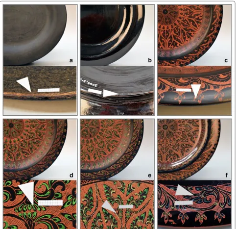
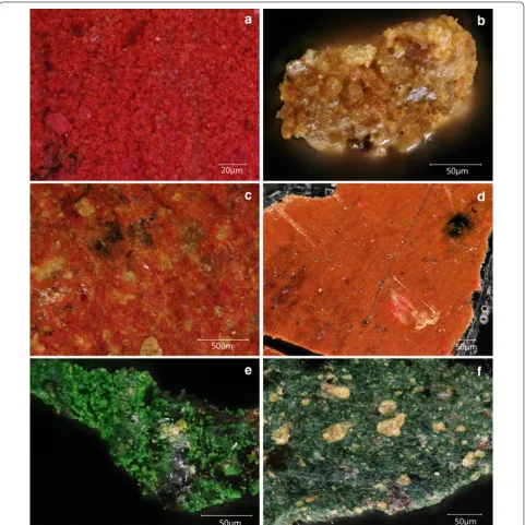
Six objects from the British Museum’s collection of Bur-mese lacquerware were selected based on chronological significance and place of production. The objects are all produced using the yun (incised) technique. Object 1 is actually a series of nine plates (registration numbers OA2000,0330.4-12, diameter 26 cm) that were made in the late twentieth century to illustrate the technique itself, as it is practised today in Bagan, the most impor-tant lacquer-producing centre in Myanmar. These plates show the nine stages necessary to produce this type of object, therefore representing a deconstructed cross sec-tion and providing a unique opportunity to investigate the materials utilised in each of the layers. The first plate represents the coiled split bamboo armature covered with a rough layer of lacquer (thayo) (Fig. 1a). Another layer of refined lacquer is then applied (Fig. 1b) and, at this point, the plate is ready for engraving. Once engraved, the red colour is applied (Fig. 1c), followed by further engrav-ings and applications of colour (green and then yellow, Fig. 1d, e). When the decoration is complete, the overall surface is polished (Fig. 1f). Six of these plates (objects 1a–f) were sampled, representing the different materials, i.e. raw lacquer, refined lacquer, red paint, green paint, yellow paint and final coating. Details of the plates and sampling areas are shown in Fig. 1, and full images of the plates are present in Additional file 1: Figure S1.
Object 2 (registration number OA1996,0501.50, Addi-tional file 1: Figure S2) is a rectangular wooden tray (length 56 cm; width 37 cm), showing yellow, red, orange/ pink and green colours on a black ground. All colours were sampled. The object was produced in the 1920s by the artist Ba Htet in the workshop of Ko Aye Hpay in Bagan. Object 3 (registration number OA1998,0723.140, Additional file 1: Figure S3) is approximately contem-porary to object 2. It is a Gaduganan betel box (height 15 cm; diameter 19.2 cm). The colours are black and red, which were both sampled. Unlike object 2, this object was acquired in the Shan States.
Object 4 (registration number OA1998,0723.206, Addi-tional file 1: Figure S4) is dated to the mid-late nine-teenth century from Bagan. It is a water bowl (height 18.8 cm; diameter 23.8 cm) showing exquisite decora-tion in green, yellow and red colours on a black ground, all of which were sampled. Object 5 (registration number OA1996,0501.52, Additional file 1: Figure S5) was a cof-fer (height 16.5 cm; width 22 cm; length 39 cm) dated to the late eighteenth century-early nineteenth century also
from Bagan and also showing the three main colours, which were all sampled. Object 6 (registration number OA1998,0723.24, Additional file 1: Figure S6) is a box (height 24 cm; diameter 37 cm), possibly for clothes, dec-orated in black on red ground. The interior surface was painted orange. The black, red and orange colours were all sampled. It is the oldest example of Burmese lacquer-ware in the BM collection (eighteenth century). The exact place of production is not known.
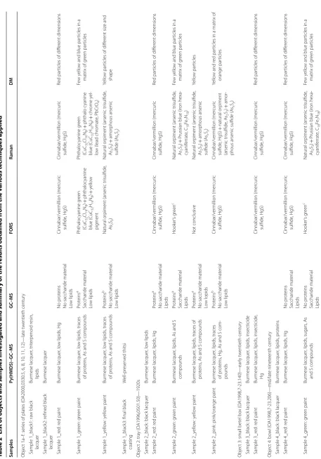
Additional details about all the objects are present in [1] and Table 2 presents a list of them together with the samples taken and a summary of the results.
Analytical approach
The selected objects underwent a visual examination to identify the exact number of colours applied and possible sampling locations, mostly at the edge of already dam-aged areas. The non-invasive analysis by FORS was then conducted followed by a sampling campaign (micro-samples of c. 50–100 µg were removed from the surface using a micro-scalpel). Black areas were sampled once, as these samples were only analysed by Py(HMDS)– GC–MS. Coloured areas were sampled twice: one set of samples underwent GC–MS analysis, whereas the other set was earmarked for microscopic investigation using digital microscopy, Raman analysis and finally Py(HMDS)–GC–MS.
Fibre optic reflectance spectroscopy (FORS)
Fibre optic reflectance spectra were recorded using an Avantes (Apeldoorn, The Netherlands) AvaSpec-ULS2048XL-USB2 spectrophotometer equipped with an AvaLight-HAL-S-IND tungsten halogen light source. The detector and light source were connected with a fibre optic bundle to an FCR-7UV200-2-1.5 × 100 probe. The spectral range considered was between 300 and 900 nm. As per the features of the monochromator (slit width 50 μm, grating of UA type with 300 lines/mm) and of the detector (2048 pixels), the best spectra resolution was 2.4 nm. Spectra were referenced against the WS-2 refer-ence tile provided by Avantes. The diameter of the inves-tigated area on the sample was approximately 1 mm. The instrumental parameters were as follows: 50 ms integra-tion time, 20 scans for a total acquisiintegra-tion time of 1 s for each spectrum. The whole system was managed by the software AvaSoft 8 for Windows™. Spectra were acquired in reflectance mode (R) and then transformed into appar-ent absorption spectra by plotting the log(1/R).
Digital microscopy (DM)
Z250R/W, an automated stage VHX-S 550E and LED reflected illumination.
Raman spectroscopy
A Jobin–Yvon LabRam Infinity spectrometer was used with a green laser (532 nm) with maximum power of 2.4 mW at the sample, a liquid nitrogen cooled CCD detector and an Olympus microscope system. The instrument was
calibrated using a silicon standard, with a wavenumber deviation of 3 cm−1. The experimental conditions were:
integration time between 5 and 10 s, averaging of up to 50 scans, spectral range between 135 and 2600 cm−1 and
a fine grating of 1800 g/mm. The analyses were carried out on the samples, trying to target differently coloured particles.
Table
2
List of objec
ts and samples in
vestiga
ted and summar
y of the r
esults obtained fr
om the v arious t echniques applied Samples Py(HMDS)– GC–MS GC–MS FORS Raman DM Objec t 1a–f : ser
ies of plat
es (
O
A2000,0330.5, 6, 8, 10, 11, 12)—lat
e t
w
entieth centur
y
Sample 1_black1: ra
w black lacquer Bur mese lacquer , tr iter penoid r esin, lipids
Sample 1_black2: r
efined black lacquer Bur mese lacquer Sample 1_r ed: r ed paint Bur mese lacquer , lo w lipids , Hg No pr ot eins No sacchar ide mat er ial Lo w lipids Cinnabar/v er million (mer cur ic sulfide , HgS) Cinnabar/v er million (mer cur ic sulfide , HgS) Red par
ticles of diff
er ent dimensions Sample 1_g reen: g reen paint Bur mese lacquer , lo w lipids , traces of pr ot eins , A
s and S compounds
Pr ot eins a Sacchar ide mat er ial Lo w lipids Phthaloc yanine g reen (Cu C32 Cl16 N8 ) + phthaloc yanine blue (C uC 32 H16 N8 ) + yello w pig ment Phthaloc yanine g reen (Cu C32 Cl16 N8 ) + phthalo cyanine blue (C uC32 H16 N8 ) + chr ome yel ‑ lo
w (lead chr
omat e, PbCrO 4 ) Fe w y ello
w and blue par
ticles in a
matr
ix of g
reen par ticles Sample 1_y ello w : y ello w paint Bur mese lacquer , lo w lipids , traces of pr ot eins , A
s and S compounds
Pr ot eins b No sacchar ide mat er ial Lo w lipids Natural or
piment (arsenic tr
isulfide , As2 S3 ) Natural or
piment (arsenic tr
isulfide , As2 S3 ) + amor phous arsenic sulfide (A sx Sy ) Yello w par
ticles of diff
er
ent siz
e and
shape
Sample 1_black3: final black coating
W ell ‑pr eser ved thitsi Objec
t 2: tra
y (
O
A1996,0501.50)—1920s
Sample 2_black
: black lacquer
Bur mese lacquer , lo w lipids Sample 2_r ed: r ed paint Bur mese lacquer , lipids , Hg Pr ot eins a No sacchar ide mat er ial Lipids Cinnabar/v er million (mer cur ic sulfide , HgS) Cinnabar/v er million (mer cur ic sulfide , HgS) Red par
ticles of diff
er ent dimensions Sample 2_g reen: g reen paint Bur mese lacquer , lipids , A
s and S
compounds Pr ot eins a Sacchar ide mat er ial Lipids Hook er ’s gr een c Natural or
piment (arsenic tr
isulfide , As2 S3 ) + P
russian blue (ir
on hexa ‑ cyanif er rat e, C18 Fe7 N18 ) Fe w y ello
w and blue par
ticles in a
matr
ix of g
reen par ticles Sample 2_y ello w : y ello w paint Bur mese lacquer , lipids
, traces of
pr
ot
eins
, A
s and S compounds
Pr ot eins a No sacchar ide mat er ial Lo w lopids Not conclusiv e Natural or
piment (arsenic tr
isulfide , As2 S3 ) + amor phous arsenic sulfide (A sx Sy ) Yello w par ticles Sample 2_pink
: pink/orange paint
Bur mese lacquer , lipids , traces of pr ot eins , Hg , A
s and S com
‑ pounds Pr ot eins b No sacchar ide mat er ial Lo w lipids Cinnabar/v er million (mer cur ic sulfide , HgS) Cinnabar/v er million (mer cur ic sulfide , HgS) + natural or piment (arsenic tr isulfide
, A
s2 S3 ) + amor ‑
phous arsenic sulfide
(A
sx Sy
)
Yello
w and r
ed par
ticles in a matr
ix of
orange par
ticles
Objec
t 3: small bet
el bo x ( O A1998,7 ‑23.140)— ear ly t w entieth centur y Sample 3_black
: black lacquer
Bur mese lacquer , lipids , insec ticide Sample 3_r ed: r ed paint Bur mese lacquer , lipids , insec ticide , Hg Cinnabar/v er million (mer cur ic sulfide , HgS) Cinnabar/v er million (mer cur ic sulfide , HgS) Red par
ticles of diff
er
ent dimensions
Objec
t 4: bo
wl ( O A1998,7 ‑23.206)—mid/lat e ninet eenth centur y Sample 4_black
: black lacquer
Bur mese lacquer , lipids , pr ot eins Sample 4_r ed: r ed paint Bur mese lacquer , lipids , Hg No pr ot eins Sacchar ide mat er ial Lipids Cinnabar/v er million (mer cur ic sulfide , HgS) Cinnabar/v er million (mer cur ic sulfide , HgS) Red par
ticles of diff
er ent dimensions Sample 4_g reen: g reen paint Bur mese lacquer , lipids , sugars , A s
and S compounds
No pr ot eins Sacchar ide mat er ial Lipids Hook er ’s gr een c Natural or
piment (arsenic tr
isulfide , As2 S3 ) + P
russian blue (ir
on hexa ‑ cyanif er rat e, C18 Fe7 N18 ) Fe w y ello
w and blue par
ticles in a
matr
ix of g
reen par
Table 2 (c on tinued) Samples Py(HMDS)– GC–MS GC–MS FORS Raman DM Sample 4_y ello w : y ello w paint Bur mese lacquer , lipids , Hg , A s and S compounds No pr ot eins No sacchar ide mat er ial Lipids Yello w pig ment + cinnabar/v er mil ‑ lion (mer cur ic sulfide , HgS) Natural or
piment (arsenic tr
isulfide , As2 S3 ) + amor phous arsenic sulfide (A sx Sy ) + cinnabar/v er mil ‑ lion (mer cur ic sulfide , HgS) Yello
w and r
ed par
ticles in an orange
matr
ix
Objec
t 5: coff
er ( O A1996,5 ‑1.52)— ear ly ninet eenth centur
y or bef
or
e
Sample 5_black
: black lacquer
Bur mese lacquer , lipids Sample 5_r ed: r ed paint Bur mese lacquer , lipids
, traces of
pr ot eins , sugars , Hg Pr ot eins a Sacchar ide mat er ial Lo w lipids Cinnabar/v er million (mer cur ic sulfide , HgS) Cinnabar/v er million (mer cur ic sulfide , HgS) Red par
ticles of diff
er ent dimensions Sample 5_g reen: g reen paint Bur mese lacquer , lipids , A
s and S
compounds Not det er mined aminoacidic frac tion No sacchar ide mat er ial Lipids Hook er ’s gr een c Natural or
piment (arsenic tr
isulfide , As2 S3 ) + P
russian blue (ir
on hexa ‑ cyanif er rat e, C18 Fe7 N18 ) Fe w y ello
w and blue par
ticles in a
matr
ix of g
reen par ticles Sample 5_y ello w : y ello w paint Bur mese lacquer , lipid , Hg , A
s and S
compounds Not det er mined aminoacidic frac tion No sacchar ide mat er ial Lipids Yello w pig ment + cinnabar/v er mil ‑ lion (mer cur ic sulfide , HgS) Natural or
piment (arsenic tr
isulfide , As2 S3 ) + amor phous arsenic sulfide (A
sSxy
) + cinnabar/v er mil ‑ lion (mer cur ic sulfide , HgS) Yello
w and r
ed par
ticles in an orange
matr
ix
Objec
t 6: lar
ge bo x ( O A1998,0723.24)— eight eenth centur y Sample 6_black
: black lacquer
Bur mese lacquer , lipids , pr ot eins Sample 6_r ed: r
ed paint (
ex ter nal par t) Bur mese lacquer , lipids , pr ot eins , Hg Pr ot eins a No sacchar ide mat er ial Lipids Cinnabar/v er million (mer cur ic sulfide , HgS) Cinnabar/v er million (mer cur ic sulfide , HgS) Red par
ticles of diff
er
ent dimensions
Sample 6_orange: orange paint (int
er nal par t) Bur mese lacquer , lipids , pr ot eins , phthalat es Pr ot eins b No sacchar ide mat er ial Lipids Not conclusiv e Cinnabar/v er million (mer cur ic sulfide , HgS) Red par
ticles in an orange matr
ix
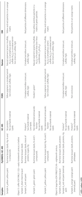
a L
OD < v alue < LOQ
b V
alue > LOQ (see “ O ther or ganic ma ter ials ” sec tion)
c T
he r
eflec
tanc
e spec
trum ma
tches the pr
ofile of Hooker
’s g
reen [
58
], which is r
epor
ted as a mix
tur
e of P
russian blue and gamboge
. T
his w
as not c
onfir
med b
y the other t
echniques
, but the t
er
m Hooker
’s g
reen is her
e
used t
o under
line tha
t this w
ould ha
ve been the a
ttr
Py(HMDS)–GC–MS
Analytical pyrolysis was performed using 1,1,1,3,3,3-hex-amethyldisilazane (HMDS, chemical purity 99.9%, Sigma Aldrich Inc., USA) as a silylation agent for the in situ thermally-assisted derivatisation of pyrolysis products. The instrumentation consisted of a 1500 CDS Pyroprobe 1000 Series filament pyrolyser (CDS Analytical, USA) (platinum coil) coupled to a gas chromatograph 6890 (Agilent Technologies, USA) equipped with an HP-5MS fused silica capillary column (30 m × 0.25 mm i.d., 0.25 µm thickness, Agilent Technologies, USA). The GC was coupled with an Agilent 5973 Mass Selective Detec-tor operating in electron impact mode (EI) at 70 eV. Pyrolysis temperature was 550 °C and the interface tem-perature was 275 °C. Similar amounts (c. 50–100 μg) of sample and HMDS (2 µL) were inserted into the centre of the pyrolysis quartz tube with quartz wool and then put into the filament coil. The GC injector was used with a split ratio of 1:10 at 300 °C. Chromatographic condi-tions were as follows: initial temperature 36 °C, 10 min isothermal, 10 °C min−1 up to 280 °C, 2 min isothermal;
20 °C min−1 up to 310 °C, 20 min isothermal. Helium
(purity 99.995%) was used as carrier gas with a constant flow of 1.0 mL min−1.
GC–MS
A microwave MLS-1200 MEGA (Milestone Microwave Laboratory System), High Performance Microwave Digestion with Exhaust Module EM-45/A was used for the hydrolysis and the saponification of the sample frac-tions prior to GC–MS analysis [42].
The GC–MS system was the same described in the pre-vious section, apart from the injector, which was a PTV injector (Agilent Technologies).
A combined procedure that allows the separation and characterisation in the same micro-sample of three dif-ferent fractions corresponding to saccharide, proteina-ceous and lipid-resinous materials was used for the GC–MS analysis. The analytical procedure was based on the ammonia extraction of proteins and polysaccharide materials from the sample in order to separate them from lipid and resinous materials. Proteinaceous and saccha-ride fractions were separated by a monolithic sorbent tip featuring a C4 stationary phase and were subsequently hydrolysed and purified. Lipids and resins were subjected to saponification. The detailed operating conditions and analytical procedure are described elsewhere [42].
Proteinaceous materials were identified based on the percentage composition of 11 determined amino acids, which were subjected to principal component analysis (PCA), together with a data-set of 121 reference sam-ples of animal glue, egg and casein [43, 44]. The first two principal components accounted for 96.3% of the data.
The presence and absence of specific sugars (aldoses and uronic acids) were used for the identification of the sac-charide content by decisional schemes described in the literature [45, 46].
The quantitative determination of amino acids, aldoses and uronic acids, aliphatic mono- and dicarboxylic acids was performed by using standard solutions, building cali-bration curves, and evaluating daily recoveries. Running blanks of the procedure highlighted a low level of con-tamination. The detection limit (LOD) and the quantita-tion limit (LOQ) of amino acids, aldoses, uronic acids, and fatty and dicarboxylic acids were calculated. At a statistical significance level of 0.05, the LODs and LOQs obtained for the proteinaceous material were 0.19 µg and 0.30 µg respectively; for the glycerolipids 0.35 µg and 0.50 µg respectively; and for the saccharide material 0.01 µg and 0.02 µg respectively.
Results Pigments Red
The red colour was present in all the objects under inves-tigation. The reflectance spectra acquired by FORS of all the red areas produced the same steep sigmoid-shaped spectrum with an inflection point at ca. 590 nm, which is typical of cinnabar/vermillion (mercuric sulfide, HgS)2 (Fig. 2). The literature reports the inflection point to be more towards 600 nm [41, 47], but a contribution due to the lacquer medium might be the reason for this slight shift. The apparent absorption spectra showed a reversed sigmoid-shaped spectrum with an inflection point at ca. 580 nm (Fig. 2).
Samples (1_red, 2_red, 3_red, 4_red, 5_red and 6_red) were taken from all the red areas and underwent digital microscopy examination. Generally, it was not always possible to completely eliminate the shiny effect pro-duced by the high reflective properties of the lacquer itself, but this did not affect the interpretation of the images in most cases. The samples showed a very fine (low-µm scale) and relatively homogeneous distribution of red particles (Fig. 3a), but irregular edges and bigger particles were generally present. This pointed towards the use of natural cinnabar or vermillion obtained through a dry process rather than a wet process [48].
The Raman spectra acquired on the micro-samples confirmed HgS to be the red pigment (Fig. 4a). The main absorption band at ca. 255 cm−1 was clearly visible in all
2 Cinnabar refers to the natural mineral form of HgS and vermillion refers to
spectra, but the detection of the two minor bands at ca. 285 and 345 cm−1, which complete the signature
finger-print of cinnabar/vermillion [11], was dependent on the quality of the analysed spot.
The extraction of the ion with m/z (mass/charge ratio) 202 (Additional file 1: Figure S7a) from the chromato-grams obtained by Py(HMDS)–GC–MS of all red sam-ples revealed the presence of a broad peak, which can be assigned to molecular mercury (Hg), as confirmed by the isotopic distribution ranging from 196 to 204 m/z. This is a reduction product of HgS, which can also be used as indi-cation of the presence of cinnabar or vermillion [27, 49].
Yellow
Yellow decoration was present in objects 1, 2, 4 and 5. The yellow areas in object 1 produced reflectance spec-tra with a simple sigmoid shape and an inflection point at ca. 480 nm, suggesting the presence of natural orpi-ment (arsenic trisulfide, As2S3) (Fig. 2) [41, 47]. None of
the other yellow areas from the other objects produced such clear spectra. For object 2, a much flatter spectrum was obtained, whereas for objects 4 and 5 a red contribu-tion was present in the spectra (Fig. 2), with an inflection
point at ca. 590 nm, which suggested a mixture of a yel-low pigment with cinnabar/vermillion.
These observations were clarified when digital micros-copy was applied to the micro-samples taken from these yellow areas. Samples 1_yellow and 2_yellow only con-tained yellow particles (Fig. 3b), whereas samples 4_yel-low and 5_yel4_yel-low revealed the presence of yel4_yel-low and red particles (Fig. 3c). Raman spectroscopy enabled sin-gle particles to be targeted. The red particles were again identified as HgS. Most of the yellow particles produced the signature spectrum of natural orpiment (As2S3), as
indicated by the absorption bands at ca. 155, 180, 205, 294, 312, 356 and 385 cm−1 [50] (Fig. 4b). However, some
particles in these samples, especially from samples 1_yel-low, 2_yellow and 4_yel1_yel-low, showed a much less resolved spectrum with a broad band centred at ca. 343 cm−1
(Fig. 4c). This was consistent with the presence of amor-phous arsenic sulfide [50]. The spectra for these particles also showed minor bands at ca. 233 and 495 cm−1, which
samples showed mixed features between natural orpi-ment and amorphous arsenic sulfide. These observations can be taken as indication of the production of amor-phous arsenic sulfide by sublimation of natural orpiment [50, 51].
Also in the case of the yellow samples, Py(HMDS)– GC–MS provided an indication for the presence of arse-nic sulfide pigments. In fact, peaks attributed to As4
(m/z 299.6), As4S3 (395.6 m/z), As4S4 (m/z 427.6),
arse-nous acid (H3AsO3; MW = 342 u), arsenic acid (H3AsO4;
MW = 358 u) and arsenolite (As4O6) were generally
detected in the yellow samples and could be extracted by using the ion with m/z 207 (Additional file 1: Figure S7b). Some of these compounds have already been reported as indicators of the presence of arsenic sulfide pigments using Py–GC–MS [13, 52].
Pink/orange
Object 2 has areas with a pink/orange colouration. The reflectance spectra (Fig. 2) highlighted the presence of cinnabar/vermillion, as observed for the red areas. How-ever, the digital microscope images of the corresponding sample 2_pink clearly showed the presence of red and yellow particles (Additional file 1: Figure S2), which were identified by Raman as cinnabar/vermillion and a mix-ture of natural orpiment and amorphous arsenic sulfide.
The internal part of object 6 showed an orange coloura-tion. FORS spectra were not conclusive for the identi-fication of the pigment (Fig. 2). Digital microscopy of sample 6_orange revealed a few red particles (identified
as cinnabar/vermillion by Raman) (Fig. 3d) embedded in a homogeneous orange matrix, which only produced intense fluorescence in the Raman spectra. Further dis-cussion about this sample is presented in “Discussion” section.
Green
Green decoration was present in objects 1, 2, 4 and 5. The reflectance spectra from the green areas of object 1 showed a relatively sharp band with maximum at ca. 525 nm and at least two minor bands at ca. 660 and 700 nm (Fig. 2), which are reported to be characteristic of phthalocyanine blue (CuC32H16N8) and
phthalocya-nine green (CuC32Cl16N8), respectively, therefore
point-ing towards a possible mixture of these two synthetic pigments [53]. The sharp maximum, which is common to both pigments, is generally reported to be centred at lower wavelengths (490–500 nm) [47, 53], but mixtures with yellow pigments can result in a shift towards the observed wavelength [53]. The apparent absorption spec-tra confirmed these observations, showing an absorption feature related to a yellow pigment (ca. 420 nm), and at least three bands at ca. 620, 680 and 735 nm (Fig. 2) indi-cated a mixture of phthalo blue and phthalo green [53].
Observations under the digital microscope of sam-ple 1_green showed a distribution of bright green parti-cles with a few yellow and blue partiparti-cles visible in some areas (Fig. 3e). When the yellow particles were targeted by Raman spectroscopy, chrome yellow (lead chromate, PbCrO4) was identified thanks to the typical strong
absorption band at 843 cm−1 and the series of absorption
bands at ca. 296, 313, 338, 361, 380 and 404 cm−1 [40, 54, 55] (Fig. 4d). Blue particles were found to be composed of phthalocyanine blue, whose spectrum showed a com-plex array of absorption bands (1531, 1480, 1455, 1433, 1345, 1144, 1040, 953, 750, 684 and 594 cm−1) [11, 40, 56, 57] (Fig. 4f). Different absorption bands were obtained in the Raman spectra recorded on the green particles (1541, 1510, 1481, 1448, 1393, 1342, 1310, 1286, 1204, 1187, 1083 and 688 cm−1) (Fig. 4e), which enabled the
phthalo-cyanine green to be identified [40, 57].
On the other hand, the reflectance spectra from the green areas of objects 2, 4 and 5 showed a broad band centred at ca. 560 nm with a dropping off after 580 nm (Fig. 2). This was consistent with the so-called Hooker’s green pigment, originally produced as a mixture of Prus-sian blue (iron hexacyanoferrate, C18Fe7N18) and
gam-boge (yellow gum resin produced from various trees growing in south Asia) [58]. The digital microscopy images of samples 2_green, 4_green and 5_green showed large yellow particles in a matrix of blue and green par-ticles (Fig. 3f). The yellow particles targeted by Raman were all identified as natural orpiment and/or amorphous Fig. 4 Raman spectra obtained for samples a 4_red (red particle—
cinnabar/vermillion), b 2_yellow (yellow particle—orpiment), c
2_yellow (different yellow particle—amorphous arsenic sulfide), d 1_ green (yellow particle—chrome yellow and phthalocyanine green), e
1_green (green particle—phthalocyanine green and chrome yellow),
arsenic sulfide. The Raman spectra obtained from the blue particles enabled the identification of Prussian blue, mainly from its characteristic absorption band at ca. 2158 cm−1 [11] (Fig. 4g). The spectra from the green
areas also showed the presence of Prussian blue and an intense background fluorescence. Peaks ascribable to gamboge were not detected by Raman. Fourier transform infrared (FTIR) spectroscopy in transmission mode and high pressure liquid chromatography mass spectrometry (HPLC–MS) after extraction were also applied to these samples with the aim to highlight the presence of gam-boge, but this resin was not detected. It is likely that the particular combination of thitsi, orpiment and Prussian blue resulted in a FORS spectrum similar to Hooker’s green, but the green colour appeared to be created as a mixture of arsenic sulfide pigments and Prussian blue.
Py(HMDS)–GC–MS of sample 1_green showed the unusual presence and high relative abundance of some aromatic compounds, such as styrene, indene and naph-thalene, which may indicate the presence of phthalo blue and phthalo green pigments [59]. Nevertheless, chlorin-ated compounds, which are usually reported as pyroly-sis products of green phthalocyanines [59], were not detected in the adopted conditions. The pyrograms of samples 2c, 4c and 5c showed the presence of the As and S pyrolysis products, as observed for the yellow samples.
Burmese lacquer
All samples contained Burmese lacquer (thitsi), confirm-ing the use of the Melanorrhoea (Gluta) usitata tree as the source of the lacquer, and therefore the results of the analyses were in agreement with the geographical ori-gin of these objects. The identification was achieved by Py(HMDS)–GC–MS (Figs. 5a and 6a), which enabled the detection of the thitsi markers ω-phenylalkyl cat-echols (ω-CT) and phenols (ω-Ph) in their trimethyl-silyl (TMS) forms. Furthermore, pyrolytic profiles of the mono-TMS alkylcatechols (CT) and alkylphenols (Ph) were extracted using the ions with m/z 179 and 180 respectively (Fig. 5b) [20]. These profiles highlighted the presence of other catechols and phenols, in particular 3-pentadecylcatechol (CT-15) and 3-pentadecylphenol (Ph-15), which are naturally present in the lacquer sap [18], but also other short-chain alkylcatechols and alkyl-phenols, which derive from the pyrolytic cleavage of the alkyl chains. When comparing the profiles of all analysed samples with each other, the molecular markers for thitsi were shown in high relative abundance in all samples from object 1 (late twentieth century), but they showed a progressive reduction in their relative abundance from object 2 to object 6 (early twentieth century to eighteenth century). On the other hand, for objects 4, 5, and 6—the oldest amongst the sampled objects—CT-15 and Ph-15
were predominant in the corresponding pyrolytic profiles with comparison to ω-CT and ω-Ph. This observation is important, as these two molecules (CT-15 and Ph-15) are considered the molecular markers for urushi (Rhus vernicifera) [32] and the distinction between the two lac-quers or their mixture in old objects can become difficult if interpretation is only based on the profile/presence of alkylcatechols and alkylphenols.
The pyrolytic profiles of other compounds were there-fore investigated to characterise possible lacquer mix-tures or distinguish aged thitsi from urushi. Helpfully, the pyrolysis of thitsi produces a characteristic Gaussian-like profile of alkylbenzenes (B), which was extracted using the ion with m/z 91 (Fig. 5c). Undecenylbenzene (B11:1) is usually the most abundant pyrolysis product, derived from the cleavage at the benzylic position of 3-and 4-(12-phenyldodecyl)catechols in the polymeric network [20], although in some cases dodecylbenzene (B12) has a higher relative abundance. All the samples showed this characteristic pyrolytic profile, which allowed us to rule out urushi being mixed with thitsi for objects 4, 5 and 6, as urushi produces a very different profile of alkylben-zenes [32]. Aliphatic hydrocarbons (C), whose pyrolytic profile was extracted using the ion with m/z 57 (Fig. 5d), also represent an important category of pyrolysis prod-ucts, but they are generally not helpful in distinguish-ing between thitsi and urushi, as tetradecene (C14:1) and pentadecane (C15) are the most abundant aliphatic hydrocarbons produced in the pyrolysis of both types of lacquer [20, 32].
For aged Burmese lacquer other classes of pyrolysis products are also reported, such as alkylphenylketones (K), alkylphenylcarboxylic acids (Ph-A) and alkyl-oxo-phenylcarboxylic acids (Ox-A) [8] (Fig. 6). The formation of the alkylphenylketones is attributed to the oxidation of the benzylic position of the alkylphenylcatechols and phenols, whereas an oxidative cleavage of the catechol ring has been proposed to explain the formation of alkyl-phenylcarboxylic acids and alkyl-oxo-alkyl-phenylcarboxylic acids [8]. For the objects under investigation, these oxi-dation products were almost absent in all the samples from object 1, but their relative abundance progressively increased in objects 2 to 6.
and 3-pentadecylcatechol. A ratio between these two val-ues was then calculated, according to the formula
This ratio was referred to as ωCT/CT ratio.
With regards to the oxidation products, alkylphenylk-etones (K) and alkylbenzenes (B) were considered and the most abundant peaks in each profile were chosen as representatives. These corresponded to 1-phenylunde-cen-1-one and undecenylbenzene (K11:1/B11:1) in most
(2-ωPh10+3-ωPh10+2-ωPh12+3-ωPh12
+3-ωCT10+3-ωCT12)/(Ph15+CT15).
cases or 1-phenyldodecan-1-one and dodecylbenzene (K12/B12) in a few cases. This ratio was referred to as the K/B ratio. The results of these calculations are shown in Fig. 7.
As mentioned above, the meaning of the K/B ratio is straightforward, as this is directly related to the oxida-tion of the benzylic posioxida-tion in alkylphenylcatechols and phenols. It therefore appears that there is a clear corre-lation between the oxidation of the lacquer and the age of the object. On the other hand, the ωCT/CT ratio can be considered as an indirect index of the same process. As alkylphenylcatechols and phenols undergo oxida-tion at the benzylic posioxida-tion, they become more prone 15.0 20.0 25.0 30.0 35.0 40.0 45.0
Retention time (min) 0
100
Re
la
bundanc
e
0 100
Re
la
bundan
ce
0 100
Re
la
bundance
0 100
Re
la
bundance
a
b
c
d
C1
5
C1
4
C1
3
C1
2
C1
1
C1
0
C9 C1
6
C1
7
B1
2
B11:
1
B1
0
B9:1
B8
B7
B6
B5
B4
B3
Benzoi
c
acid
3-ωC
T1
2
3-ωPh1
2
2-ωPh12
3-ωC
T1
0
3-ωPh1
0
2-ωPh10
CT
15
Ph15
CT
7
CT
6
CT
8
CT
5
CT
9
Fig. 5 Chromatographic profiles obtained by Py(HMDS)–GC–MS of sample 1_black2 (late twentieth century. a Total ion chromatogram; b
15.0 20.0 25.0 30.0 35.0 40.0 0
100
Re
la
bundance
0 100
Re
la
bundance
0 100
Re
la
bundanc
e
0 100
Re
la
bundance
Retention time (min)
B1
2
B1
1
B1
0
B9
B8
B7
B6
B4
B3 B5
Ph-A
8
Ph-A
9 Ph-A 10 Ph-A1
1
Ph-A12
Ph-A13
K1
2
K1
1
K1
0
K9
K8
K7
K6
K2
K5 Ox-A10 A11 A12
Ox-A1
3
A6 A7 A8 A9 A10 A12 A13 A1
4
A1
5
A1
6
A1
7A1 8 Ph-A11
Ph-A12 Ph-A13
0 100
Rel
abundanc
e
Benz
oi
c
ac
id
Ph
15
CT
15
3-ωPh12
3-ω
CT10
3-ωPh10
CT
7
acid
CT
8 acid
CT
7
CT
8 CT
9
a
b
c
d
e
Fig. 6 Chromatographic profiles obtained by Py(HMDS)–GC–MS of sample 5_black (early nineteenth century or before). a Total ion chromatogram;
b extracted ion chromatogram of the ion with m/z 179, showing CT7‑acid) 7‑(2,3‑dihydroxyphenyl)heptanoic acid (2TMS), CT8‑acid)
to subsequent degradation and, considering the radical nature of the oxidation, they can evolve in different ways within the polymeric network [8]. Consequently, their relative abundance is reduced. By contrast, alkylcatechols and phenols do not have a benzylic position suitable for such oxidation. They are therefore more stable and the result is an increase in their relative abundance. Also in this case, a direct trend with the age of the object was expected and obtained.
Other organic materials (resins, lipids, proteins and polysaccharides)
Py(HMDS)–GC–MS analysis of sample 1_black1 (raw lacquer) revealed the presence of a triterpenoid dammar-like resin, as suggested by the detection of 20,24-epoxy-25-hydroxy-dammaran-3-one and other dammarane and oleanane derivatives [60].
GC–MS analyses highlighted a very low level of lipid material, which was around the detection limit (LOD) of the analytical procedure, for all the samples from object 1 and for samples 2_yellow and 2_pink. By con-trast, the presence of a lipid material was highlighted in all the other samples. The distinction between sicca-tive, semi-siccative and non-siccative oils in this kind of analysis is generally based on the ratio between the chromatographic areas of azelaic and palmitic acids (A/P value) and the sum of the chromatographic areas of the aliphatic dicarboxylic acids expressed as a percentage (ΣD%) [36]. These values for the samples containing an amount of lipid material above the LOQ are shown in Additional file 1: Table S1, and an example of a chro-matographic profile of the fatty acid content is shown in
Fig. 8a. The A/P value is usually > 1 and the ΣD% > 40% in siccative oils [36], but all the samples investigated showed lower values, which were more in agreement with a non-siccative lipid binder or with the lipid components of egg (whole or yolk). Regarding sample 4_black and all the samples from object 5, a very high value of the palmitic/ stearic (P/S) ratio (ca. 4, Additional file 1: Table S1) was interesting to notice. This is a parameter generally used to differentiate among oils [9, 61] and such high value has sometimes been considered indicative of perilla oil [7, 9]. Regarding object 3, a lipid material was detected on the basis of the presence of azelaic acid in the pyrograms, but GC–MS analysis was not possible for this object, there-fore no further considerations can be made.
of the relative amino acid percentage content to be per-formed, as described in “Experimental section”. The rela-tive amino acid percentage content for these samples is shown in Additional file 1: Table S2, and an example of a chromatographic profile of the amino acid content is shown in Fig. 8c. The results of the PCA (Fig. 9) high-lighted that sample 1_yellow and 6_orange are located close to the egg cluster, suggesting the presence of egg as binder. However, a shift towards lower values of the first principal component (PC1), and therefore towards the animal glue cluster, may suggest the presence of ani-mal glue as well. This is also supported by the presence of hydroxyproline in the amino acidic profile of these
samples, which is considered a marker for animal glue [43, 44]. Hydroxyproline was also found in all the other samples showing a content of proteins below the LOQ. Sample 2_pink, instead, was well located in the egg clus-ter (Fig. 9).
The GC–MS analysis of sample 1_green revealed the presence of some saccharide material. The profile, shown in Fig. 8b, is not in agreement with any source present in the literature [46], but further considerations are pre-sented in “Discussion” section. Samples 4_red and 4_ green also showed the presence of a saccharide material. The saccharide profiles of the samples showed the pres-ence of mannose and galactose that allows us to hypoth-esise the use of a gum, such as locust bean or guar gum [46].
Finally, the pyrogram of sample 6_orange was actu-ally dominated by peaks attributed to phthalates. These are usually indicative of an alkyd resin, which could have been used as a previous treatment, but no record is available.
Discussion
The results led to new insights about the materials used in this traditional decorative technique. The use of cinna-bar/vermillion and arsenic sulfide pigments, which were present in all the corresponding red and yellow areas of the objects investigated, is in agreement with historical records [1, 4, 5]. Nevertheless, evidence of production of amorphous arsenic sulfide by sublimation of natural orpiment was found [50, 51, 62], indicating that natural orpiment was not the only arsenic sulfide pigment used. Moreover, green is usually reported to be a mixture of orpiment and indigo (Table 1), but no indigo was found in the green areas of these objects. By contrast, Prus-sian blue was identified in a mixture with natural orpi-ment and/or amorphous arsenic sulfide. As Prussian blue has been available since the beginning of the eighteenth Fig. 8 a Total ion chromatogram of the lipid‑resinous fraction
of sample 6_red; b chromatogram obtained in the single ion monitoring mode of the saccharide fraction of sample 1_green; c
chromatogram obtained in the single ion monitoring mode of the amino acidic fraction of sample 2_pink. I.S.1 = internal standard for injection; I.S.2 = internal standard for derivatisation
century [63], its presence is in line with the date of these objects, highlighting the availability of the pigment in Myanmar at least from the beginning of the nineteenth century. It has also been found in Japanese lacquerware from the late nineteenth century [11]. A completely dif-ferent green formulation was found in the modern plates (object 1, late twentieth century), where phthalocyanine green, phthalocyanine blue and chrome yellow were identified, showing the integration of modern synthetic pigments in this traditional decorative technique. At least one other example of phthalocyanine blue is reported in twentieth century Vietnamese lacquerware [11] and some reference to mass-produced chemical colours appear in written records (Table 1).
Another discrepancy between literature and evidence collected during this study was revealed by a sample from object 2 that accidentally contained multiple paint layers (Fig. 10). This sample allowed the study of the stratigra-phy by digital microscopy without the need to prepare an embedded cross section. In fact, although useful for interpreting the distribution of materials in lacquerware [7], cross sections were avoided in this work mainly to minimise the number of samples taken and because the series of nine plates (object 1) represents a deconstructed cross section in itself. While clearly showing the three traditional colours applied, the sample shown in Fig. 10 revealed that the traditional order of application was not respected in this case, as red is applied on the top of yel-low and green layers, whereas red, green and then yelyel-low is the order traditionally reported [1, 4, 5].
In terms of the techniques adopted for the identifica-tion of the pigments used, this work shows that the non-invasive FORS can provide valuable information about the pigments. However, the correct interpretation of pig-ment mixtures is not always straightforward and yellow
colourants do not always produce useful spectra. For example, most green would have been wrongly identified as Hooker’s green (Prussian blue and gamboge) based on the FORS spectra. On the other hand, Raman spec-troscopy offers the possibility to target single particles and therefore identify mixtures of pigments with high accuracy. However, samples have to be taken in order to reduce the interference related to the background fluo-rescence [11].
The analysis of the lacquer by Py(HMDS)–GC–MS resulted in the novel formulation of two parameters, such as the ωCT/CT ratio and the K/B ratio, which are related to the oxidative degradation of the lacquer. Clear trends correlated oxidative degradation with the age of the objects, establishing a direct line between the two. Nev-ertheless, the fact that the two trends are not completely specular suggests that age is not the only parameter involved in lacquer degradation processes. Photo-oxi-dation is reported to justify lacquer degraPhoto-oxi-dation [9], but the actual contribution of this phenomenon is difficult to predict if the exact history of the objects is not known. On the other hand, it has been demonstrated that the addition of siccative oil to lacquer hastens the oxidation of the lacquer, as a radical-rich environment is created [8, 14]. These observations suggest that more research is needed to pin down the causes of Burmese lacquer deg-radation, although a simple contribution from natural ageing appears evident.
lipids. A reason for this might be that the autoxidative process of drying oils is affected by the presence of lac-quer, resulting in a drastic decrease in the A/P and ΣD% values compared to values of oils that cure on their own [14]. Although this has only been ascertained for mix-tures of urushi and tung oil, it is reasonable to believe that other drying oils and Burmese lacquer behave simi-larly. This makes the A/P and ΣD% values less reliable in these systems and the presence of siccative oil cannot be completely excluded. As an additional complication, the presence of egg, which was identified in a few cases, is known to alter the lipid profile, making the A/P and ΣD% values even more unreliable.
Vegetable gums are also reported and their use appears to be related to sealing a layer before applying the next one [1, 4, 5]. Saccharides were detected in four out of the six objects, in either the red or green samples. The iden-tification of the gum source was not straightforward. In one case (sample 1_green) tragacanth gum was sus-pected due to the presence of fucose, which is considered a marker of this gum [46], but the gum from the neem/ tama tree (Azadirachta indica) is often mentioned and contains traces of xylose, arabinose, fucose, glucuronic acid and galactose [64]. A reference sample of this gum was not available and the saccharide profile of sample 1_ green is not in complete agreement with the reference in the literature [64]. However, studies on the artificial age-ing of paint layers containage-ing pigments showed that both qualitative and quantitative changes occur in the saccha-ride composition depending on the ageing conditions and the pigment present [46].
Proteins are not explicitly mentioned as materials used in the yun technique (Table 1), but “secret ingredients” are often mentioned, whose nature was not shared with foreigners [1, 4–6]. Nevertheless, proteins were identified in several samples (four out of six objects) and the results pointed towards egg or animal glue as possible sources. The identification was made on the basis of a database that considers the most common proteinaceous materi-als in the European tradition (egg, milk and animal glue). Materials from South East Asia should be considered, but their amino acidic profile is not available in the literature. However, Asian polychrome objects studied in the litera-ture show that the materials used by the Asian artists are mainly egg, milk and animal glue [65–67], as confirmed in some cases by the use of proteomics. The risk of con-tamination is difficult to rule out: the use of bare hands to apply materials is reported, as well as spittle used in the thayo. Moreover, no precise records of conserva-tion treatments are available and these could represent a source of animal glue, although care was taken to sample in areas that appeared original.
Conclusions
This study presents the first systematic investigation of a selection of Burmese lacquered objects decorated with the yun technique and the first application of a multi-analytical approach based on digital microscopy, FORS, Raman, GC–MS and Py(HMDS)–GC–MS to such objects.
The results of this study shed new light on the materials traditionally used for this decorative technique and, when compared with the written records, new findings were highlighted, such as the use of Prussian blue instead of indigo to be mixed with yellow (natural or artificial orpi-ment) in green pigment formulations. Other synthetic pigments have been found on modern objects, such as chrome yellow and phthalocyanine blue and green, high-lighting an evolution of the materials. Further exploring pigment production/technology, including the differ-ence between mineral cinnabar and synthetic vermillion, natural and synthetic orpiment, and use of Prussian blue, promises to be an interesting avenue for future research.
A special focus has been the identification of organic materials usually mixed with Burmese lacquer. Interest-ing mixtures of proteins, lipids and vegetable gums were detected and, although the identification was not unequiv-ocal in most cases, it appeared that these materials were specifically used with certain pigments. Other analytical techniques, such as MS-based proteomics and lipidomics or MALDI-ToF analysis might be useful in the future to better identify the natural sources of these materials.
These findings also represent an important source of information for conservators entrusted with the care of these objects. Characterisation of materials enables the formulation of appropriate collections care strategies, can explain deterioration patterns, and provides guid-ance for planning interventive conservation treatments. For example, the confirmation of orpiment as a yellow colourant is valuable as this pigment is prone to fading. Additionally, a better understanding of the components in Burmese lacquerware lays the foundations for future comparisons among the varying lacquer traditions within the country and with lacquer forms from regions sur-rounding it. Since the lacquer traditions in the region can share stylistic features, analyses of materials will augment knowledge of the extent of cross-cultural exchanges and the movement of information and objects over time.
![Table 1 Summary of the available information about the materials and manufacturing process of Burmese lacquered objects decorated with the ]6–4, 1according to written sources [](https://thumb-us.123doks.com/thumbv2/123dok_us/839346.2078649/3.595.65.529.78.731/summary-available-information-materials-manufacturing-lacquered-decorated-according.webp)