Turkish Journal of Fisheries and Aquatic Sciences18: 1325-1329(2018)
www.trjfas.org ISSN 1303-2712 DOI: 10.4194/1303-2712-v18_11_09
SHORT PAPER
© Published by Central Fisheries Research Institute (CFRI) Trabzon, Turkey in cooperation with Japan International Cooperation Agency (JICA), Japan
A Survey of Some Bacterial Fish Pathogens on Whiting (Merlangius
merlanguseuxinus) in Eastern Black Sea Coast, Turkey
Introduction
Akcaabat, which is located in the Southeastern of Black Sea, is one of the important places in terms of aquaculture activities in Turkey. This town has a significant potential for both aquaculture activities and fisheries. Whiting (Merlangius merlanguseuxinus) as well as Anchovy (Engraulis encrasicolus), Sprat (Sprattus sprattus) and Horse mackerel (Trachurus trachurus) areamong the most important fish species economically in Turkey. It is approximately obtained 14000 tons via fisheries per year (TUIK, 2015; Kasapoğlu & Düzgüneş, 2017).
In recent years, there has started been an important awareness regarding the transmission of the diseases between the cultured and wild fish stocks. Some scientific studies indicate that several diseases were affecting wild fish populations before the aquaculture industry existed (Olivier, 2012). The studies related to fish diseases such as pseudomoniasis,pasteurellosis, motile Aeromonas septicemia and lactococcosis generally focused on the reared fish species in Turkey (Korun & Timur, 2005; Onuk et al. 2015; Ture & Alp, 2016). On the other hand, Nishizawa et al. (2006) investigated to detection of viral hemorrhagic septicemia virus from
wild turbot (Psetta maxima) in a Turkish coastal area. Fish pathogenic bacteria described in cultured fish are also present in wild fish populations. However, they seldom cause mortality due to the lack of stressful conditions and stock density in natural environments (Toranzo, Magarinos & Romalde, 2005). In spite of efforts to improve culture techniques, bacterial diseases are still one of the most crucial problems in marine aquaculture. Therefore the success of sustainable aquaculture depends on efficient fish health management (Kusuda & Kawai, 1998).
The aim of this study was to investigate the bacterial fauna and infection prevalence of the whiting collected near from the Akcaabat port, Turkey. This study is first to provide data on bacterial fauna in whiting collected from the Eastern Black Sea coast. Our results can be beneficial for determining the suitability of the area for aquaculture activity.
Material and Methods
Sampling and Microbiological Examination
The study area is located near to Akcaabat port, Turkey. A total of 180 fish (Merlangius merlanguseuxinus) were monthly sampled at the
Mustafa Ture
1,*, D. Selim Misir
1, Cemil Altuntas
1, Ilyas Kutlu
11 Central Fisheries Research Institute, 61250 Yomra, Trabzon, Turkey.
* Corresponding Author: Tel.: +90.462 3411053; Fax:+90.462 3411152 ; E-mail: [email protected]
Received 21 September 2017 Accepted 16 January 2018
Abstract
In the present study, a total of 180 whiting(Merlangius merlanguseuxinus) were caught in the Eastern Black Sea coast of Turkey and investigated through for six month period for their bacteria. The bacterial agents isolated from fish were identified by Analytical Profile Index (API 20NE, 20E and rapid ID 32 Strep). Fifty-two bacteria were further confirmed by 16S rRNA gene sequencing. Nine bacterial species, Pseudomonas luteola, Staphylococcus equorum, Vibrio anguillarum, Pseudomonas putida, Acinetobacter johnsonii, Pseudomonas protogens, Oceanisphaera profunda, Pseudomonas fluorescens,and Serratia fonticolawere identified. This study is the first report on the bacteria of whitingin Turkey and provides significant information in terms of bacterial fish pathogens of this area.
period December 2016 and May 2017. All fish samples were caught by using different types of gill nets. The length and weight of fish varied between 14.3-17.7cm and 24.28-40.64g respectively. The temperature of water varied between 7.11°C and 11.55°C during sample collection were measured by CTD profile. Fish were examined for bacterial pathogens (Austin & Austin, 2012). For this purpose, fish samples were aseptically collected and transported to the laboratory (Central Fisheries Research Institute, Turkey, Laboratory of Fish Diseases) per month. 30 fish were sampled each sampling. The body surfaces of the fish were cleaned with ethyl alcohol before accessing internal organs. After that, liver and head-kidney of fish were streaked onto Nutrient agar and Tryptic soy agar (NA and TSA, Merck) and incubated at 25°C for 3 days. Following incubation, typical colonies were selected from the plate and streaked onto same media to check the purity of bacteria. Pure colonies were biochemically characterized by following biochemical tests: Gram staining, cytochrome oxidase, catalase, and motility. Analytical Profile Index (API 20NE, 20E and rapid ID 32 Strep) were performed to identify for bacteria species (Capkin, Terzi & Altinok, 2015). Isolates were stored in Nutrient broth (NB, Merck) supplemented with 15-20% glycerol at −80°C.
PCR Amplification and Sequencing of Bacteria
Genomic DNA of Gram-positive bacteria was extracted for the PCR assay by QIAamp DNA kit (Qiagen), according to the manufacturer’s instructions. DNA was extracted from Gram-negative bacteria using a boiling technique with minor modifications described by Capkin et al. (2015). RNA/DNA calculator (Spectrophotometer, Biorad) was used to measure optical density at 260 and 280 nm. All strains were also identified by DNA sequencing of their 16S rRNA genes. A forward primer fDl (AGAGTTTGATCCTGGCTCAG) and
reverse primer rP2
(ACGGCTACCTTGTTACGACTT) were used for
PCR amplification (Weisburg, Barns, Pelletier & Lane, 1991). The universal primers were synthesized for a less conserved region of the small subunit 16S rRNA gene sequence of all bacteria. DNA amplification was performed with PCR master mix (Qiagen) in a thermocycler (Applied Biosystems) as described by Weisburg et al. (1991). The PCR products were subjected to electrophoresis in 1.5% (w/v) agarose gel prepared with 1×TBE (Tris-Borate-EDTA) buffer and run at 100 V for 30 min. The stained DNA bands were viewed by UV transillumination. The sizes of the PCR products were determined with the migration of 100-bp DNA ladder (Bio Basic).
Sequencing reaction was doneon a 10 μl scale using the BigDye® Terminator v3.1 Cycle Sequencing Kit (Applied Biosystems), according to the manufacturer's instructions. 1 μl of 125 mM of EDTA and 30 μl of 100% ethanol were added to each sequencing reaction, and the mixture was vortexed. After centrifugation, the supernatant was aspirated and discarded. Next, after drying the precipitate, 10 μl of Hi-Di Formamide was added to each reaction. ABI PRISM 3500 Genetic analyzer and POP-7 polymer were used as the separation machine and matrices. The sequence data were analyzed with ABI Prism DNA Sequencing Analysis Software v5.1. The quality value score 20 (QV20) was used as an indicator of sequencing quality. The derived nucleotide sequences were analyzed and aligned with the BioEdit Sequence Alignment Editor (NCBI). The 16S rRNA gene sequences of isolates have been submitted to the GenBank databases under relevant accession numbers.
Results
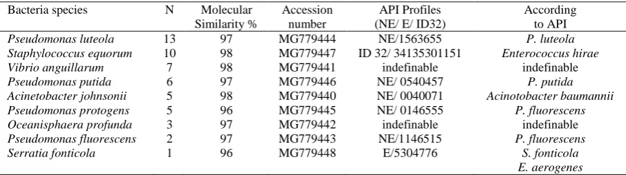
Different bacterial species were isolated during the screening of 180 whiting for six months. There were no significant clinical signs of the collected fish. A total of 52 bacterial isolates (9 species) were identified to species level (Table 1). Two bacteria species were only identified by molecularly (sequencing). These bacteria species are Vibrio
Table 1. Bacteria isolated from whiting and their molecular and biochemical identification rates
Bacteria species N Molecular Similarity %
Accession number
API Profiles (NE/ E/ ID32)
According to API
Pseudomonas luteola 13 97 MG779444 NE/1563655 P. luteola
Staphylococcus equorum 10 98 MG779447 ID 32/ 34135301151 Enterococcus hirae
Vibrio anguillarum 7 98 MG779441 indefinable indefinable
Pseudomonas putida 6 97 MG779446 NE/ 0540457 P. putida
Acinetobacter johnsonii 5 98 MG779440 NE/ 0040071 Acinotobacter baumannii
Pseudomonas protogens 5 96 MG779445 NE/ 0146555 P. fluorescens
Oceanisphaera profunda 3 97 MG779442 indefinable indefinable
Pseudomonas fluorescens 2 97 MG779443 NE/1146515 P. fluorescens
Serratia fonticola 1 96 MG779448 E/5304776 S. fonticola
E. aerogenes
anguillarumand Oceanisphaera profunda.The other bacteria isolated from the fish were identified by both biochemically (API) and molecularly. These bacteria species are Pseudomonas luteola, Staphylococcus equorum, Pseudomonas putida, Acinetobacter johnsonii, Pseudomonas protogens, Pseudomonas fluorescensand Serratia fonticola. Pseudomonas luteolawas noted as the most common bacterial isolates in whiting. The identification of all strains was also confirmed by DNA sequencing of their 16S rRNA genes. 9 bacterial isolates have the expected 1500-bp PCR amplification product were shown Figure 1.
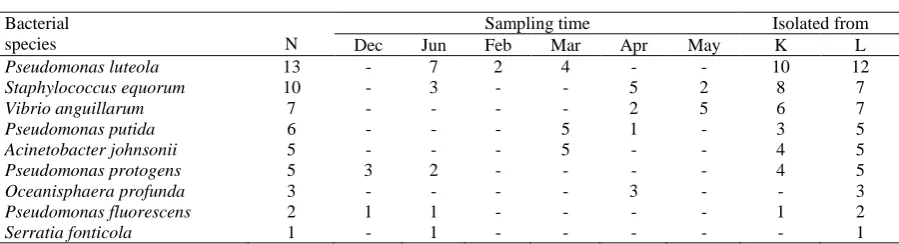
The 16S rRNA gene sequences ofP. luteola (GenBank accession number MG779444), S. equorum (GenBank accession number MG779447), V. anguillarum (GenBank accession number MG779441), P. putida (GenBank accession number MG779446), A. johnsonii (GenBank accession number MG779440), P. protogens (GenBank accession number MG779445), O. profunda (GenBank accession number MG779442), P. fluorescens (GenBank accession number MG779443), and S. fonticola (GenBank accession number MG779448) strains have been deposited in GenBank databases (Table 1). 16S rRNA gene sequences of P. luteola, S. equorum, V. anguillarum, P. putida, A. johnsonii, P. protogens, O. profunda, P. fluorescens, and S. fonticola strains were demonstrated to have >96% similarity with reference strains (Accession numbers: NZ JRMB01000004.1, NZ CP013114.1, NC 015633.1, NC 002947.4, NZ CP010350.1, NC 021237.1, NZ CP021377.1, NC 016830.1, NZ CP011254.1) from Genbank. Also, the number of isolated bacteria at different sampling times and sampling organs were shown in Table 2.
Discussion
The current study is the first report on the
bacterial fauna of the whiting captured from the Eastern Black Sea coast of Turkey. This study provides new information about its bacterial fauna and determining the compatibility of this area for mariculture.
In this study, Pseudomonas luteolafound to be the most prevalent bacteria in whiting, followed by Staphylococcus equorum, Vibrio anguillarum,andPseudomonas putida. Pseudomoniasisis the main disease caused by Pseudomonas species in fish. It is found in microbial flora of both freshwater and saltwater fish. These bacteria are generally considered as a possible opportunistic pathogen and may lead to secondary infections. Species of Pseudomonas causes numerous diseases including hemorrhagic septicemia, ulcerative disease, red spot disease and gill diseases (Austin & Austin, 2012). P.putida, P. luteola, P. aeruginosa, P. fluorescens, P. chlororaphisand P. anguillicepticafound to be the pathogenic species of Pseudomonas genusin Turkey (Ozturk & Altinok, 2014). In the present study, determination of a high number of strains belonging to Pseudomonas genus may indicate a considerable risk for aquaculture activities.
Vibriosis is a term for a group of well-known fish diseases and has a wide distribution and host range all over the world. Vibriosis, red-pest, and salt-water furunculosis are diseases caused by Vibrio anguillarum(Listonella anguillarum). The bacteria are Gram-negative, motile, rod-shaped and grow between at 5-40°C. They may behaveas opportunistic agents or be primer pathogens (Austin & Austin, 2012). V. anguillarum was first isolated from sea bream (Sparus aurata) in Turkey (Candan, 1991). It has subsequently been reported in many fish species including sea bass (Dicentrarchus labrax), salmonid spp. (Salmo salar andOncorhynchus mykiss) and red porgy (Pagrus pagrus) in Turkey due to the uncontrolled fish transfer (Ozturk & Altinok, 2014).In this study,
Vibrio anguillarumstrains were isolated from seven whiting especially in spring samplings. Consequently, this bacterial agent is still posing a risk for aquaculture facilities in Eastern Black Sea.
Acinetobacter johnsoniiis a fish pathogenic bacteria belonging to Acinetobacter genus which are widely distributed in the environment. This bacteria from this genus are Gram-negative, non-motile, rods. The bacteria are precisely aerobic and grow at a temperature from 20ºC to 37ºC. These bacteria are also considered as a possible opportunistic pathogen (Kozinska, Pazdzior, Pekala& Niemcezuk, 2014). Oceanisphaera profundais a bacteria isolated from marine bottom samples. The bacteria is Gram-negative, aerobic, halophilic and grow between at 4-42°C (Romanenko et al. 2003). Staphylococcus equorum has frequently been isolated from fermented food products especially cheeses and sausages. They may be as exceptional opportunistic pathogens in some clinical situations. Moreover, they have emerged as causative agents of nosocomial infection (Irlinger et al. 2012). According to the available literature, Oceanisphaera profunda and Staphylococcus equorumhave not been isolated from any fish species. In this study, Staphylococcus equorum, Acinetobacter johnsoniiand Oceanisphaera profundastrains were isolated from ten, five and three fish samples respectively.
In the present study, all bacterial isolates were tried to identified by both API and 16S rRNA gene sequencing methods. However, there is a poor relationship between the phenotypic and genotypic identification methods. According to both identification methods, bacteria of P. luteola, P. putida, P. fluorescensand S. fonticola were identified correctly. It is believed that DNA sequencing method was more discriminative than the other methods.
In conclusion, the current study was to identify bacteria isolated from whiting which found in the mentioned area. In terms of bacterial fish pathogens, current case of this area has been revealed by the present study. Before performing aquaculture activities, the presence of these pathogens should be taken into consideration. Bacteria belonging to Pseudomonas genus were noted as the most common
bacteria. To our knowledge, in fish, thepresence of Staphylococcus equorum and Oceanisphaera profundais the first report for Turkey.
Acknowledgement
This study was funded by Central Fisheries Research Institute. Samples were collected while conducting field studies for a project named “Investigation of Gillnet and Effect in Black Sea Fisheries" TAGEM /HAYSUD/2015/A11/P-09/02.
References
Austin, B., & Austin, D.A. (2012). Bacterial fish pathogens; diseases of farmed and wild fish. Springer, New York, London.
Candan, A. (1991). Çipura (Sparus aurata l. 1758) yetiştiriciliğinde mevsimsel olarak görülen hastalık etkenlerinin tespit ve tedavi yönteminin geliştirilmesi. PhD Thesis. İstanbul: İstanbul Üniversity.
Capkin, E., Terzi, E., & Altinok, I. (2015). Occurrence of antibiotic resistance genes in culturable bacteria isolated from Turkish trout farms and their local aquatic environment, Dis Aquat Org 114: 127-137. https://dx.doi.org/ 10.3354/dao02852.
Irlinger, F., Loux, V., Bento, P., Gibrat, J.F., Straub, C., Bonnarme, P., Landaud, S., & Christophe Monnet, C. (2012). Genome Sequence of Staphylococcus equorumsubsp. equorumMu2, Isolated from a French Smear-Ripened Cheese, Journal of Bacteriology, 194 (18) 5141-5142.
https://dx.doi.org/10.1128/JB.01038-12 J.
Kasapoglu, N., & Duzgunes, E. (2017). The Common Problem in the Black Sea Fisheries: By-catch and Its Effects on the Fisheries Economy. Turkish Journal of Fisheries and Aquatic Sciences, 17: 387-394. https://dx.doi.org/ 10.4194/1303-2712-v17_2_18 Korun, J., & Timur, G. (2005). The Fırst Pasteurellosis Case
in Cultured Sea Bass (Dıcentrarchus Labrax L.) at Low Marine Water Temperatures in Turkey. The Israeli Journal of Aquaculture – Bamidgeh 57(3), 197-206.http://hdl.handle.net/10524/19146
Kozinska, A., Pazdzior, E., Pekala, A., & Niemczuk, W. (2014). Acinetobacter johnsonii and Acinetobacter lwoffii-the emerging fish pathogens, Bull Vet Inst Pulawy 58: 193-119.
https://dx.doi.org/10.2478/bvip-2014-0029.
Table 2. Number of isolated bacteria at different sampling times and sampling organs
Bacterial
species N
Sampling time Isolated from
Dec Jun Feb Mar Apr May K L
Pseudomonas luteola 13 - 7 2 4 - - 10 12
Staphylococcus equorum 10 - 3 - - 5 2 8 7
Vibrio anguillarum 7 - - - - 2 5 6 7
Pseudomonas putida 6 - - - 5 1 - 3 5
Acinetobacter johnsonii 5 - - - 5 - - 4 5
Pseudomonas protogens 5 3 2 - - - - 4 5
Oceanisphaera profunda 3 - - - - 3 - - 3
Pseudomonas fluorescens 2 1 1 - - - - 1 2
Serratia fonticola 1 - 1 - - - 1
Kusuda, R., & Kawai, K. (1998). Bacterial diseases of cultured marine fish in Japan. Fish Pathol 33: 221−227 http://dx.doi.org/10.3147/jsfp.33.221 Nishizawa, T., Savas, H., Isidan, H., Ustundag, C.,
Iwamoto, H., & Yoshimizu, M. (2006). Genotyping and pathogenicity of viral hemorrhagic septicemia virus from free-living turbot (Psetta maxima) in a Turkish coastal area of the Black Sea. Applied and Environmental Microbiology, 72 (4): 2373-2378. https://dx.doi.org/10.1128/aem.72.4.2373-2378.2006 Olivier, G. (2012). Disease interactions between wild and
cultured fish- Perspectives from the American Onuk, E.E., Tanrıverdi, C.Y., Coban, A.Y., Çiftci, A.,
Didinen, B.I., Altun, S., Sogut, M., & Deveci, A. (2015). Detection of the first QnrS gene positivity in aquatic Aeromonas spp. isolates in Turkey.
Mikrobiyol Bul. 49 (1): 114-123. https://dx.doi.org/10.5578/MB.8839
Ozturk, R.C., & Altinok, I. (2014). Bacterial and Viral Fish Diseases in Turkey, Turkish Journal of Fisheries and Aquatic Sciences 14: 275-229. https://dx.doi.org/ 10.4194/1303-2712-v14_1_30.
Romanenko, L.A., Schumann, P., Zhukova, N.V., Rohde,
M., Mikhailov, V.V., & Stackebrandt, E. (2003).
Oceanisphaera litoralis gen. nov., sp. nov., a novel halophilic bacterium from marine bottom sediments.
International Journal of Systematic and Evolutionary Microbiology, 53: 1885–1888.
https://dx.doi.org/10.1099/ijs.0.02774-0.
Toranzo, A.E., Magarinos, B., & Romalde, J.L. (2005). A review of the main bacterial fish diseases in mariculture systems, Aquaculture, 246, 37–61. https://dx.doi.org/10.1016/j.aquaculture.2005.01.002 TUIK, (2015). Turkish Statistical Institute,
http://www.turkstat. gov.tr/Start.do, Access date: 27.07.2017.
Ture, M., & Alp, H. (2016). Identification of Bacterial Pathogens and Determination of Their Antibacterial Resistance Profiles in Some Cultured Fish in Turkey.
J VET RES., 60, 141-146.
http://dx.doi.org/10.1515/jvetres-2016-0020.
Weisburg, W.G., Barns, S.M., Pelletier, D.A., & Lane, D.J. (1991). 16S ribosomal DNA amplification for phylogenetic study. Journal of Bacteriology, 173 (2) 697-703.