J
ACEKK
URCZ1, E
WAN
IENARTOWICZ1, J
OANNAS
ŁONINA1, J
ERZYG
ARCAREK1,
K
RZYSZTOFM
OROŃ1The Usefulness of CT−angiography in Detecting
Anatomical Variants of Arteries Arising from
the Abdominal Aorta and Aortic Arch
Przydatność angiografii TK w wykrywaniu odmian anatomicznych
tętnic odchodzących od aorty brzusznej i łuku aorty
1Department of Radiology, Silesian Piasts University of Medicine in Wrocław, Poland
Adv Clin Exp Med 2007, 16, 6, 751–760 ISSN 1230−025X
ORIGINAL PAPERS
© Copyright by Silesian Piasts University of Medicine in Wrocław
Abstract
Background.The evaluation of arteries originating from the abdominal aorta and aortic arch using CT−angiogra− phy plays a major role in the diagnostics and treatment of many conditions. The method is particularly meaning− ful in diagnostics before potential stentgraft implantation or angiosurgery, follow−up evaluation of patients after endovascular or angiosurgical procedures, management before hepatic transplantation and liver resection, patients with renal hypertension caused by renal artery stenosis, and patients with intermittent claudication or postprandial abdominal angina.
Objectives.The goals of the study were to assess the prevalence of anatomical variants of arteries arising from the abdominal aorta and aortic arch and their clinical aspects and to evaluate the diagnostic potential and available techniques of modern CT−angiography in visualizing the selected arterial system.
Material and Methods.CT−angiographies were performed in 240 consecutive patients enrolled between January 2004 and May 2007. The examinations were carried out using a 10−row CT unit with 3−mm slice thickness (pri− mary reconstruction by means of thin slices with slice thickness of 1 mm). Contrast medium was injected with flow rate 4 ml/sec in a volume of 2 ml/kg patient body weight. Initially raw axial images of CT−angiographies were eval− uated and then the following secondary two−dimensional and three−dimensional reconstructions were employed: coronal and sagittal reformations, multiplanar reformation, curved planar reformation, maximum intensity projec− tion, and the volume rendering technique.
Results.A common prevalence of anatomical variants of arteries arising from the abdominal aorta was demon− strated and their frequencies were estimated. The analyzed material revealed the following kinds of anomalies: renal arteries: early division of the renal artery and the presence of unilateral or bilateral additional renal arteries; hepatic arteries: different variants of hepatic arterial supply; celiac trunk: different anomalies of the origins from the abdominal aorta; aortic arch: rare presence of anatomical variants of large arteries arising from the aortic arch. Conclusions.CT−angiography enables rapid and accurate imaging of the abdominal arteries and the arteries com− mencing at the aortic arch, including detection of vascular anatomical variants, which is of great value before planned surgery and interventional radiological procedures as well as in the diagnostics of internal conditions. Various techniques of reconstruction are helpful in the visualization of arterial branches, which makes evaluation of the abdominal arterial system easier (Adv Clin Exp Med 2007, 16, 6, 751–760).
Key words:diagnostic imaging, spiral computed tomography, angiography, aorta.
Streszczenie
Wprowadzenie. Ocena tętnic odchodzących od aorty brzusznej i łuku aorty za pomocą angiografii TK odgrywa dużą rolę w diagnostyce wielu schorzeń. Omawiana technika obrazowania ma szczególne znaczenie w kwalifikacji do leczenia wewnątrznaczyniowego lub angiochirurgicznego oraz kontroli po przeprowadzonym leczeniu; u pac− jentów przed przeszczepem lub resekcją wątroby; u chorych z podejrzeniem zwężenia tętnicy nerkowej; u pacjen− tów z objawami chromania przestankowego i/lub poposiłkowej anginy brzusznej.
The recently noticeable rapid technical devel− opment of diagnostic imaging methods has result− ed in increased accuracy of vessel visualization by means of superb spatial, contrast, and temporal resolution. Therefore, the imaging of not only large lumen vessels, but also of even thinner ves− sels has become possible and the presence of vari− ous, formerly undetectable, arterial anatomical variants (i.e. diverse arterial renal or liver supply) can easily be visualized. Modern diagnostic modalities play a significant role in the diagnosis of different arterial−associated internal conditions (such as arterial hypertension of renal origin or postprandial abdominal angina) for an optimal patient assessment before planned surgery, when it is extremely important to evaluate the feasibility of the intervention as well as to facilitate and shorten the duration of the interventional proce− dure. Modern computed−assisted imaging tech− niques allow achieving the visualization of arterial vessels in a very short time without the necessity of invasive and complication−related conventional arteriography, which can therefore be performed almost exclusively during therapeutic intravascu− lar interventions, for instance during percutaneous transluminal angioplasty (PTA) or stent implanta− tion. Additionally, in contrast to traditional inva− sive arteriography, modern methods such as com− puted tomographic angiography (angio−CT) or magnetic resonance angiography (angio−MR) enable the use of reprocessing techniques aimed at obtaining diverse two−dimensional (2D) or three− dimensional (3D) vessel reconstructions which make the evaluation of vessels more accurate and easier to interpret. In particular, the introduction of spiral (helical) multirow (multidetector, multi− slice) computed tomography (SMCT) has con− tributed to the considerable progress in the evalu−
ation of the arterial system. The short duration of such an examination in combination with the pos− sibility of rapid vessel evaluation result in quick diagnosis, which is of great importance in the case of emergent patients who require prompt surgical or intravascular intervention. Particularly angio− CT is a short examination and is quite widely available in Poland. Angio−CT can also be per− formed as an elective screening or follow−up pro− cedure (i.e. in patients with ultrasound−detected small aortic aneurysms or as surveillance after stentgraft implantation). Non−ionic contrast media of low osmotic pressure are at present commer− cially available and their common use is indicated as they are less toxic and cause adverse reactions much more seldom than ionic hyper−osmotic−pres− sure contrast agents. Therefore, angio−CT may be performed in most of the population, even in patients with advanced age or coexisting diseases. In case of absolute contraindications (i.e. allergy to iodine), gadolinium−based contrast medium may be used. The average population age has been increasing in developed countries, which is why the number of artery−related diseases and organ transplantations is still going to increase. Therefore, a tool for prompt and reliable imaging diagnosis is required.
Material and Methods
Two hundred forty consecutive patients (184 males and 56 females, median age: 67 years) in whom CT−angiography of the thoracic and/or abdominal aorta was performed for various reasons between January 2004 and May 2007 were includ− ed in this retrospective study. The patients were referred for elective angio−CT or under emergency
Drugim celem przeprowadzonego badania była ocena możliwości diagnostycznych nowoczesnej angiografii TK w obrazowaniu wybranych tętnic.
Materiał i metody. Ocenie poddano 240 kolejnych chorych, u których w okresie między 01.2004 a 05.2007 z róż− nych przyczyn wykonano angio−TK aorty brzusznej i/lub piersiowej. Badania wykonano spiralnym aparatem 10−rzędowym (grubość warstw 3 mm, rekonstrukcja cienkimi warstwami grubości jednego milimetra). Środek kon− trastowy podawano z prędkością 4 cm3/s w ilości 2 cm3/kg masy ciała. Wyjściowo badania oceniano w surowych
obrazach poprzecznych, następnie za pomocą uzyskanych wtórnie technik dwu− oraz trójwymiarowych: rekonstruk− cji wielopłaszczyznowej, rekonstrukcji po krzywej, projekcji maksymalnej intensywności oraz rekonstrukcji objęto− ściowej.
Wyniki. W wykonanych badaniach wykazano dużą częstość występowania wariantów anatomicznych tętnic odchodzących od aorty brzusznej oraz małą w przypadku tętnic odchodzących od łuku aorty. W analizowanym materiale autorzy stwierdzili częste występowanie odmian anatomicznych w obrębie tętnic nerkowych, tętnic zaopatrujących wątrobę oraz pnia trzewnego. Określono częstość występowania poszczególnych wariantów tętnic odchodzących od aorty brzusznej i łuku aorty.
Wnioski. Badanie angio−TK pozwala na szybkie i dokładne określenie przebiegu naczyń tętniczych, w tym na wyodrębnienie naczyniowych wariantów anatomicznych, co ma ogromne znaczenie przed planowanym zabiegiem chirurgicznym, wewnątrznaczyniowym, a także w diagnostyce chorób internistycznych. Dostępne techniki rekon− strukcyjne są bardzo przydatne w wizualizacji naczyń tętniczych jamy brzusznej oraz naczyń odchodzących od łuku aorty i znacznie ułatwiają ich ocenę (Adv Clin Exp Med 2007, 16, 6, 751–760).
conditions with different initial diagnoses, includ− ing suspicion of the presence or evaluation of pro− gression of a thoracic/abdominal aortic aneurysm or aortic dissection [1] as well as for assessment of the morphology of aneurysmal and arterial anatom− ical conditions before potential qualification for stentgraft implantation [2], vascular prosthesis surgery (i.e. Y−prosthesis), or a peripheral arterial by−pass procedure. CT−angiography was also car− ried out to diagnose the stenoses of arteries origi− nating from the aorta, i.e. to detect steal subclavian syndrome (SSS) caused by obstruction of the prox− imal portion of (usually the left) subclavian artery, to confirm renal hypertension caused by renal artery stenosis [3], and to diagnose patients with clinical symptoms of intermittent claudication, postprandial abdominal angina, Lerich’s syn− drome, Takayasu disease [4–6], and potential com− plications after intravascular minimally invasive or angiosurgical procedures [7–9].
In total, 216 abdominal and 72 thorax CT−angiographies were evaluated. The examina− tions were conducted using a 10−row CT unit with a 3−mm slice thickness reconstructed with thin sections (every millimeter). Contrast medium (Iomeron 350, iodine concentration: 350 mg/ml) was injected into the peripheral vein of the upper limb by means of a power injector with a flow rate of 4 ml/sec in a volume dependant on the patient’s body weight (2 ml/kg). The scan delay was set automatically using bolus triggering with the region of interest (ROI) placed in the proximal part of the ascending aorta (angio−CT of the tho− racic aorta) or in the distal part of the descending thoracic aorta (CT−angiography of the abdominal aorta). The scanning was initiated as soon as the contrast enhancement in the ROI exceeded 160 Hounsfield units (HU). The raw axial images were initially evaluated to get a first look, but also to confirm the optimal quality of the study (i.e. to exclude the presence of substantial artifacts). During assessment, the cine−mode was often used to obtain a “dynamic” image. Subsequently, vari− ous reconstruction techniques were employed, including not only two−dimensional, i.e. coronal and sagittal reformations, multiplanar reformatting (MPR), and curved planar reformatting (CPR), but also three−dimensional, i.e. maximum intensity projection (MIP) and volume rendering (VR). Thus all the available technical means were used to perform an optimal evaluation of the CT−angio− graphic examinations. Each examination was assessed independently by two radiologists and the results were compared (and analyzed once more in case of any divergences) so as to minimize the risk of misinterpretation.
Results
The common prevalence of anatomical vari− ants of arteries commencing at the aorta was demonstrated, especially in arteries originating from the proximal portion of the abdominal aorta. The anatomical variants of large−lumen arteries arising from the aortic arch, although also present, were identified much more seldom. With reference to the abdominal aorta, the anomalies of the fol− lowing arteries were shown: renal arteries, with unilateral or bilateral additional arteries or early division of renal artery; hepatic arteries, with var− ious variants of hepatic arterial supply; and the celiac trunk, with different anomalies of the ori− gins from the abdominal aorta.
Renal Arteries
The major renal arteries arose from the abdominal aorta almost always (212/216, 98.1%) at the level of the lower border of the body of Th12 or at the intervertebral space between the 1st and
2ndlumbar vertebrae (only in 4 patients at the level
of L2), in 97.2% (210/216) of cases 1–2 cm distal− ly to the commencement of the superior mesen− teric artery (SMA), and in 6 cases just at the level of the SMA. The major right renal artery (RA) usually originated (156/216, 72.2%) 1–2 cm prox− imal to the origin of the major left RA, in 19.4% (42/216) of the patients both arteries arose at the same level and in 8.3% (18 cases) the origin of the left RA was localized higher than that of the right. The main and additional RAs had their origin in 98% of cases at the lateral aspect of the abdominal aorta, the right RA commencing more anteriorly and the left RA more posteriorly.
arteries occurred on each side with the same fre− quency. Bilateral duplication of the RA was observed 7/216 patients (3.2%) (Figs. 4 and 5).
The site of origin of additional arteries from the abdominal aorta was highly differentiated; according to this feature, one can divide the origin of the additional RA into four groups: 1) proximal
to the major RA (18/78 duplications, 23% of cases) supplying the upper pole of the kidney or taking its course into the renal hilum), 2) close to the origin of the major RA (the most commonly observed variant, with 32/78 duplications, 41% of cases), the additional artery usually accompanying the major RA, 3) distal to the major RA (26/78 duplications, 33.3% of cases, the second most commonly variant; in the analyzed cases the addi− tional RA always passed anteriorly to the inferior vena cava (IVC) supplying the lower pole of kid− ney), and 4) which was very rare, arising from the common iliac artery (CIA) and, in these cases, always supplying the lower pole of the kidney (2/78, 2.6%).
The renal region of supply of the additional arteries was also very diverse and in 41/78 cases (52.6% of patients with RA duplication) the sec− ond smaller artery accompanied the major RA into the renal hilum; in rest of the cases (37/78, 47.4%) they supplied blood to either the upper (16 cases) or, more often, the lower (21 cases) pole of the kidney.
Unilateral triplication of the RA was shown episodically (3/216 patients, 1.4% of cases). In these patients the presence of one dominant and two small additional vessels was observed. In one
Fig. 1. Early division of the right renal artery with two arterial branches accompanying the renal vein into the renal hilum
Ryc. 1. Wczesny podział prawej tętnicy nerkowej, której gałęzie biegną wzdłuż odpowiedniej żyły do wnęki nerki
Fig. 2. Small−lumen additional left renal artery in a patient with diffuse calcified atheromatic changes in the arterial walls
Ryc. 2. Drobna dodatkowa lewa tętnica nerkowa u pacjenta z rozsianymi zwapniałymi zmianami miażdżycowymi w ścianach tętnic
Fig. 3. Large additional left renal artery arising from the wall of an aneurysm in the lower segment of the abdominal aorta; risk of significant left renal ischemia in case of abdominal stentgraft implantation
patient, unilateral RA triplication in combination with contralateral RA duplication was visualized.
Hepatic Arteries
The analyzed material allowed the detection of considerable diversity of the hepatic arterial supply. The typical arterial supply exclusively by the prop− er hepatic artery (PHA) was demonstrated in 60.2% of cases (130/216) (Fig. 6). The most prevalent vari− ant of hepatic arterial supply (19.4% of cases, 42/216) was the left hepatic artery (LHA), which did not arise from the PHA but was a branch of the left gastric artery (LGA; 32/42, 76.2% of cases) or, depending on the caliber of the LGA, the LHA divided into the LGA on its way to the left lobe of the liver (10/42, 23.8% of cases). The second most common vascular anomaly, observed in 13% of the patients (28/216), was the right hepatic artery (RHA) originating not from the PHA, but from the proximal part of the SMA. In 2.3% of the patients (5/216) the LHA arose from the more proximal part of the common hepatic artery (CHA), in three cases this variant being accompanied by another vascular anomaly in the form of the RHA originating from the proximal portion of the SMA (Fig. 7).
In 5.5% of the analyzed patients (12/216), a triple arterial hepatic supply was demonstrated
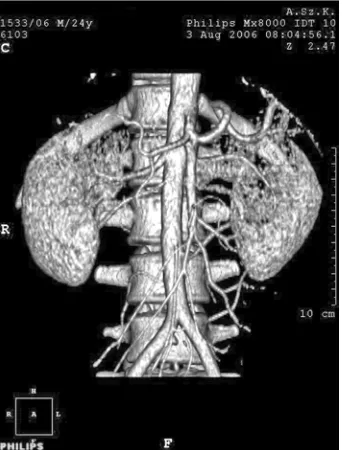
Fig. 4. Maximum intensity projection image of bilater− al duplication of renal arteries with the right additional artery originating close to the major right renal artery and the left renal artery arising much more distally, at the level of the inferior mesenteric artery
Ryc. 4. Obustronne zdwojenie tętnic nerkowych, przy czym prawa dodatkowa tętnica nerkowa odchodzi od aorty nieco poniżej głównej prawej tętnicy nerkowej, a lewa dodatkowa tętnica nerkowa znacznie bardziej dystalnie – na poziomie odejścia tętnicy krezkowej dolnej. Obraz w projekcji maksymalnej intensywności
Fig. 5. The same patient as in Fig. 4, but this image was obtained using the volume rendering reconstruc− tion technique. Additional renal arteries in the patient are excellently visible
Ryc. 5. Ten sam przypadek co na ryc. 4, ale obraz wy− konano w technice rekonstrukcji objętościowej (Volu− me Rendering). Zarówno główne, jak i dodatkowe tęt− nice nerkowe są doskonale widoczne
Fig. 6. The typical arterial supply of the liver with the common hepatic artery dividing into the gastroduo− denal artery and the proper hepatic artery, with the lat− ter then dividing into the left and right hepatic arteries
with the following origins: the RHA from the SMA, the middle hepatic artery (MHA), which was the prolongation of the PHA, and the LHA from the LGA (Fig. 8). In another 12 patients (5.5%), a double arterial supply was demonstrated with the RHA arising from the SMA and the LHA directly from celiac trunk; in these cases the PHA was absent and the CHA prolonged into the gas− troduodenal artery (GDA). No cases of the LHA originating from the splenic artery, left gastroepi− ploic artery, or abdominal aorta, described in liter− ature, were observed. The length of the CHA dif− fered (average: 3.5 cm) and, due to this parameter, one can distinguish between early, normal, and late division of the CHA into the PHA and the GDA (short, medium, and long CHA). The length of the PHA also showed considerable variety (average: 2.5 cm), with early, normal, and late division into the left and right branches of the HA (short, medium, and long PHA).
Celiac Trunk
With reference to the celiac trunk (celiac artery), it was usually short (approximately 3 cm long) and of large lumen [10]; however, longer variants (reaching even 5 cm in length) were observed in 11.1% (24/216) of patients. The celiac trunk always had its origin at the anterior or ante− rio−left aspect of the abdominal aorta and nearly
always at the level of the intervertebral space between the 12ththoracic and 1stlumbar vertebrae
(only in 2 cases was its origin located half a verte− bral body lower). In most cases the typical tri−divi− sion of the celiac trunk was noted, but in about 30% of cases the LGA arose more proximally from the main stem of the celiac trunk. The most commonly observed anomaly of the celiac trunk was the separate origin of the LGA: from the ante− rior wall of the abdominal aorta close and usually a bit proximal to the origin of the celiac trunk, with the presence of a hepato−splenic trunk (20/216, 9.3%) or from a proximal part of the CHA (1.9%, 4/216). In 3.7% of the patients (8/216) the only source of hepatic arterial blood supply was the SMA; in these cases the presence of a gastro− splenic trunk was demonstrated. Only one case, described in the literature, of a separate origin of the splenic artery with the formation of a gastro− hepatic trunk was demonstrated. Also, in one case all three arteries originated separately from the anterior wall of the abdominal aorta (Fig. 9). No case of a common celiac−mesenteric trunk was found in the present study.
Fig. 7. Liver supplied by two sources: the right hepat− ic artery (RHA) originates from the superior mesen− teric artery (SMA), whereas the left hepatic artery arises from the common hepatic artery (LHA) and runs along the course of the proper hepatic artery (CHA); CT division – division of the celiac trunk
Ryc. 7. Zaopatrzenie tętnicze wątroby z dwóch źródeł – prawa tętnica wątrobowa (RHA) odchodzi z tętnicy krezkowej górnej (SMA), lewa tętnica wątrobowa (LHA) odchodzi z tętnicy wątrobowej wspólnej (CHA) i przebiega w lokalizacji tętnicy wątrobowej właściwej; CT division – podział pnia trzewnego
Fig. 8. A rare case of the liver supplied simultaneously by three sources: the superior mesenteric artery (AMS) divides into the right hepatic artery, the prolongation of the proper hepatic artery is the middle hepatic artery, and the left hepatic artery (LGA) arises from the left gastric artery
Superior Mesenteric Artery
The superior mesenteric artery (SMA) always had its commencement 1.0–1.5 cm distally to the celiac trunk and took its course mainly at an acute angle to the abdominal aorta. However, in patients with a large amount of retroperitoneal fatty tissue (approximately 30% of the analyzed cases), the SMA ran in its proximal course anteriorly and then curved caudally. Thus the right angle of the SMA’s origin does not necessarily indicate the presence of retroperitoneal lymphadenopathy. The origin of the SMA was always at the anterior aspect of the abdominal aorta in its midline; only in 6 cases (2.8%) was it precisely at the level of the RA, and in the rest of the cases it was proximal to the origin of the RA (about half of a lumbar verte− bra higher).
The inferior mesenteric artery (IMA) appears to be the most stable artery, the origin of which is predominantly located at the anterior or anterior− left aspect of the abdominal aorta at the level of the lower part of the 3rdlumbar vertebra about 5 cm
above the aortic bifurcation. No anatomical vari− ants of the IMA were observed in this study.
With reference to the aortic arch, one case (1/72, 1.4%) of a right−sided aortic arch with
a mirrored origin of the large arteries from the aortic arch and with a coexisting heart−defect (confirmed subsequently in cardiac ultrasound) was detected (Figs. 10 and 11). Three cases (3/72, 4.2%) of a common origin of the brachiocephalic trunk (innominate artery) and the left common carotid artery (CCA) were observed (Fig. 12) as well as 2 cases (2.8%) of a left CCA arising from the proximal part of the brachiocephalic trunk. The origin of the left vertebral artery directly from the aortic arch between the origin of the left CCA and the left subclavian artery was observed in 1 case (1/72, 1.4%). What is of particular interest was one case of arteria lusoria [11, 12] (the right subclavian artery arising from the aortic arch as its last branch and directing on to the right) that was demonstrated with a retroesophageal location of the vessel, without, however, the dysphagia so typical of this anomaly. Generally, 5 cases of an aortic arch with two large originating arteries were observed (5/72, 6.9%) and in 2 patients, four large vessels commencing at the aortic arch were demonstrated.
Discussion
Knowledge of the anatomical variants of the arterial vessels arising from the abdominal aorta and the aortic arch facilitates and, in some cases, is
Fig. 9. All three arteries that typically originate from celiac trunk are direct branches of the abdominal aorta. No celiac trunk was identified in this study.
GDA – gastroduodenal artery
Ryc. 9. Wszystkie trzy tętnice odchodzące typowo od pnia trzewnego, w tym przypadku odchodzą bezpośred− nio z przedniej ściany aorty brzusznej. Pnia trzewnego nie uwidoczniono w badaniu.
GDA – tętnica żołądkowo−dwunastnicza
Fig. 10. Right−sided aortic arch in multiplanar recon− struction (MPR). The proximal part of the descending aorta is visible on the right side of the vertebral col− umn, on the right of the esophagus
essential to undertake optimal surgical [13], angio− surgical [14], or minimally invasive intravascular intervention. Understanding the anatomical loca− tions of liver−supplying arteries is extremely helpful in evaluating liver transplant donors [15, 16] and recipients [17], in performing lobar or segmental resection of the liver [18], in performing palliative or preoperative embolization of liver neoplasms, in super−selective arterial chemotherapy of hepatic malignant lesions [19], and in the minimally inva− sive treatment of hemobilia [20] and bilhemia.
Knowledge about the number and anatomical locations of renal arteries is helpful not only in the treatment of renal hypertension caused by athero− matic stenoses or fibromuscular dysplasia of the renal arteries [21, 22], but also in the assessment of living kidney donors [23–26] and in planning endovascular treatment of abdominal aortic aneurysms [27, 28], where the detection of an additional renal artery supplying a significant part of the renal parenchyma is a contraindication for performing an endovascular procedure due to the risk of renal parenchymal necrosis and subsequent renal insufficiency. As in the case of liver malig− nancies, renal malignancies can also be treated by means of local super−selective chemotherapy, which allows a reduction or even elimination of the adverse effects of cytostatic agents. The accu− racy of local chemotherapy depends to a large extent on knowledge of the arterial supply of the organ. Arteriovenous fistulas that can be a conse− quence of iatrogenic intervention (i.e. renal biop− sy) or trauma can be effectively treated on the basis of knowledge of renal arterial localization.In conclusion, knowledge about the presence of an arterial anatomical anomaly significantly facili− tates or even makes feasible the performance of diagnostic and, most of all, therapeutic procedures on arteries arising from the aorta, which enables increasing the rate of successful interventions and shortens the duration of the procedure, including shortening the patient’s and staff’s exposure to radiation.
In some centers, CT−angiography has replaced the traditional calibration angiography and is used as the only measurement tool in the evaluation of aortic aneurysms before potential qualification for thoracic or abdominal stentgraft implantation. CT−angiography is superior to traditional angiog− raphy as it is noninvasive and enables the perfor− mance of multiple two− and three−dimensional reconstructions, thanks to which the measure− ments are more accurate. Additionally, CT−angiog− raphy shows not only the channel of flow, but also the thrombus in the aneurysm (and, consequently, the real size of the aneurysm) and allows accurate measurements of the aneurysmal neck and com−
Fig. 11. The same case seen in the volume rendering reconstruction. The brachiocephalic trunk originates as the first branch of the aortic arch from its anterior wall, takes its course on to the left, and divides into the left common carotid artery and the left subclavian artery. The right common carotid artery originates as the second branch and the right subclavian artery as the most distally commencing branch
Ryc. 11. Ten sam przypadek w rekonstrukcji objętościo− wej (Volume Rendering). Pień ramienno−głowowy od− chodzi jako pierwszy z przedniej ściany łuku aorty i przechodzi na stronę lewą, gdzie dzieli się na lewą tęt− nicę szyjną wspólną i lewą tętnicę podobojczykową. Ja− ko druga z łuku odchodzi prawa tętnica szyjna wspólna, najbardziej dystalnie zaś prawa tętnica podobojczykowa
Fig. 12. The most common variant of arteries originat− ing from the aortic arch: the left common carotid artery (LCCA) arises not directly from the aortic arch, but from the proximal part of the brachiocephalic trunk; truncus b–c – brachiocephalic trunk
mon iliac arteries, which is critical in preventing migration of the stentgraft. The next advantage of CT−angiography is the possibility of performing noninvasive follow−up examinations in patients who have had intravascular procedures.
CT−angiography, if properly carried out, allows prompt and precise visualization of the arterial localization and the arterial supply of the various organs and, regardless of the equipment’s manu− facturer, offers a broad scope of reconstruction techniques that are definitely useful in visualizing arterial vessels and constitute significant facilita− tion in the assessment of the examination.
In this study, each available reconstruction technique (except virtual angioscopy) was used; however, it must be stressed that the starting point in assessing each examination should be the eval− uation of the raw axial images followed by thick axial sections and coronal, sagittal, as well as oblique reformations. Afterwards one should use other two−dimensional reconstruction methods, i.e. multiplanar reformatting (MPR) and, in partic− ular, curved planar reformatting (CPR), a method that is extremely useful in vessel assessment (the course of the artery, wall changes, the vessel’s lumen, measurements before interventions). CPR is more accurate in vessel−tracking, which is why it
was the favored method of artery evaluation in this study in spite of the fact that the modality of refor− matting is more time−consuming and relatively dif− ficult to carry out. Therefore it is advantageous to possess semi−automated software that makes CPR more time−efficient. As the next step, three−dimen− sional reconstructions are performed, first the max− imum intensity projection (MIP), which is an excellent time−efficient method for tiny vessel visualization (particularly after the invention of semi−automated bone removal utilities), and then the volume rendering technique (VR), which is helpful in visualizing larger arteries. However, one must be cautious as semi−automated bone removal was occasionally accompanied by elimination of some parts of the arteries, which resulted in mis− visualization of the arterial vessels.
In conclusion, evaluation of CT−angiography should begin with raw data viewing followed by CPR, MIP, and finally VR techniques. According to the personal experience of the authors, CPR and MIP establish the most reliable tools for the eval− uation of arteries. Additionally it should not be forgotten that there is still the possibility of manip− ulating the window level and the window width during interpretation of CT−angiography, which is often of great value.
References
[1] Jeudy J, White ChS: Evaluation of acute chest pain in the emergency department: utility of multidetector com− puted tomography. Semin Ultrasound CT MR 2007 Apr, 28 (2), 109–114.
[2] Pasławski M, Krzyżanowski K, Złomaniec J: Diagnostic value of multiplanar CT reconstructions in the assess− ment of abdominal aortic aneurysms. Ann Univ Mariae Curie Sklodowska [Med] 2004, 59 (1), 91–98.
[3] Fleischmann D: Multiple detector−row CT angiography of the renal and mesenteric vessels. Eur J Radiol 2003 Mar, 45, Suppl 1, 79–87.
[4] Cocho D, Marti−Fabregas J, Llobet JM, Marti−Vilalta L: Percutaneous transluminal angioplasty of the subcla− vian artery in Takayasu disease: results of long−term follow−up. Neurologia 2005 Oct, 20 (8), 419–421.
[5] Park JH: CT angiography of Takayasu arteritis: comparison with conventional angiography. J Vasc Interv Radiol 1997 May–Jun, 8 (3), 393–400.
[6] Sada E, Kohno M, Iwamasa K, Hasegawa H, Mochizuki T, Yamauchi T, Fujita S: Clinical usefulness of mul− tiplanar reconstruction images obtained by multi−slice computed tomographic angiography for early−stage Takayasu’s arteritis. Mod Rheumatol 2004 Jul, 14 (3), 245–249.
[7] Waasdorp E, Van’t Hullenaar C, van Herwaarden J, Kelder H, van de Pavoordt E, Overtoom T, Moll F, de Vries JP: Renal Function After Endovascular Aortic Aneurysm Repair: A Single−center Experience with Transrenal Versus Infrarenal Fixation. J Endovasc Ther 2007, 14 (2), 130–137.
[8] Cotroneo AR, Iezzi R, Giancristofaro D, Santoro M, Pierro A, Spigonardo F, Storto ML: Endovascular abdominal aortic aneurysm repair and renal complications: a comparison between suprarenal and infrarenal fixa− tion of stent grafts. Radiol Med (Torino), 2007, 112 (2), 252–263.
[9] Hiramoto JS, Reilly LM, Schneider DB, Sivamurthy N, Rapp JH, Chuter TA: Long−term outcome and rein− tervention after endovascular abdominal aortic aneurysm repair using the Zenith stent graft. J Vasc Surg 2007, 45, (3), 461–465.
[10] Savastano S, Teso S, Corra S, Fantozzi O, Miotto D: Multislice CT angiography of the celiac and superior mesenteric arteries: comparison with arteriographic findings. Radiol Med (Torino) 2002, 103 (5–6), 456–463. [11] Czekajska−Chehab E, Uhlig S, Staskiewicz G, Mazur−Stazka E, Torres A, Gaweda K, Drop A: Arteria luso−
ria in patients with a normal and a right−sided aortic arch diagnosed with multi−slice computed tomography. Folia Morphol (Warsz), 2007 Feb, 66 (1), 74–77.
[13] Maeda H, Okabayashi T, Kobayashi M, Morishita S, Nishimori I, Ito S, Sugimoto T, Akimori T, Onishi S, Araki K: Usefulness of multi−detector row computed tomography for accurate preoperative assessment of pan− creatic adenocarcinoma: report of a case. West Afr J Med 2006, 25, 3, 242–245.
[14] Simoni G, Perrone R, Cittadini G Jr, De Caro G, Baiardi A, Civalleri D: Helical CT for the study of abdom− inal aortic aneurysms in patients undergoing conventional surgical repair. Eur J Vasc Endovasc Surg 1996, 12, 3, 354–358.
[15] Bogetti JD, Herts BR, Sands MJ, Carroll JF, Vogt DP, Henderson JM: Accuracy and utility of 3−dimension− al computed tomography in evaluating donors for adult living related liver transplants. Liver Transpl 2001, 7, 8, 687–692.
[16] Byun JH, Kim TK, Lee SS, Lee JK, Ha HK, Kim AY, Kim PN, Lee MG, Lee SG: Evaluation of the hepatic artery in potential donors for living donor liver transplantation by computed tomography angiography using mul− tidetector−row computed tomography: comparison of volume rendering and maximum intensity projection tech− niques. J Comput Assist Tomogr 2003, 27, 2, 125–131.
[17] Boraschi P, Donati F, Cossu MC, Gigoni R, Vignali C, Filipponi F, Bartolozzi C, Falaschi F: Multi−detector computed tomography angiography of the hepatic artery in liver transplant recipients. Acta Radiol 2005, 46, 5, 455–461.
[18] Takeshita K, Furui S, Ban S, Harasawa A, Kohtake H, Yamauchi T, Sasaki Y, Shirai T, Kikuchi Y: Three− dimensional images of hepatic tumors and hepatic vessels obtained by helical computed tomography. Nippon Igaku Hoshasen Gakkai Zasshi 1996, 56, 11, 744–746.
[19] Hildebrandt B, Pech M, Nicolaou A, Langrehr JM, Kurcz J, Bartels B, Miersch A, Felix R, Neuhaus P, Riess H, Dorken B, Ricke J: Interventionally implanted port catheter systems for hepatic arterial infusion of chemothera− py in patients with colorectal liver metastases: A phase II−study and historical comparison with the surgical approach. BMC Cancer (on−line) 2007, 7, 69.
[20] Garcarek J, Kurcz J, Jędrzejewska−Orchowska A, Cader J, Moroń K, Nienartowicz E, Błachut K, Sajewicz Z, Waszczuk E, Bladowska J, Paradowski L: Haemobilia – etiology, diagnostics and treatment. Gastroenterol Pol 2007, 14, 2, 77–82.
[21] Zeller T: Renal artery stenosis. Curr Treat Options Cardiovasc Med 2007, 9, 2, 90–98.
[22] Fraioli F, Catalano C, Bertoletti L, Danti M, Fanelli F, Napoli A, Cavacece M, Passariello R: Multidetector− row CT angiography of renal artery stenosis in 50 consecutive patients: prospective interobserver comparison with DSA. Radiol Med (Torino) 2006, 111, 3, 459–468.
[23] Ayuso JR, Openheimer F, Ayuso C, Alvarez−Vijande R, Gutierrez R, Lacy A, Alcaraz A, Nicolau C: Living donor kidney transplantation: helical CT evaluation of candidates. Actas Urol Esp 2006, 30, 2, 145–151. [24] Hänninen EL, Denecke T, Stelter L, Pech M, Podrabsky P, Pratschke J, Ricke J, Schindler R, Neuhaus P,
Felix R, Tullius SG: Preoperative evaluation of living kidney donors using multirow detector computed tomog− raphy: comparison with digital subtraction angiography and intraoperative findings. Transpl Int 2005, 18, 10, 1134–1141.
[25] Laugharne M, Haslam E, Archer L, Jones L, Mitchell D, Loveday E, Lear P, Thornton M: Multidetector CT angiography in live donor renal transplantation: experience from 156 consecutive cases at a single centre. Transpl Int 2007, 20, 2, 156–166.
[26] Mishra A, Ehtuish EF: The pattern of renal vessels in live related potential donors pool. A multislice computed tomography angiography review. Saudi Med J 2006, 27, 6, 841–844.
[27] Lee WA: Endovascular abdominal aortic aneurysm sizing and case planning using the TeraRecon Aquarius work− station. Vasc Endovascular Surg 2007, 41, 1, 61–67.
[28] Yau FS, Rosero EB, Clagett GP, Valentine RJ, Modrall GJ, Arko FR, Timaran CH: Surveillance of small aortic aneurysms does not alter anatomic suitability for endovascular repair. J Vasc Surg 2007, 45, 1, 96–100.
Address for correspondence:
Jacek Kurcz
Department of Radiology
Silesian Piasts University of Medicine M. Skłodowskiej−Curie 68
50−369 Wrocław Poland
Tel: +48 71 795 08 98 Mobile: +48 698 626 063 E−mail: [email protected]
Conflict of interest: None declared