This is an open access journal, and articles are distributed under the terms of the Creative Commons Attribution-Non Commercial-ShareAlike 4.0 License, which allows others to remix, tweak, and build upon the work non-commercially, as long as appropriate credit is given and the new creations are licensed under the identical terms.
© 2018 Journal of Advanced Pharmacy Education & Research | Published by SPER Publication 62 .
Correlation between Glasgow coma score and bispectral index in
patients with mild and moderate traumatic brain injury
Jarineshin H
1*, Estabraghnia H
2, Feizi A
3, Fekrat F
41*Associate professor of Anesthesiology, Depatment of Anesthesia, Faculty of Medicine, Hormozgan University of Medical Sciences, Bandarabbas, Iran, 2MSC in Nursing ICU, Shahid Mohammadi Hospital, Bandarabbas, Iran, 3Assistant Professor of Nursing, Nursing and Midwifely Faculty, Urmia University of Medical Sciences, Urmia, Iran, 4Anesthetist, Anesthesia, Critical Care and Pain Management Research Center, Bandarabbas, Iran.
Correspondence: Hashem Jarineshin, Anesthesiology, Critical Care and Pain Management Research Center, Hormozgan University of Medical Sciences, Bandar Abbas, Iran. E_mail: [email protected]
ABSTRACT
Background: Monitoring the level of consciousness in patients with traumatic brain injury (TBI) has a key role in the patients’ care, treatment, and prognosis. Currently the Glasgow Coma Scale (GCS) is the most commonly used tool for measuring the level of consciousness in these patients. The Bispectral index (BIS) is a novel tool for measuring consciousness in patients with brain injury. We aimed to assess the correlation between GCS and BIS scores in patients with mild and moderate TBI.
Methods: In this descriptive correlational study, 47 patients with mild (GCS=13-15) and moderate (GCS=9-12) TBI were enrolled using the simple randomization method. We included 18-60 year-old patients admitted to Shahid Mohammadi Hospital, Hormozgan, Southern Iran, who were examined by a neurologist during the study period. We measured their level of consciousness using the GCS for 6 hours with one-hour intervals. Simultaneous with GCS measurement, the patients’ BIS was also monitored and recorded every 15 minutes. Pearson’s correlation coefficient and the linear regression model were used. We also determined the specificity and sensitivity of the BIS through ROC curve analysis.
Results: Of the 47 studied patients, 40 (85.1%) were men. The mean±SD age of the patients was 28.8±10.2 years (range:18-59 years).We found a significant relationship between BIS and GCS scores in our patients (r=0.372, P=0.014).Moreover, BIS could be used to differentiate patients with mild and moderate TBI using the ROC curve (P=0.004).
Conclusion: The simultaneous and consistent alterations between the two scores could indicate these measurements’ relations to the severity of trauma and depth of coma. However, the changeable nature of BIS scores for each GCS score could reduce its application for predicting the depth of coma and severity of trauma. We suggest that the BIS device be used as an assisting aid to consistently monitor the level of consciousness in patients with mild and moderate TBI.
Keywords: Glasgow coma score, traumatic brain injury, level of consciousness, bispectral index, intensive care unit
Introduction
Trauma is an important and ever increasing health hazard worldwide, taking the lives of thousands of individuals each day. Millions of people are annually hospitalized for long periods due to trauma, most of whom will never be able to
lead a normal life and attend to previous daily tasks and recreations [1].
Traumatic brain injury (TBI) is a main health problem worldwide which is estimated to be the leading cause of mortality and morbidity by the year 2020[2, 3]. TBI is one of the causes of morbidity and mortality in individuals below 45 years of age causing 40% of all mortalities in the United States [4]. According to existing statistics, 1.5 million Americans suffer from TBI each year. Of these, about 230 000 are hospitalized due to TBI, 52 000 die, and 80 000 continue life severely disabled. An estimated 56.3 billion dollars is spent annually on TBI in the United States [5].
In Iran, traumas and accidents are the second leading cause of mortality and morbidity in different age groups following cardiovascular complications. TBI is the first leading cause of mortality in individuals below 40 years old and the leading Access this article online
Website: www.japer.in E-ISSN: 2249-3379
How to cite this article:Jarineshin H, Estabraghnia H, Feizi A, Fekrat F.
Correlation between glasgow coma score and bispectral index in patients with mild and moderate traumatic brain injury. J Adv Pharm Edu Res 2018;8(4):62-66.
cause of disease burden in all age groups and both sexes [6]. There are no accurate statistics regarding brain injury in Iran; however, in a study Iran, the researchers found that most patients with trauma were young with a mean age of 34.7 years, and 53.7% of the mortalities were related to road accidents. The most important site of trauma leading to hospitalization and death in these patients was the head [7]. Various indices are used in the neurological assessment of patients with head trauma such as determining the level of consciousness. The accurate measurement of this index in patients with brain injury is crucial for diagnosis and prompt treatment [8]. To assess the level of consciousness in patients with brain injury various scales have been developed the most common of which is the Glasgow Coma Scale (GCS). This scale is used by the medical team to correctly report the patient’s level of consciousness [9]. However, in recent decades the GCS has been criticized and many studies have pointed out its advantages and disadvantages [8-12]. For instance, opening the eyes could be considered as being completely conscious, while a person’s eyes could be open without being completely conscious, as is the case of patients with vegetative State [9]. Moreover, the GCS does not have the necessary sensitivity for predicting the patients’ recovery, especially with respect to higher scores such as 13-15. Also, there might be other factors that could interfere with determining the GCS score such as injuries to the eyes, ears, and spinal cord as well as the inability to speak in another language [11]. The application of the GCS could also be limited based on user experience and preference, inability of some patients to respond to questions, and its inconsistency [10]. In the first six hours following trauma, the GCS does not have adequate specificity because of its instability [9, 13]. On the other hand, other conditions besides trauma can reduce the GCS score such as shock, hypoxia, alcohol, post-epileptic seizure states, and use of narcotics and Sedative drugs. In such conditions, the GCS cannot be used to differentiate between trauma-induced decline in consciousness and other factors accompanying trauma [8]. On the other hand, GCS cannot be used for patients needing prompt monitoring because the patient wakes up during GCS measurement and this stimulation might be harmful for the patients [10]. Previous studies have shown that inexperienced personnel make serious mistakes when measuring the GCS score and report its score 4-5 scores more or less than the actual score on average [12]. Considering the mentioned problems, it is necessary to use a different faster and more reliable method to assess the level of consciousness in clinical examinations as well as examining patients with TBI [1]. The bispectral index (BIS) is a parameter derived from the EEG and is used as a quantitative tool to measure brain cortex activity and consequently the level of consciousness [11]. Since its introduction in 1996, it has been gaining increasing popularity as a reliable index for determining the effect of analgesics and Sedatives on the brain [14]. Currently studies on the relationship between GCS and BIS scores in patients with TBI is scares [15] and their results are controversial [11]; some studies have found a correlation between these two
indices [15], while others have not [11]. Therefore, we aimed to assess the correlation between GCS and BIS scores in patients with mild and moderate TBI.
Patients and Methods
This study was approved by Anesthesiology, Critical Care and Pain Management Research Center, and Deputy for Research and Medical Ethics Committee of Hormozgan University of Medical Sciences. Implementation of research project and possible side effects were explained to the patients’ legal representatives or relatives, and a written informed consent was obtained for each patient prior to the intervention in accordance with the Helsinki declaration.
In this descriptive correlational study, 47 patients with mild (GCS=13-15) and moderate (GCS=9-12) TBI were enrolled using the simple randomization method. We included 18-60 year-old patients admitted to Shahid Mohammadi Hospital, Hormozgan, Southern Iran, who were examined by a neurologist during the study period and were not on analgesics during the study or for at least 6 hours before entering the study. Patients with lesions, bruises, edema, hematoma, or stitches in the forehead (the site where the sensor was to be placed), history of diabetes, seizure, renal, cardiac or hepatic failure, dialysis, fever (>38ºc), and hemodynamic instability were excluded from the study.
After selecting the patients and obtaining their written informed consent, we measured their level of consciousness using the GCS for 6 hours with one-hour intervals. Simultaneous with GCS measurement, the patients’ BIS was also monitored by the BIS device (Model BISXP2000, Aspect Medical Systems Inc. USA) and recorded every 15 minutes. The BIS measurements can range from 0 to 100. The first BIS was recorded each hour before determining the GCS for that hour because the painful stimulation created for determining the GCS could also alter the BIS. Ultimately, 24 BIS measurements were recorded for each patient. Simultaneous with BIS recordings, the signal quality index (SQI) and Electromyogram (EMG) were monitored and recorded. A BSI with low SQI (less than 50%) or high EMG (more than 50%) was considered as unreliable and was excluded from the study. The reliable BIS measurements were added and a total score was obtained for each patient and its mean was calculated. Ultimately, these numbers along with related GCS scores were analyzed.
Jarineshin H, et al.: Correlation between glasgow coma score and bispectral index
Results
Of the 47 studied patients, 40 (85.1%) were men. The mean±SD age of the patients was 28.8±10.2 years (range: 18-59 years). The main cause of brain injury and hospitalization was road accidents (83%), followed by falls (8.5%) and beating (8.5%). Moreover, the most common trauma to the brain was diffuse axonal injury (34%) followed by epidural hemorrhage (23.4%). We found a significant relationship between BIS and GCS scores in our patients (r=0.372, P=0.014, figure 1). The regression line equation was as follow:
y = 2.972x + 25.27
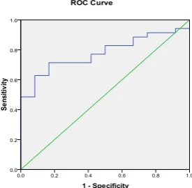
We also obtained the specificity and sensitivity of the BIS using the ROC curve and considering the GCS as the gold standard. For this reason, the patients were divided into mild and moderate trauma groups with respect to the GCS scores and the ROC curve was drawn. We found that BIS could be used to differentiate patients with TBI based on the severity of trauma (figure 2). The surface below the ROC curve was 77.9% which was statistically significant (P=0.004). Table 1 summarizes the results obtained through the ROC curve. As shown, as the BIS increases, its sensitivity decreases while its specificity increases. It seems that the cut-off point of 61.70 (specificity=83.3%, sensitivity=71.4%) is the optimum point for differentiating patients with mild and moderate brain injury. At this cut-off point, the BIS can identify 71.4% of the patients with mild TBI and 83.3% of those with moderate TBI (Table 1).
Figure 1. There was a significant relationship between BIS and GCS scores of the TBI patients in ICU (r=0.372, P=0.014). Figure 2. According to ROC curve analysis the BIS could be used to differentiate patients with TBI based on the severity of trauma. The surface below the ROC curve was 77.9% which was statistically significant (P=0.004).
Table 1. The numerical values of ROC curve are shown in detail. The sensitivity and specificity of BIS in relation to GCS reveals that the cut-off point of 61.70 (specificity=83.3%, sensitivity=71.4%) is the optimum point for differentiating patients with mild and moderate brain injury.
Figure 1: Correlation between BIS and GCS scores
Figure 2:
ROC curve for predicting the cut-off point for BIS
Table 1: Sensitivity and specificity of BIS scores according to the ROC curve Positive if Greater
Than or Equal Toa Sensitivity Specificity 1 -
34.3333 1.000 1.000
35.9167 .971 1.000
36.7500 .943 1.000
41.5000 .943 .917
46.7500 .914 .917
47.7500 .914 .833
48.1000 .914 .750
48.6000 .886 .750
49.1000 .886 .667
49.5167 .857 .667
50.7500 .829 .667
52.2500 .829 .583
53.2500 .829 .500
54.0333 .800 .500
54.8667 .771 .500
55.9167 .771 .417
57.5000 .743 .417
59.4500 .714 .417
60.7000 .714 .333
61.2000 .714 .250
61.7000 .714 .167
62.8750 .686 .167
63.8750 .657 .167
64.1000 .629 .167
65.2250 .629 .083
66.6250 .600 .083
67.5000 .543 .083
y = 2.9722x + 25.279 R² = 0.1258
0 20 40 60 80 100
0 5 10 15 20
BIS
GCS
69.0000 .514 .083
70.8333 .486 .083
71.8333 .486 .000
72.1667 .457 .000
72.3667 .429 .000
72.8667 .400 .000
74.3667 .371 .000
75.7000 .343 .000
76.5000 .314 .000
77.8750 .286 .000
79.2083 .257 .000
79.7333 .229 .000
80.6500 .200 .000
81.7500 .171 .000
82.7500 .143 .000
83.5500 .114 .000
83.9667 .086 .000
84.6667 .057 .000
85.5000 .029 .000
87.0000 .000 .000
Discussion
We found a significant linear relationship between GCS and BIS scores. However, we obtained different BIS scores for a specific GCS score which could reduce the predictive value of this index for differentiating between patients with mild and moderate TBI and determining the depth of coma. Our results are similar to several previous studies. In one study, Gill and colleagues found a significant correlation between BIS and GCS scores in patients with reduced levels of consciousness (GCS≤14, r=0.387, P=0.165) [16]. In another consistent study on 29 patients with mild to moderate TBI undergoing craniotomy, the researchers also found a significant correlation between the two indices [15]. Similar to our study, various amounts of BIS scores were obtained for each GIS score in the two previously mentioned study. Other researchers assessed the possibility of using BIS for measuring the level of consciousness in patients with TBI and assessed the correlation between BIS and GCS, Richmond Agitation-Sedation Scale (RASS) and Reaction Level Scale (RLS). Considering the significant correlation found in the mentioned study, the researchers concluded that BIS could be used as an objective tool for consistent monitoring of the level of consciousness in patients with brain injury [17].
Although we found a significant correlation between BIS and GCS scores, the obtained amounts were low. This weak
correlation could be attributed to the fact that each one of these tools measures different elements of brain function; BIS measures the frontal lobe function while GCS often measures movement disorders [18]. Various factors can interfere with the correct interpretation of BIS scores. BIS reduces during normal sleep [19, 20]. We also encountered this problem in our study which was inevitable. Moreover, complications such as cerebral ischemia and hypothermia further reduce BIS [19, 21]. We excluded patients with hypothermia but we could not control for cerebral ischemia and its severity differed among the patients.
Other factors leading to alterations in BIS are the signal artifacts produced by other medical instruments such as warming blankets, high-frequency ventilators, suctions, surgical tools, pacemakers, and defibrillators. As the distance of these instruments to the BIS device become lesser the possibility of signal artifacts become higher [22]. In our study, the patients used floating mattresses, pulse oximetries and cardiac monitoring and ventilators and portable suctions were close to the patients’ beds. SQI and EMG are diagrams that help medical personnel interpret the reliability of the BIS [22]. In our study, a BSI with low SQI (less than 50%) or high EMG (more than 50%) was considered as unreliable and was excluded from the study. Previous researchers have made no mention of this in their studies.
We found that BIS could be used to differentiate patients with TBI based on the severity of trauma according to ROC curve analysis. Inconsistently, other researchers have found that this index cannot adequately differentiate patients with TBI since they had obtained a surface area of 61-73% under the ROC curve [16]. This difference could be attributed to the higher patients’ homogeneity in our study. In our study all patients suffered from TBI, while in the mentioned study, the researchers had included patients with reduced levels of consciousness due to drug toxicity, seizure, etc. Moreover, the mean age of the patients in the mentioned study was 42 years (range: 14-93 years) [16] while the mean age of our patients was 28.8 years (range:18-59). Another difference between our study and the mentioned study was that we excluded BIS scores obtained from low-quality signals (SQI<50%) or high muscular signals (EMG>50%), while the researchers in the mentioned study had not.
Conclusion
Jarineshin H, et al.: Correlation between glasgow coma score and bispectral index
consistently monitor the level of consciousness in patients with mild and moderate TBI.
Abbrevations
ICU: Intensive Care Unit, BIS: Bispectral Index, GCS: Glasgow Coma Scale, TBI: Traumatic Brain Injury, ROC: Receiver Operating Characteristic, EEG: Electroencephalography, SQI: signal quality index, EMG: Electromyogram, SPSS: Statistical Package for the Social Sciences
Competing interests
The authors thereby declare no related financial interests with any company or organization in regard to this article and that there is no conflict of interest in this study.
Acknowledgement
We would like to express our gratitude to the representatives’ and relatives’ of the patients who allowed us to apply this study in the Intensive Care Unit of Shahid Mohammadi Hospital and also to its staff.
Author details
Anesthesiology, Critical Care and Pain Management Research Center, Hormozgan University of Medical Sciences, Bandar Abbas, Iran.
References
1. Behdad A, Hosseinpour M, Adaryani MR. Assessment of Chest Trauma in Patients Admitted to Academic Medical Centers of Isfahan. Feyz Journals of Kashan University of Medical Sciences. 2008;11(5):43-6.
2. Puvanachandra P, Hyder AA. The burden of traumatic brain injury in Asia: a call for research. Pak J Neurol Sci. 2009;4(1):27-32.
3. Hyder AA, Wunderlich CA, Puvanachandra P, Gururaj G, Kobusingye OC. The impact of traumatic brain injuries: a global perspective. NeuroRehabilitation-An Interdisciplinary Journal. 2007;22(5):341-54.
4. Miller RFL, Johns RA, Savarese JJ, Wiener-Kronish JP, Young WL. Miller's Anesthesia. 7 ed. Philadelphia: Elsevier; 2010.
5. Bell SE, Hlatky R. Update in the treatment of traumatic brain injury. Current treatment options in neurology. 2006;8(2):167-75.
6. Moghisi A, Afsarim N. A comprehensive guide to a safe community. 1 ed. Tehran: Andishand Publication; 2007. 7. Karbakhsh M, Zandi N, Rouzrokh M, Zarei M-R. Injury
epidemiology in Kermanshah: The National Trauma Project in Islamic Republic of Iran. East Mediterr Health J 2009;15(1):57-64.
8. Winn. HR. Youmans Neurological Surgery. 6 ed. New York: ElsevierSaunders; 2011.
9. Bordini AL, Luiz TF, Fernandes M, Arruda WO, Teive HA. Coma scales: a historical review. Arq Neuropsiquiatr. 2010;68(6):930-7. doi:10.1590/S0004-282X2010000600019.
10. Hsia S-H, Wu C-T, Wang H-S, Yan D-C, Chen S-C. The use of bispectral index to monitor unconscious children. Pediatric neurology. 2004;31(1):20-3.
11. Haug E, Miner J, Dannehy M, Seigel T, Biros M. Bispectral Electroencephalographic Analysis of head‐
injured Patients in the Emergency Department. Academic emergency medicine. 2004;11(4):349-52.
12. Rowley G, Fielding K. Reliability and accuracy of the Glasgow Coma Scale with experienced and inexperienced users. The Lancet. 1991;337(8740):535-8.
13. Greenberg MS. Hand book of Neurosurgery. 7 ed. New York: Thieme medical Publisher 2010.
14. Kelley SD. Monitoring level of consciousness during anesthesia and sedation. A Clinicians Guide to the Bispectral. Newton, MA, USA: Aspect Medical System, Inc 2003.
15. Paul DB, Rao GU. Correlation of Bispectral Index with Glasgow Coma Score in mild and moderate head injuries. Journal of clinical monitoring and computing. 2006;20(6):399-404.
16. Gill M, Green SM, Krauss B. Can the bispectral index monitor quantify altered level of consciousness in emergency department patients? Academic emergency medicine. 2003;10(2):175-9.
17. Cho JH, Cheong SH, Kim HS, Kim SH, Cho KR, Lee SE et al. Bispectral index monitoring to assess the level of consciousness in patients with brain injury. Korean Journal of Anesthesiology. 2009;57(2):185-9.
18. Gill M, Green SM, Krauss B. A study of the bispectral index monitor during procedural sedation and analgesia in the emergency department. Annals of emergency medicine. 2003;41(2):234-41.
19. Rosow C, Manberg PJ. Bispectral index monitoring. Anesthesiol Clin North America. 2001;19(4):947-66, xi. 20. Sleigh JW, Andrzejowski J, Steyn-Ross A, Steyn-Ross M.
The bispectral index: a measure of depth of sleep? Anesthesia & Analgesia. 1999;88(3):659-61.
21. England M. The changes in bispectral index during a hypovolemic cardiac arrest. Anesthesiology. 1999;91(6):1947-9.