DOI
10.17219/acem/68845
Copyright
© 2018 by Wroclaw Medical University This is an article distributed under the terms of the Creative Commons Attribution Non-Commercial License (http://creativecommons.org/licenses/by-nc-nd/4.0/)
Address for correspondence
Przemysław Janas
E-mail: [email protected]
Funding sources
None declared
Conflict of interest
None declared
Received on January 1, 2017 Reviewed on January 15, 2017 Accepted on February 7, 2017
Abstract
Telocytes are emerging cell population localized in the stroma of numerous organs, characterized by a distinc-tive morphology – small cell body with very long, slender prolongations, termed telopodes. Those cells can be found in the whole female reproductive system: in the vagina, uterus, oviducts and ovaries, mammary glands and also in the placenta. In our review, we aim at complete and transparent revision of the current knowledge of telocytes’ localization and function, enriched by the analysis of the possible future direction of development of their clinical applications. The function of telocytes in the reproductive system has not been fully elucidated yet; however, many researchers point at their role in the regulation of local microenviron-ment, myogenic contractile mechanism, bioelectrical signaling, immunomodulation and regulation of blood flow. Additionally, previous research suggests that telocytes might act as sex hormone level sensors and are connected with pregnancy maintenance. As the morphology and number of those cells change under pathological conditions, such as pre-eclampsia, endometriosis and ovarian failure, there is a chance that they may contribute to therapy of abovementioned conditions. The impact of telocytes on stem cells and angiogenesis has been proven in many organs, and may be useful in regenerative medicine of the female reproductive system. A recently found connection between the proliferation rate of breast cancer cells and stromal cells like telocytes might be a step forward to the management of mammary gland neoplasms. Key words: ovary, uterus, placenta, telocytes, telopodes
Telocytes in the female reproductive system:
An overview of up-to-date knowledge
Przemysław Janas
A–D, Iwona Kucybała
B–D, Małgorzata Radoń-Pokracka
E,F, Hubert Huras
E,FDepartment of Obstetrics and Perinatology, Jagiellonian University Medical College, Kraków, Poland
A – research concept and design; B – collection and/or assembly of data; C – data analysis and interpretation; D – writing the article; E – critical revision of the article; F – final approval of the article
Introduction
Definition
Telocytes (TCs) are a new type of cells located in the stroma of several organs. Their most characteristic feature is the presence of extraordinarily long, slight prolongations called telopodes (Tps).1 As far as the female reproductive system is concerned, TCs were described in the mammary gland, vagina, uterus, uterine tube, and placenta.2–6
A brief history
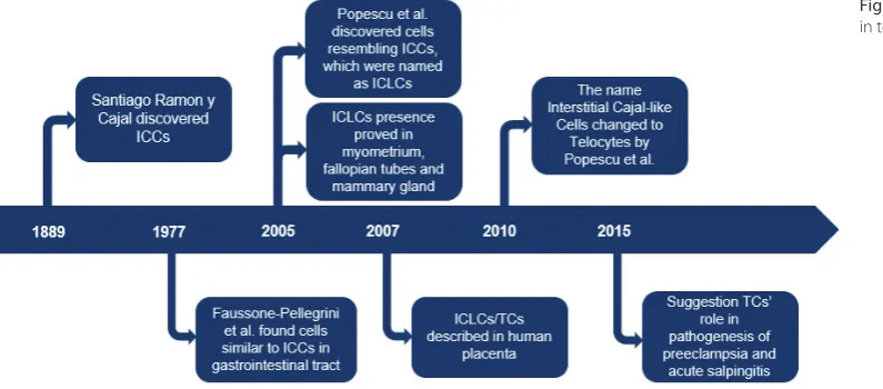
The history of TCs derives from Interstitial Cajal Cells (ICCs) discovered by the Spanish neuroanatomist and Nobel Prize winner Santiago Ramon y Cajal. In 1889, Cajal found a new type of cells located within the muscle layer of the intestine, between nerve ganglia and smooth muscle cells.1
About a half a century later, Faussone-Pellegrini et al. proved the existence of cells similar to ICCs in the gas-trointestinal tract with the use of electron microscopy.7 In 2005, Popescu et al. found interstitial cells resembling ICCs in the specimens from a mammary gland.2 They called them Interstitial Cajal-like Cells (ICLCs). A re-examination of the muscular coat of the gut performed by Pieri et al. showed that ICLCs are functionally and morphologically different from ICCs.8 As a result of these studies, in 2010 Popescu suggested that the name “Inter-stitial Cajal-like Cells” should be changed to “Telocytes” to clearly distinguish these 2 cell populations.
In the female reproductive system, TCs were first de-scribed in 2005, and they were found within the wall of the uterus.4,9 Further research proved their existence also in the vagina, uterine tubes and placenta (Fig. 1).3,6,10
The name “Telocytes” derives from ancient Greek – the word “telos” refers the object of huge potential.11 Nowa-days, thanks to TCs’ morphology and functions, they arouse huge interest among researchers as potential con-tributors to intercellular communication, cancerogenesis and a target for regenerative medicine purposes.12–14 In our
review, we are trying to summarize the current knowledge of tissue distribution, identification methods and potential functions of TCs in female reproductive system.
General aspects of telocytes
Cell phenotype
TCs are small cells with 1–5 long and thin prolongations named telopodes (Tps).11 The cell body is rather small, its average length oscillates around 9.39 ±3.26 μm. It contains big, heterochromatic nucleus and a thin, perinuclear rim of cytoplasm including few organelles, mainly mitochon-dria.11,15 The shape of the cell body is influenced by the actual number of Tps, ranging from piriform, through spindle and triangular to stellate.16 Tps are up to 1000 μm long, which makes them one of the longest structures in the body, except for some axons.15,17 As they are composed of dilated segments, named podoms, and thin podomeres, their shape is termed as moniliform.15,16 Podoms contain organelles responsible for protein synthesis and intercel-lular signaling, namely rough and smooth endoplasmic re-ticulum, Golgi apparatus, mitochondria, and caveolae.12,16 Considering the small size of TCs’ body and even more narrow Tps (around 0.5 μm wide podoms), it would be ut-terly difficult to distinguish them from other cells in light microscopy, thus the golden standard for identifying TCs is transmission electron microscopy (TEM).16,18
Localization
Until now, TCs have been identified in the female repro-ductive system, placenta, mammary gland, cardiovascular system, urinary system, gastrointestinal tract, liver and pancreas, trachea, lungs and pleura, spleen, bone marrow, dura mater, choroid plexus, meninges, trigeminal ganglion, testicles and prostate, skin, skeletal muscles, and eyes.16,19
Cell immunophenotype
Since numerous cells, for instance fibroblasts, neurons or pericytes, have a morphology closely resembling that of telocyte, it became apparent that there is a need for finding specific markers for TCs. Until now, TCs have been confirmed for expression of CD34, c-Kit, vimentin, PDGFR-α and -β, caveolin-1, CD44, Sca-1, Nanog and Oct4.16,17,20
Nevertheless, those markers are not perfect. There are a lot of discrepancies between studies in terms of CD34 and c-Kit expression in TCs in different tissues. From all abovementioned markers, CD34 is considered as the most reliable for TCs, although Suciu et al. reported that only 70% of the cells in the placenta, which have TC’s mor-phology, express CD34.20,21 Moreover, the second most frequently used marker, c-Kit, is not expressed by TCs in gastrointestinal tract.22 That inconsistency might be caused by imperfections in technical procedures or the existence of tissue-specific subtypes of TCs.18,21 Another difficulty is connected with the expression of those mark-ers in cells with similar morphology – CD34 is expressed by endoneurial fibroblasts, while vimentin together with PDGFR-β are present in fibroblasts, pericytes and neu-rons.18 In order to minimize the risk of misidentification, at least double immunofluorescence staining should be used in order to detect TCs. All things considered, a uni-versal marker for TCs should be found – expressed in every tissue and characteristic only for them.
Intercellular communication
TCs establish both homo- and heterocellular junctions together with connections with extracellular matrix.20 Moreover, they also communicate through extracel-lular vesicles, which is a form of juxtacrine/paracrine signaling.23
Homocellular junctions are formed between 2 Tps or between Tps and TC’s body.24 In most cases, they are connected by simple apposition of 2 plasma membranes, but there also exist more complex forms of linkage – punc-ta adhaerentia minima, processus adhaerens, recessus adhaerens, and manubria adhaerentiva. Their primary function is to maintain the integrity of 3-dimensional network of TCs during changes of shape of the tissue. Second presumptive function is linked with catenins, which are important components of adherens junctions, as they might play role in mechanosensing. The last type of homocellular contact is gap junction (nexus), which allows the exchange of ions and small molecules between adjacent cells.20
Heterocellular junctions typically have the morphology of point contacts, nanocontacts, planar contacts or simple apposition of plasma membranes.20 TCs are connected through them with fibroblasts, myofibroblasts, pericytes, endothelial cell, neurons, stem cells, macrophages, mast
cells, lymphocytes, plasma cells, Schwann cells, cardio-myocytes and smooth muscle cells.16,21,24,25 Distinctive type of heterocellular junctions is stromal synapse, which links TCs with immune cells and cardiac stem cells.24,26
Extracellular vesicles shedded by TCs can be divided into 3 groups: exosomes (from endosomes), ectosomes (from plasma membrane) and multivesicular cargo (multiple, tightly packed endomembrane-derived vesicles).23 Vesicles mainly contain proteins, lipids, miRNAs, mRNAs, and mtDNA.12 Hence, they are crucial for intercellular signal-ing and they even might be connected with a modifica-tion of the post-transcripa modifica-tional activity of recipient cells.27 Fibronexuses and focal adhesions were found between TCs and components of extracellular matrix.20
Electrophysiological properties
of telocytes
Since TCs form an abundant network interconnecting numerous types of cells in the interstitium, a hypothesis was postulated that TCs may be involved in bioelectrical signaling. Thus far, it has been confirmed that TCs ex-press: T-type calcium channels (Cav3.1 and Cav3.2), small-conductance calcium-activated potassium channels (SK3), large-conductance calcium-activated potassium channels (BKCa), inwardly rectifying potassium channels (IKir) and calcium-dependent hyperpolarisation-activated chloride inward channels.28,29,30,31
L-type calcium channels, transient outward potassium channels (Ito) and ATP-sensitive potassium channels (KATP) were not detected in TCs in previous studies.30,31
Physiological functions
of telocytes
Telocytes
in female reproductive system
Telocytes in vagina
Proto-oncogene c-Kit positive cells with long processes, which nowadays might be classified as TCs, were found in muscular layer of the human vagina. Shafik et al. sug-gested their potential role in generating slow waves re-sulting in the contractility of smooth muscle cells in the vagina.3 However, their presence and role in that organ should be thoroughly assessed in the future.
Telocytes in uterus
In the human uterus, TCs were observed in the inter-stitial space of the endometrium and myometrium.38,39 Endometrial TCs were detected in the stroma of the stra-tum functionalis and strastra-tum basalis around the endome-trial glands, while myomeendome-trial TCs form a 3-dimensional network intermingling with smooth muscle bundles.1,38 That location in the myometrium may suggest that TCs might be involved in myogenic contractile mechanism during sperm transport prior to fertilization, embryo im-plantation and delivery.1
The quantity of TCs in the endometrium and the myo-metrium correlates with the reproductive state.40 Imma-ture rat uteruses present the lowest density of TCs com-pared to pregnant and postpartum ones.41 The number
of endometrial TCs in pregnant state increases, while there is a reduction of their number in the myometrium.40 Hence, changes in the quantity of TCs in the myometrium may be associated with the prevention of preterm deliv-ery.25 Moreover, the highest number of myometrial TCs were found in the postpartum uteri, which may be related with its involution.40
Additionally, the morphology of Tps is different in preg-nant and non-pregin preg-nant myometrium – in non-pregin preg-nant uterus podomers are wider and podoms are thinner com-pared to pregnant ones (Table 1).31
The most useful markers used for the identification of TCs in the uterus are CD34 and PDGFR-α/PDGFR-β in double immunolabelling.25 PDGFR-α/β is mostly ex-pressed at TC’s cell body, while Tps are intensely positive for CD34.28 TCs in uterus are also positive for α-SMA, CD44, vimentin, Sca-1, and c-Kit.41 Nevertheless, the expression of c-Kit here is questionable. Yang et al. detected various types of TCs in uterine samples: c-Kit(–)/vimentin(+), c-Kit(+)/ vimentin(+) and c-Kit(+)/CD34(+). As a consequence, they suggested the presence of various subpopulations of TCs in uterus which may perform disparate functions.42
The comparison of markers applicable to the detection of TCs in female reproductive system is shown in Table 2. TCs also express connexin43, a gap junction protein which might play an essential role in decidua maturation. A de-crease in the expression of that protein is linked with re-current pregnancy loss.41,43
Furthermore, the expression of estrogen receptor alpha (ERα) and progesterone receptor A (PR-A) were confirmed on the surface of uterine TCs.44 This suggests that TCs may be the sensors of sex hormone levels. TCs also express these receptors in uterine tubes and upper lamina propria of the human urinary tract.45,46
Additionally, human uterine TCs express 2 types of channels at their cell membrane: T-type calcium chan-nels (Cav3.1, Cav3.2) and small-conductance calcium-ac-tivated potassium channels (SK3).25
The presence of Cav3.1 and Cav3.2 channels were con-firmed both in cell body and Tps of TCs. Expression of Cav3.1 and Cav3.2 channels correlates with the reproduc-tive state: Cav3.1 expression in pregnant and non-pregnant Table 1. The variation in morphology of Tps and extracellular vesicles
between non-pregnant and pregnant myometrium31
Parameter Non-pregnant myometrium myometriumPregnant
Podom thickness [nm] 268.6 ±8.27 316.38 ±17.56
Podomer gauge [nm] 81.94 ±1.77 75.53 ±1.81
Number of exosomes/
shedded microvesicles 26/89 20/168
Diameter of extracellular vesicles [nm]
58–405 average 151
65–362 average 170
Table 2. Expression of markers on TCs in individual parts of female reproductive system
Organ Marker
CD34 c-kit vimentin PDGFR-α PDGFR-β ERα PR-A Additional markers
Vagina n/d + n/d n/d n/d n/d n/d n/d
Uterus + +/– + + + + + α-SMACD44
Sca-1
Uterine tubes + – + n/d n/d + + n/d
Ovaries + n/d + + + n/d n/d n/d
Mammary gland + +/– + n/d n/d n/d n/d CD10
Placenta + + + n/d n/d n/d n/d caveolin1
state is equal, while Cav3.2 channels are mostly detected in pregnant myometrium.25 Moreover, in non-pregnant state Cav3.1 were strongly expressed in Tps, while Cav3.2 were observed only in cell body. The differences in expres-sion of these channels suggest that TCs may play role in de-tection of mechanical stretching of the pregnant uterus.28 SK3 channels were observed in the uterus of several spe-cies (for instance in human, mice or rat). Their expression is also correlated with pregnant or non-pregnant state. The density of SK3 channels in non-pregnant myometrium is elevated where they are present on TCs and vascular endothelium, in contrast to pregnant state with decreased number of channels, which are only located on vascular endothelium.47 Downregulation of SK3 channels during pregnancy probably reduce contractility of the uterus. Changes in the expression of SK3 channels could also be caused by different stage of the hormonal cycle (expres-sion of channels influenced by sex hormones) or diseases connected with hormonal imbalance.29
Uterine TCs create connections with other cells and components of extracellular matrix (for instance collagen or elastic fibres). Heterocellular junctions between TCs and smooth muscle cells, nerve endings and blood vessels were additionally observed.48 TCs in uterus can also com-municate by shedding membrane microvesicles: exosomes and ectosomes. No differences were noticed in number and diameter of shedded vesicles in pregnant myometrium compared with non-pregnant (Table 1).25 Moreover, Diaz-Flores et al. proved that TCs have endocytic properties, which suggest existence of bidirectional information ex-change between TCs and neighbouring cells.49
Additionally, TCs establish contacts with immune cells, namely lymphocytes, eosinophils, basophils, plasma cells, and macrophages.38 Chi et al. confirmed that TCs are able to activate peritoneal macrophages, which may result in increased amount of IL-6, IL-10, IL1R1, TNF-α and iNOS. Pathologically high levels of these proteins could lead to implantation failure, immunologically-mediated abortion or endometriosis.50
TCs may also be useful in uterine regenerative medicine. Recent studies revealed that TCs from pregnant and non-pregnant myometrium have different reactivity to low-level laser stimulation (LSSS). Tps from pregnant uterus are more susceptible to extension after using LSSS compared to Tps from non-pregnant uterus. These differences are probably caused by variations in TCs’ cytoskeleton structure (up-reg-ulation of integrins) in pregnant and non-pregnant state.51
Telocytes in uterine tubes
TCs are located in lamina propria and muscular layer of uterine tubes. Their Tps create a 3-dimensional net-work between smooth muscle cells (SMCs), nerve end-ings and blood vessels. Close relations between TCs and SMCs may suggest that TCs participate in uterine tube contractility.52
TCs in the uterine tube present a typical cell phenotype. They shed 3 types of extracellular vesicles: exosomes (di-ameters: 60–100 nm), ectosomes (100–250 nm) and mi-crovesicles (250–350 nm).52 Similarly to the uterus, they express on their surface PR-A and ERα receptors, which might be connected with the control of peristaltic move-ments in the uterine tube due to changes in estrogen (ac-celeration of contractions) and progesterone (de(ac-celeration of contractions) levels.1
TCs are connected with other cells located in the uter-ine tubes tissue. Heterocellular junctions with fibrocytes, pericytes, SMCs, nerve endings, mast cells and stem cells were observed.42 Yang et al. claimed that Tps are located in close vicinity to lymphocytes and plasma cells, which, according to their study, may suggest that TCs are involved in the stimulation of plasma cells to antibodies synthesis. However, the aforementioned property requires further investigation.53
Yang et al. proved that TCs’ quantity and ultrastructure dramatically change in rat model of acute salpingitis. TCs retrieved from salpingitis-affected uterine tubes presented numerous abnormalities, such as: loss of organelles, cy-toplasmic vacuolization, dilatation of rough endoplasmic reticulum and loss of intercellular junctions. Addition-ally, the number of TCs was significantly decreased.53 The declined quantity of TCs, which is probably caused by the overproduction of iNOS, COX-2, LPO and estra-diol, damages those cells and were also observed in pelvic endometriosis and tubal ectopic pregnancy.40,42,54 Never-theless, TCs of nearly normal appearance can be found even in endometriosis-affected uterine tube. Presumably, that is the reason why some women in this state have only reduced fertility instead of complete infertility.42
Telocytes in ovary
TCs were detected in the stroma of mice ovaries and they were positive for CD34, vimentin, PDGFR-α and -β. Thus far, the function of TCs in ovaries has not been deter-mined; however, there is an assumption that they might be responsible for maintaining the local microenvironment.55 Liu et al. found statistically a significant decrease in the number of TCs in ovaries affected by cyclophosphamide-induced premature failure compared to healthy con-trols. As a consequence, they might be used as a mark-er of declining ovarian functions caused by the intake of cyclophosphamide.55
Telocytes in mammary gland
gland ducts, predominantly by their Tps located perpen-dicularly to long axes of those structures.56
In mammary gland, TCs are positive for CD34 and vi-mentin.57 However, there is an incongruity between the studies in terms of expression of c-kit – Gherghiceanu et al. along with Mou et al. confirmed the presence of that mark-er, while Petre et al. negated its expression.56–58 Addition-ally, TCs in mammary gland might be partially positive for CD10.58
TCs are connected by stromal synapses with stromal immune cells, such as plasma cells, lymphocytes, mast cells and macrophages. Contacts with fibroblasts were also observed.56 The direct link between TCs and endothelial cells or pericytes have not been found.13
Apart from the possible involvement of TCs in the or-ganization of properly functioning structure of mammary gland, they may play an important role in the modulation of an immune system, thanks to their connections with immune cells.56
As accurate arrangement of stroma has a beneficial effect on maintaining local microenvironment, any alterations in the function of TCs’ network may lead to an increase in the risk of neoplastic process in the tissue.13 Mou et al. investigated the influence of TCs and other stromal cells on the growth dynamic of breast cancer.57 They observed that TCs establish membrane-to-membrane connections with breast cancer cells in co-culture and presumably par-ticipate in the formation of neoplastic cell clusters. There was an increase in the proliferation index and a reduction in the apoptosis ratio among breast cancer cells accompa-nied by stromal cells, compared to isolated breast cancer cell culture.57 Furthermore, the number of heterocellular junctions formed by TCs is diminished in neoplastic tis-sue.13 Summing up, TCs along with other stromal cells may contribute to neoplasm development and survival.57 As a consequence, TCs might emerge as a novel target in breast cancer therapy.
Telocytes in placenta
Placental TCs were detected by Suciu et al. in the large and peripheral stem villi, where they are located just be-neath the trophoblast. Their Tps are orientated parallelly to the basement membrane and circularly or longitudinally to blood vessels.21,40 They are positive here for: c-kit, CD34, vimentin, caveolin1, VEGF and iNOS.25
TCs create heterocellular contacts with mast cells, myo-fibroblasts, SMCs and specific placental macrophages, called Hofbauer cells (HBCs).25 However, the function of TCs in human term placenta is still unknown. The pres-ence of junctions between TCs and HBCs suggest their possible contribution to immune surveillance. Consid-ering the fact that placenta is not an innervated organ, Bosco et al. suggested that TCs may be crucial for signal transduction resulting in blood flow in foetal vessels and aetiopathogenesis of pre-eclampsia.59
Conclusions
TCs are a unique cell population, located in the stroma of numerous organs, also in the female reproductive sys-tem. Their role specific for that system can be connected with their involvement in muscular layer contractility, pregnancy maintenance, immunomodulation and tissue regeneration. Alterations of their number in the female re-productive system might be connected with pre-eclampsia, endometriosis or acute salpingitis and further research on that subject may lead to a turning point in TCs-related treatment of those conditions.
References
1. Varga I, Urban L, Kajanová M, Polák Š. Functional histology and pos-sible clinical significance of recently discovered telocytes inside the female reproductive system. Arch Gynecol Obstet. 2016;294:417–422. 2. Popescu LM, Andrei F, Hinescu ME. Snapshots of mammary gland
interstitial cells: Methylene-blue vital staining and c-kit immunopos-itivity. J Cell Mol Med. 2005;9:476–477.
3. Shafik A, El-Sibai O, Shafik I, Shafik AA. Immunohistochemical iden-tification of the pacemaker Cajal cells in the normal human vagina.
Arch Gynecol Obstet. 2005;272:13–16.
4. Ciontea SM, Radu E, Regalia T, et al. C-kit immunopositive intersti-tial cells (Cajal-type) in human myometrium. J Cell Mol Med. 2005;9: 407–420.
5. Urban L, Miko M, Kajanova M, et al. Telocytes (interstitial Cajal-like cells) in human Fallopian tubes. Bratisl Lek Listy. 2016;117:263–267. 6. Suciu L, Popescu LM, Gherghiceanu M. Human placenta: De visu
demonstration of interstitial Cajal-like cells. J Cell Mol Med. 2007;11: 590–597.
7. Faussone-Pellegrini MS, Cortesini C, Romagnoli P. Ultrastructure of the tunica muscularis of the cardial portion of the human esophagus and stomach, with special reference to the so-called Cajal’s intersti-tial cells. Arch Ital Anat Embriol. 1977;82:157–177.
8. Pieri L, Vannucchi MG, Faussone-Pellegrini MS. Histochemical and ultrastructural characteristics of an interstitial cell type different from ICC and resident in the muscle coat of human gut. J Cell Mol
Med. 2008;12:1944–1955.
9. Duquette RA, Shmygol A, Vaillant C, et al. Vimentin-positive, c-kit-negative interstitial cells in human and rat uterus: A role in pacemak-ing? Biol Reprod. 2005;72:276–283.
10. Shafik A, Shafik AA, El Sibai O, Shafik IA. Specialized pacemaking cells in the human Fallopian tube. Mol Hum Reprod. 2005;11:503–505. 11. Popescu LM, Faussone-Pellegrini MS. TELOCYTES – A case
of ser-endipity: The winding way from Interstitial Cells of Cajal (ICC), via Interstitial Cajal-Like Cells (ICLC) to TELOCYTES. J Cell Mol Med. 2010;14:729–740.
12. Edelstein L, Fuxe K, Levin M, Popescu BO, Smythies J. Telocytes in their context with other intercellular communication agents. SeminCell
Dev Biol. 2016;55:9–13.
13. Mihalcea CE, Moroşanu AM, Murăraşu D, et al. Particular molecular and ultrastructural aspects in invasive mammary carcinoma. Rom
J Morphol Embryol. 2015;56:1371–1381.
14. Bei Y, Zhou Q, Sun Q, Xiao J. Telocytes in cardiac regeneration and repair. Semin Cell Dev Biol. 2016;55:14–21.
15. Kostin S. Myocardial telocytes: A specific new cellular entity. J Cell
Mol Med. 2010;14:1917–1921.
16. Mirancea N. Telocyte – A particular cell phenotype. Infrastructure, relationships and putative functions. Rom J Morphol Embryol. 2016; 57:7–21.
17. Chang Y, Li C, Lu Z, Li H, Guo Z. Multiple immunophenotypes of car-diac telocytes. Exp Cell Res. 2015;338:239–244.
18. Kostin S. Cardiac telocytes in normal and diseased hearts. Semin Cell
Dev Biol. 2016:55:22–30.
19. Cretoiu SM, Popescu LM. Telocytes revisited. Biomol Concepts. 2014;5: 353–369.
21. Suciu L, Popescu LM, Gherghiceanu M, et al. Telocytes in human term placenta: Morphology and phenotype. Cells Tissues Organs. 2010;192:325–339.
22. Ibba-Manneschi L, Rosa I, Manetti M. Telocyte implications in human pathology: An overview. Semin Cell Dev Biol. 2016;55:62–69. 23. Fertig ET, Gherghiceanu M, Popescu LM. Extracellular vesicles release
by cardiac telocytes: Electron microscopy and electron tomography.
J Cell Mol Med. 2014;18:1938–1943.
24. Gherghiceanu M, Popescu LM. Cardiac telocytes – their junctions and functional implications. Cell Tissue Res. 2012;348:265–279. 25. Cretoiu D, Cretoiu SM. Telocytes in the reproductive organs: Current
understanding and future challenges. Semin Cell Dev Biol. 2016;55: 40–49.
26. Popescu LM, Fertig ET, Gherghiceanu M. Reaching out: Junctions between cardiac telocytes and cardiac stem cells in culture. J Cell MolMed. 2016;20:370–380.
27. Cismasiu VB, Popescu LM. Telocytes transfer extracellular vesicles loaded with microRNAs to stem cells. J Cell Mol Med. 2015;19:351–358. 28. Cretoiu SM, Radu BM, Banciu A, et al. Isolated human uterine telo-cytes: Immunocytochemistry and electrophysiology of T-type cal-cium channels. Histochem Cell Biol. 2015;143:83–94.
29. Rosenbaum ST, Svalø J, Nielsen K, Larsen T, Jørgensen JC, Bouche-louche P. Immunolocalization and expression of small-conductance calcium-activated potassium channels in human myometrium. J Cell
Mol Med. 2012;16:3001–3008.
30. Sheng J, Shim W, Lu J, et al. Electrophysiology of human cardiac atri-al and ventricular telocytes. J Cell Mol Med. 2014;18:355–362. 31. Cretoiu SM, Cretoiu D, Marin A, Radu BM, Popescu LM. Telocytes:
Ultrastructural, immunohistochemical and electrophysiological char-acteristics in human myometrium. Reproduction. 2013;145:357–370. 32. Edelstein L, Smythies J. The role of telocytes in morphogenetic bio-electrical signaling: Once more unto the breach. Front MolNeurosci. 2014;7:41
33. Yang C, Xiao J. Editorial. Telocytes in regeneration and repair. Curr Stem Cell ResTher. 2016;11:382.
34. Díaz-Flores L, Gutiérrez R, Goméz MG, Sáez FJ, Madrid JF. Behaviour of telocytes during physiopathological activation. SeminCell Dev Biol. 2016;55:50–61.
35. Xiao J, Chen P, Qu Y, et al. Telocytes in exercise-induced cardiac growth. J Cell Mol Med. 2016;20:973–979.
36. Popescu LM. The tandem: Telocytes – stem cells. Int J Biol Biomed Eng. 2011;5:83–92.
37. Enciu AM, Popescu LM. Telopodes of telocytes are influenced in vitro by redox conditions and ageing. Mol Cell Biochem. 2015;410:165–174. 38. Hatta K, Huang ML, Weisel RD, Li RK. Culture of rat endometrial
telo-cytes. J Cell Mol Med. 2012;16:1392–1396.
39. Creţoiu SM, Creţoiu D, Popescu LM. Human myometrium – the ultra-structural 3D network of telocytes. J Cell Mol Med. 2012;16:2844–2849. 40. Aleksandrovych V, Walocha JA, Gil K. Telocytes in female reproduc-tive system (human and animal). J Cell Mol Med. 2016;20:994–1000. 41. Roatesi I, Radu BM, Cretoiu D, Cretoiu SM. Uterine telocytes: A review
of current knowledge. Biol Reprod. 2015;93:10.
42. Yang XJ, Yang J, Liu Z, Yang G, Shen ZJ. Telocytes damage in endo-metriosis-affected rat oviduct and potential impact on fertility. J Cell
Mol Med. 2015;19:452–462.
43. He X, Chen Q. Reduced expressions of connexin 43 and VEGF in the first-trimester tissues from women with recurrent pregnancy loss.
Reprod Biol Endocrinol. 2016;14:46.
44. Cretoiu D, Ciontea SM, Popescu LM, Ceafalan L, Ardeleanu C. Intersti-tial Cajal-like cells (ICLC) as steroid hormone sensors in human myo-metrium: Immunocytochemical approach. J Cell Mol Med. 2006;10: 789–795.
45. Cretoiu SM, Cretoiu D, Suciu L, Popescu LM. Interstitial Cajal-like cells of human Fallopian tube express estrogen and progesterone recep-tors. J Mol Histol. 2009;40:387–394.
46. Gevaert T, De Vos R, Van Der Aa F, et al. Identification of telocytes in the upper lamina propria of the human urinary tract. J Cell Mol Med. 2012;16:2085–2093.
47. Albulescu R, Tanase C, Codrici E, Popescu DI, Cretoiu SM, Popescu LM. The secretome of myocardial telocytes modulates the activity of car-diac stem cells. J Cell Mol Med. 2015;19:1783–1794.
48. Ullah S, Yang P, Zhang L, et al. Identification and characterization of telo-cytes in the uterus of the oviduct in the Chinese soft-shelled turtle, Pel-odiscus sinensis: TEM evidence. J Cell Mol Med. 2014;18: 2385–2392. 49. Díaz-Flores L, Gutiérrez R, García MP, et al. Uptake and
intracytoplas-mic storage of pigmented particles by human CD34+ stromal cells/ telocytes: Endocytic property of telocytes. J Cell MolMed. 2014;18: 2478–2487.
50. Chi C, Jiang XJ, Su L, Shen ZJ, Yang XJ. In vitro morphology, viability and cytokine secretion of uterine telocyte-activated mouse perito-neal macrophages. J Cell Mol Med. 2015;19:2741–2750.
51. Campeanu RA, Radu BM, Cretoiu SM, et al. Near-infrared low-level laser stimulation of telocytes from human myometrium. Lasers Med Sci. 2014;29:1867–1874.
52. Yang P, Zhu X, Wang L, et al. Telocytes as a novel interstitial cells pres-ent in the magnum of chicken oviduct. Cell Transplant. 2016 [ahead of print].
53. Yang J, Chi C, Liu Z, Yang G, Shen ZJ, Yang XJ. Ultrastructure dam-age of oviduct telocytes in rat model of acute salpingitis. J CellMol Med. 2015;19:1720–1728.
54. Yang XJ, Xu JY, Shen ZJ, Zhao J. Immunohistochemical alterations of Cajal-like type of tubal interstitial cells in women with endometrio-sis and tubal ectopic pregnancy. Arch Gynecol Obstet. 2013;288:1295– 1300.
55. Liu T, Wang S, Li Q, Huang Y, Chen C, Zheng J. Telocytes as potential targets in a cyclophosphamide-induced animal model of premature ovarian failure. Mol Med Rep. 2016;14:2415–2422.
56. Gherghiceanu M, Popescu LM. Interstitial Cajal-like cells (ICLC) in human resting mammary gland stroma. Transmission electron micro-scope (TEM) identification. J Cell Mol Med. 2005;9:893–910. 57. Mou Y, Wang Y, Li J, et al. Immunohistochemical characterization
and functional identification of mammary gland telocytes in the self-assembly of reconstituted breast cancer tissue in vitro. J Cell Mol Med. 2013;17:65–75.
58. Petre N, Rusu MC, Pop F, Jianu AM. Telocytes of the mammary gland stroma. Folia Morphol (Warsz). 2015 [ahead of print].