Dr. Gite Shital Shridhar Department of Pharmaceutics,
KCT’S RGS College of Pharmacy, Anjaneri, Nashik, Maharashtra, India
Email: [email protected]
Address for correspondence
Access this article online www.japer.in
Mucoadhesive buccal drug delivery: An Overview
INTRODUCTION
Among all dosage form, oral route is more preferred to patient. The per oral route of administration of drug has disadvantages hepatic first pass metabolism and enzymatic degradation within the GI tract, that eliminate oral administration of certain classes of drugs like peptides and proteins. [1] Transmucosal routes of drug delivery offer distinct advantages over per oral administration for systemic drug delivery. Mucoadhesion is define as drug delivery system that utilize the property of bioadhesion of certain water-soluble polymers which become adhesive on hydration and hence can be used for targeting a drug to a particular region of the body for an extended period of time. [2] The transmucosal delivery of drug can involve the mucosal lining of buccal, sublingual, nasal, vaginal, rectal and ocular. Among that oral mucosa is perhaps most convenient and preferred route for drug delivery. [3] The buccal mucosa lines the inner cheek and buccal formulations are positioned in the mouth sandwiched between the
upper gingival (gums) and cheek to treat local and systemic conditions. The buccal route offers one of the potential routes for typically hydrophilic, large and unstable proteins, oligonucleotides and polysaccharides, as well as conventional small drug molecules. The oral cavity is mostly preferred site for local and systemic drug delivery. Adhesion of bioadhesive formulation leads to increase concentration gradient of drug across absorption site and therefore improve dosage form bioavailability. That has been used for local and systemic disorder to reduce dose requirement and side effects.[4]Drug Compounds having partition co-efficient in the range 40-20000 and pka 2-10 are considered optimal to be given through buccal drug delivery. [5]
Advantages of Buccal drug delivery [6]
• It avoids hepatic first-pass metabolism. • Ease of administration of dosage form. • It can be administered to unconscious patient • Fast onset of action.
• Patient compliance is more.
• Drugs which are not stable in the acidic environment and destroyed by enzymatic or alkaline environment of intestine can be administered by this route.
• It deals with a passive system of drug absorption
and does not require any activation Review
ReviewReview
Review ArticleArticleArticle Article
The main aim of buccal drug delivery of the drug as potential therapeutic agent is there instability in acidic environment, extensive first pass metabolism and low bioavailability of drug results an inadequate oral absorption. The buccal mucosa is rich in blood supply and relatively permeable. Buccal cavity was found to be most accessible site for both local and systemic delivery of drug. This review highlights that the oral mucosal drug delivery by discussing briefly the structural feature of mucosa, mechanism and theories of bioadhesion. It also contains mucoadhesive polymer, evaluation methods and some of the review on reported research work done on different types of drug by using different mucoadhesive polymers.
Keywords: Oral mucosa, Mucoadhesion, Bioadhesive polymer, Dosage form. ABSTRACT
ABSTRACT ABSTRACT ABSTRACT Gite Shital Shridhar*, Shinkar
Dattatraya Manohar, Saudagar Ravindra Bhanudas1
*Department of Pharmaceutics, KCT’S RGS College of Pharmacy, Anjaneri, Nashik, 422 213. Maharashtra, India. 1Department of Pharmaceutical Chemistry, KCT’S RGS College of Pharmacy, Anjaneri, Nashik, 422 213. Maharashtra, India.
J. Adv. Pharm. Edu. & Res.
• The buccal mucosa is highly perfuse with blood vessels and offers a greater permeability than the skin.
• A significant reduction in dose can is achieved thereby reducing dose related side effects.
Limitations of Buccal Drug Delivery System [7]
• Small dose of drug is required.
• Drug have a bitter or unpleasant taste or obnoxious odor or that irritate the mucosa cannot be administered.
• Eating and drinking may mostly restrict.
• Drug which is unstable at buccal environment cannot be given by this route.
• Drug that show passive diffusion can only
administered by this route.
• Permeability of buccal mucosa is low as compared to sublingual route.
Overview of oral mucosa
The total surface area of oral cavity 100 cm2 and is lined with mucous membranes. Other several distinct areas: the floor of mouth (sublingual), the buccal mucosa (cheeks), the gums (gingiva), the palatal mucosa and the lining of the lips. The oral mucosal tissues consist of a multi- layered epithelium enclosed with mucus. The basal lamina attaches the epithelium to a connective tissue layer; the lamina propria.The oral mucosa protects the body from external influences, such as the entry of potentially dangerous substances. The epithelium of the human oral mucosa shows several distinct patterns of maturation, related to the functional demands of the tissue. Keratinized epithelium (dehydrated, mechanically tough and chemically resistant) is found in the less flexible masticatory mucosa of the gingiva and part of the hard palate. Non- keratinized epithelium (flexible) forms the surface of the distensible lining mucosa of the soft palate, floor of mouth, lips and cheek. The buccal mucosal tissue has a multi-layered distinguished epithelium. [8] The oral mucosa mainly composed of three layers. The outermost is stratified squamous epithelium act as barrier which protects underlying tissues; intermediate lamina propria serves
mechanical support and followed by innermost layer submucosa. The epithelium of the buccal mucosa is about 40-50 cell layers thick, when compared with sublingual epithelium. The epithelial cells increase in size and become flatter as they travel from the basal layers to the superficial layers.The turnover period for the buccal epithelium has been found at 5-6 days, and this is representative of the oral mucosa as a whole. [9]
Fig. 1: Anatomy of Oral Mucosa
Mucus Composition [10]
The epithelial cells of buccal mucosa are surrounded by mucus with thickness about 40 mm -300 mm. Thickness can vary from location. Sublingual gland, parotid gland, and other salivary gland can contribute only 10 % saliva, combinely they produce mucus. Mucus is translucent, viscid secretion which form thin gel like blanket adherent to mucus surface. It is secreted by goblet cell or by special exocrine gland with mucus cell acini.
Water 95%
Glycoprotein and lipid 0.5-5%
Mineral salt 1%
Free proteins 0.5-1%
a. L-fructose b. D-galactose
c. N-acetyl-D-glucosamine d. N-acetyl-D-galactosamine e. Sialic acid [11]
Functions of mucus-
• Cell-cell adhesion • Lubrication • Bioadhesion • Protective • Barrier
Mucoadhesion
Mucoadhesion or bioadhesion is defined as the attachment of synthetic or biological macromolecules to biological tissue. When it is applied to mucosal epithelium bioadhesive interaction occurs primarily with mucus layer and this is called as mucoadhesion.
Mechanism of Mucoadhesion [12]
Mucoadhesion is two-step process describes the interaction of mucoadhesive material and mucus membrane.
Step 1: contact stage: an intimate contact occurs between mucoadhesive and mucus membrane Step 2: consolidation stage:various physicochemical interactions occur to consolidate and strengthen the adhesive joint leading to prolonged adhesion.
Fig 2: Mechanism of mucoadhesion
Theories of mucoadhesion
1. Adsorption Theory:
According to the adsorption theory, later an initial contact between two surfaces the material adheres because of surface forces acting between the atoms in the two surfaces. Two types of chemical bonds resulting from these forces can be notable. (i) Primary chemical bonds of covalent nature, which are
undesirable in mucoadhesion because their high strength may result in permanent bonds. (ii) Secondary chemical bonds contain many different forces of attraction includingvander Waals forces, electrostatic forces, hydrogen and hydrophobic bonds. [13]
2. Wetting theory:
The wetting theory applies to liquid systems or low viscosity bioadhesives. It describes the affinity to the surface in order to spread over it. The surface energy of both polymer and tissue is an important considerationto predict mucoadhesive performance. This affinity can be found by using measuring techniques such as the contact angle. This theory states that if lower the contact angle, the greater is the affinity. The contact angle should be equal or close to zero for proper speeding. The spreadability coefficient, SAB, can be calculated by taking difference between the surface energies γB and γA and the interfacial energy γAB, as specified in the equation given below.This theory explains the importance of contact angle and reduction of surface and interfacial energies to achieve good amount of mucoadhesion. [14]
S = γSG - (γSL - γLG) 3. Electronic theory:
According to this theory transfer of electrons occurs across the adhesive interface and adhering surface. This results in the establishment of the electrical double layer at the interface and a series of attractive forces responsible for maintaining contact between the two layers. [14]
4. Diffusion theory:
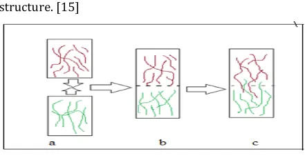
As per diffusion theory inter diffusion of polymers chains across an adhesive interface causes adhesion and is driven by concentration gradient. The polymer chains and the mucus coming in contact to a sufficient depth to create a semi-permanent adhesive bond (figure 3). The penetration depends on diffusion coefficient. The diffusion relay on molar weight and decreases rapidly as the cross-linking density increases. For good mucoadhesion some factors are Gite Shital Shridhar et al.: Mucoadhesive buccal drug delivery: An Overview
consider those are the chain flexibility, molecular weight, expanded nature of both the mucoadhesive and substrates as well as similarity in chemical structure. [15]
Fig 3: Diffusion theory
5. Fracture theory:
This is most accepted theory in case of mucoadhesion. This theory analyses the force required to detach two surfaces after adhesion. It measures the maximum tensile strength during detachment and it can be expressed by formula given below. [15]
G = (Eε/L) ½
Where E— Young’s modulus of elasticity ε — Fracture energy
L— Critical crack length when two surfaces are separated
G— Fracture strength equation details
Factors affecting to mucoadhesion
• Polymer based factors:
1. Molecular weight of polymer:
As the molecular weight of polymer is low, it can penetrate better in the mucus. The molecular weight can increases with increase in molecular weight up to 10,000 above that the mucoadhesive strength decreases. For linear polymer the bioadhesiveness improve with increase in molecular weight which depends on two things that for lower molecular weight, interpenetration is more critical and entanglement is important for higher molecular weight. [16]
2. Concentration:
For maximum bioadhesion it requires optimum concentration of bioadhesive polymer. In highly concentrated system beyond the optimum level, adhesive strength drops because coiled molecule
become separated from medium. When concentration of polymer is too low, the number of penetrating polymer chain per unit volume of mucus is also low. The concentration from 1-2.5% can show increased potential of bioadhesion. [17]
3. Flexibility of polymer chains:
For good bioadhesion it requires diffusion of polymer chains in interfacial region. So there must be requiring that polymer chain contain substantial degree of flexibility in order to achieve better adhesion. In mobility and flexibility of polymers can be related to their viscosities and diffusion coefficients. The higher flexibility of a polymer causes greater diffusion into the mucus network. [18]
4. Swelling factor:
It is an important factor that affects mucoadhesive strength of polymeric component. Hydration is required for a mucoadhesive polymer to expand and create a proper “macro-molecular mesh” of sufficient size and also to induce mobility in the polymer chains in order to enhance the interpenetration process between polymer and mucin. Polymer swelling results interpenetration by exposing the bioadhesive sites for hydrogen bonding and/or electrostatic interaction between the polymer and the mucous network. However, a critical degree of hydration of the mucoadhesive polymer exists where optimum swelling and bioadhesion occurs [19].
5. Degree of cross linking:
There are three important inter related structural parameters of polymer network are average pore size, average molecular weight and density of cross-linking. It is found that increase in density of cross linking water diffuse in polymer network at very lower rate, which shows insufficient swelling of polymer and interpenetration between polymer and mucin decreases. [19]
• Physical factors:[20]
1. pH:
pH can be inbetween 6.5-7.5. Polyanions are mostly preferred than polycations when two factors are consider, toxicity and bioadhesion. In case of carboxylic acid, pH value below pKa value is more favorable. It is suggested that within poly acrylic acid system approximately 80% of protonation of carboxylic group.
2. Applied strength:
To residence a solid bioadhesive system, it is compulsory to concern a defined strength. Whatever the polymer, carbopol 934, poly (acrylic acid / vinyl benzene poly (HEMA) or, the adhesion strength increases up to an optimum with the applied strength or with the period of its application. The pressure firstly applied to the mucoadhesive tissue contact site can impact the depth of interpenetration. The polymer used becomes bioadhesive even they don’t have interaction capacity when high pressure is applied.
• Physiological factors [21]:
1. Mucin turnover rate:
The turnover of mucin molecules is important because, the mucin turnover is expected to limit the residence time of the mucoadhesive on the mucus layer. If the adhesive strength is high, mucoadhesive are get detached from the surface due to mucin turn over. Mucin turnover results in substantial amounts of soluble mucin molecules. These molecules interact with the mucoadhesive before they have a chance to interact with the mucus layer. Presence of food can affect the mucin turnover.
2. Diseased state:
The physicochemical properties of mucus are get change during some disease state such as gastric ulcer, cold, ulcerative colitis and cystic fibrosis, bacterial and fungal infection.
Mucoadhesive Polymer
Mucoadhesive polymer which utilizes the property of bioadhesion. This can be used for targeting of drug to
particular region of the body. Mucoadhesive polymer is water soluble and water insoluble polymer which is swellable network joined by crosslinking agent. The polymer can possess sufficient polarity so that it can permit wetting by mucus and sufficient fluidity that enhance the mutual adsorption and penetration of polymer and mucus. Conventionally the mucoadhesive polymer divided into three main classes
1. Polymers that place in water become sticky and owe their mucoadhesion to stickiness.
2. Polymers that adhere through non-covalent, nonspecific interactions that is predominantly electrostatic in nature (although hydrogen and hydrophobic bonding may be involved).
3. Polymers that bind to specific receptor site on tile self-surface. [22]
Characteristics of Mucoadhesive Polymer [23]
1. Polymer must have the maximum molecular weight upto 10,000 or more to enhance adhesiveness between polymer and mucus. 2. In case of long chain polymer the chain length
must be enough long that promote interpenetration.
3. Flexibility of polymer chain must be there. 4. The polymer and its degradation products should
be nontoxic and should be non-absorbable from the gastrointestinal tract.
5. It should be non-irritant to the mucous membrane.
6. It should form a strong non-covalent bond with the mucin-epithelial cell surfaces.
7. It should adhere quickly to most tissue and should possess some site-specificity.
8. The polymer must not decompose on storage or during the shelf life of the dosage form.
9. The cost of polymer should not be high so that the prepared dosage form remains competitive. Gite Shital Shridhar et al.: Mucoadhesive buccal drug delivery: An Overview
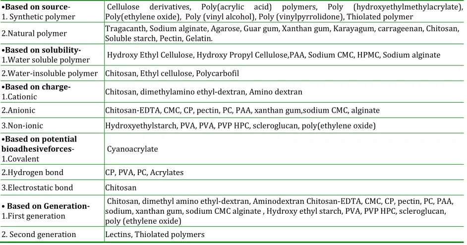
Table 1: Classification of Mucoadhesive Polymers
•Based on source- 1. Synthetic polymer
Cellulose derivatives, Poly(acrylic acid) polymers, Poly (hydroxyethylmethylacrylate), Poly(ethylene oxide), Poly (vinyl alcohol), Poly (vinylpyrrolidone), Thiolated polymer
2.Natural polymer Tragacanth, Sodium alginate, Agarose, Guar gum, Xanthan gum, Karayagum, carrageenan, Chitosan, Soluble starch, Pectin, Gelatin.
•Based on
solubility-1.Water soluble polymer Hydroxy Ethyl Cellulose, Hydroxy Propyl Cellulose,PAA, Sodium CMC, HPMC, Sodium alginate
2.Water-insoluble polymer Chitosan, Ethyl cellulose, Polycarbofil •Based on
charge-1.Cationic Chitosan, dimethylamino ethyl-dextran, Amino dextran
2.Anionic Chitosan-EDTA, CMC, CP, pectin, PC, PAA, xanthan gum,sodium CMC, alginate
3.Non-ionic Hydroxyethylstarch, PVA, PVA, PVP HPC, scleroglucan, poly(ethylene oxide) •Based on potential
bioadhesiveforces- 1.Covalent
Cyanoacrylate
2.Hydrogen bond CP, PVA, PC, Acrylates
3.Electrostatic bond Chitosan
• Based on Generation-1.First generation
Chitosan, dimethyl amino ethyl-dextran, Aminodextran Chitosan-EDTA, CMC, CP, pectin, PC, PAA, sodium, xanthan gum, sodium CMC alginate , Hydroxy ethyl starch, PVA, PVP HPC, scleroglucan, poly (ethylene oxide)
2. Second generation Lectins, Thiolated polymers
• Advantages of second generation polymer-
1. Site specific hence it called as cytoadhesive. 2. They are little or not affected by mucus
turnover rate.
3. Adhesive strength increase than normal mucoadhesive strength.
This second generation newer polymer can directly adhere to cell surface rather than mucus. They form covalent bond with mucus hence showimproved chemical interaction. This class of polymer includes lectins, thiolated polymer, polyox- WSRA, PAA-co-PEG. [24]
Mucoadhesive buccal dosage form
Based on their geometry buccal mucoadhesive dosage forms can be categorized into three types
Type I:
It is a single layer dosage form with multidirectional drug release. One of the disadvantages of this type of dosage form is that it suffers from significant loss of drug due to swallowing.
Type II:
It is a type, in which on top drug loaded bioadhesive layeran impermeable backing layeris superimposed,
creating a double-layered device and preventing drug loss from the top surface into the oral cavity.
Type III:
It is a unidirectional drug release device, from which drug loss is prevented or minimizes, since the drug is released only from the side that attaches to buccal mucosa. This can be achieved by covering every face of the dosage form, except the one that is in contact with the buccal mucosa. [25]
Fig 4: Buccal dosage forms
• Solid dosage form: 1. Buccal tablet:
oval shape. This tablet adheres to mucosa, softens and retained at site until release and/or dissolution is complete. [25] Mucoadhesive tablet can be prepared by methods such as wet granulation and direct compression. In case of buccal drug delivery, tablets are placed in buccal pouch where it can dissolve or erode hence it can be formulated with optimum pressure to produce hard tablet. Some of the examples of reported bioadhesive tablets are shown in table 2.
2. Bioadhesive microsphere:
Microsphere is an important part in case of novel drug delivery system. This mucoadhesive microsphere is mainly used for purpose of targeting to specific body cavity. Bioadhesive microspheres have benefits such as efficient absorption and enhanced bioavailability of drugs owing to a high surface-to-volume ratio, a much more close contact with the mucus layer and precise targeting of drugs to the absorption site.
3. Bioadhesive wafers:
It is theoretically novel periodontal drug delivery system that is proposed for the treatment of microbial infections linked with periodontitis. The Bioadhesive wafers with possessing adhesive properties, while the bulk layer contains of antimicrobial agents, biodegradable polymers and matrix polymers. [26]
4. Bioadhesive lozenges:
Bioadhesive lozenges may be used for delivery of drug that acts as antimicrobials, corticosteroids, local anesthetics, antibiotics and antifungal topically in the mouth. Lozenges produce a high release of drug at initial stage in the oral cavity, which rapidly drops to subtherapeutic levels, thus multiple dosing is required. A slow release bioadhesive lozenge also available which offers the potential for extended drug release with better patient compliance. [26]
• Semisolid dosage form:
1. Bioadhesive patch/ film:
Patches or film are preferred over tablet because of their comfort and flexibility. They are formulated such that it can provide contact between bioadhesive formulation and mucosa. Thickness of patch is a constraint which cannot provide control release of drug for longer period of time.In case of drug containing reservoir layer type; drug is released in controlled manner. Patches and film are mostly preferred for local action to treat oral diseases. There are many methods used for formulation of patch or films such as solvent casting method, hot melt extrusion technique, direct milling, semisolid casting, solid dispersion extrusion etc. Among that solvent casting is most popular method and widely used. [27]
2. Buccal gel and ointment:
As the advantage of dispersion gel and ointment has come in focus. They do not have accurate dosing as unit dosage form like tablet, patches or films, hence they are mostly preferred for local action where dose accuracy is less or not concern. For example local application of steroidal gel for treatment of mucosal ulceration. It has less patient acceptability than other mucoadhesive formulation. [28]
3. Medicated chewing gum:
Medicated chewing gum contains drug which after chewed, offer high amount of drug to prove local action in mouth. It can also shows absorption through systemic circulation. The medicated chewing gum for nicotine replacement therapy is available. Likewise caffeine chewing gums are also available. [29]
• Liquid dosage form:
These are available in form of solution or suspension of drug in suitable vehicle. This type of dosage form is available in market such as antibacterial mouthwashes, mouth freshener, employed for local action. Wide varieties of polymers are use from that chitosan has greatest binding capacity than other. Viscous liquid formulations are preferred to coat buccal cavity either as vehicle or as protectant. [30] Gite Shital Shridhar et al.: Mucoadhesive buccal drug delivery: An Overview
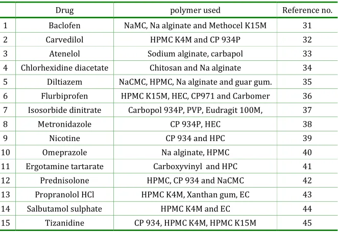
Table 2: Reported mucoadhesive drug delivery system
Drug polymer used Reference no.
1 Baclofen NaMC, Na alginate and Methocel K15M 31
2 Carvedilol HPMC K4M and CP 934P 32
3 Atenelol Sodium alginate, carbapol 33
4 Chlorhexidine diacetate Chitosan and Na alginate 34 5 Diltiazem NaCMC, HPMC, Na alginate and guar gum. 35
6 Flurbiprofen HPMC K15M, HEC, CP971 and Carbomer 36
7 Isosorbide dinitrate Carbopol 934P, PVP, Eudragit 100M, 37
8 Metronidazole CP 934P, HEC 38
9 Nicotine CP 934 and HPC 39
10 Omeprazole Na alginate, HPMC 40
11 Ergotamine tartarate Carboxyvinyl and HPC 41
12 Prednisolone HPMC, CP 934 and NaCMC 42
13 Propranolol HCl HPMC K4M, Xanthan gum, EC 43
14 Salbutamol sulphate HPMC K4M and EC 44 15 Tizanidine CP 934, HPMC K4M, HPMC K15M 45
Evaluation of buccal mucoadhesive dosage forms
[46]
Bioadhesive drug delivery devices are subjected for regular evaluation as that of conventional such that for tablet hardness, content uniformity, weight variation, thickness, in vitro dissolution, for patches and film tensile strength, film endurance, hygroscopicity and for gels and ointment viscosity, effect of aging.
1. Experimental Methodologies for Buccal Absorption/Permeability Study:
A) In Vitro Methods:
Now days, most of the in vitro studies examining drug transport across buccal mucosa have used buccal tissues from animal models. Animals are sacrificed immediately before the start of an experiment. Buccal mucosa with underlying connective tissue is surgically detached from the oral cavity, the connective tissue is then carefully removed and the buccal mucosal membrane is isolated. The membranes are then placed and stored in ice-cold (4°C) buffers (usually Krebs buffer) until mounted between side-by-side diffusion cells for the in vitro permeation experiments. B) In Vivo Methods:
It is also called as buccal absorption test. For kinetic drug absorption measurement this method can be
used. The procedure involves the swirling of a 25 mL sample of the test solution for up to 15 min by human volunteers followed by the expulsion of the solution. To calculate amount of drug absorbed, the amount of drug present in expelled volume can be determine. Some of the disadvantages are there like salivary dilution of drug and accidental swallowing of sample solution.
C) Experimental Animal Species:
Choice of animal for the experimental study is very important factor. To perform in vivo study researchers can prefer the animals depending on test to be perform. Most of animals having the keratinized buccal mucosa, but the rabbit and pig are the only animals which having non-keratinized mucosa as like humans. To study permeation of drug monkey, dog, pig animals are mostly used.
D) In Vitro Release Study:
For simulating in vivo conditions, researchers have developed different apparatus like:
• Beaker method
• Dissolution apparatus • Interface diffusion system • Modified Keshary Chien cell
Polymer characterization can be done by evaluating there mucoadhesive strength both in vivo and in vitro technique.
• In vitro evaluation:
1. Measurement of tensile strength:
This method includes measurement of force required to break bioadhesive bond between mucus membrane and polymer. For that various instruments are used that are as follows.
a) Modified physical balance or tensile tester:
A modified balance method was used to determine the in vitro mucoadhesive strength. This apparatus consist of modified physical balance. The right side of pan can be replaced by the glass slide with copper wire and also additional weight to compensate with left side weight. A teflon block of 2 cm height and 3.8 cm diameter was fabricated with an upward portion of 1.5 cm diameter and 2 cm height on one side. This was placed in beaker filled with pH 6.75buffer media, which was then positioned below right side of the balance. Rat or goat buccal mucosa was used as a model mucus membrane and for moistening purpose buffer medium 6.75 buffer media. The goat or rat buccal mucosa was obtained from local slaughter house and kept in a Krebs buffer solution during transportation. The mucus membrane was tied to Teflon block with the help of thread. The beaker was filled with phosphate buffer media to maintained viability of buccal mucosa. The one side of the tablet was attached to the glass slide of the right arm of the balance and then the beaker was elevated slowly until interaction between goat mucosa and mucoadhesive tablet was established. A 10 gm preload was placed on the slide for 5 min (preload time) to established adhesion bonding between mucoadhesive tablet and goat or rat stomach mucosa. After the finishing of preload time, preload was detached from the glass slide and water added in the plastic bottle in left side arm by peristaltic pump at a constant rate of 100 drops per minute. When mucoadhesive tablet was detached from the goat or rat buccal mucosa the addition of water was stopped. The weight of water
required to detach mucoadhesive tablet from buccal mucosa was noted as mucoadhesive strength in grams.
Force of adhesion (N) =Mucoadhesive strength 9.81/1000
Bond strength (N/m2)=Force of adhesion (N)/Surface area of tablet (m2)
b) Wilhelmy Plate Technique:
This technique conventionally used to measure dynamic contact angles and utilizes a microtensiometer and a microbalance. The instrument analyses the bioadhesive force between mucosal tissue and the formulation. This method measures the bioadhesive force between the mucosal tissue and the polymer attached to a metal wire and suspended into the microtensiometer. The mucosal tissue is placed in the tissue chamber and this chamber is raised so as to make intimate contact between the tissue and the test material. After a certain period the force of adhesion is measured by lowering the stage. By using the Cahn software system, parameters such as fracture strength, deformation to failure, and work of adhesion can be analyzed. This apparatus mainly analyze parameters such as fracture strength and deformation failure. [47]
2. Measurement of shear strength:
This is technique by which measurement of the shear stress give precise correlation to the adhesion strength. It consist of two smooth, polished plexi glass boxes were selected; on a glass plate one block was fixed with adhesive, which was fixed on leveled table. The level was adjusted with the spirit level. To the upper block, a thread was tied and it was passed down through a pulley. The length of the thread from the pulley to the pan was 12cm. At bottom side where the thread ends, a 17 g pan was attached containing weights. A recent technique involve the measurement of mucoadhesion by make use of a stainless steel rotating cylinder coated with freshly excised porcine intestinal mucosa to which polymer disc were attached. The cylinder was positioned in dissolution apparatus with rotating speed 125 rpm. It was Gite Shital Shridhar et al.: Mucoadhesive buccal drug delivery: An Overview
analysed after every 30 min for the attachment of the polymer discs. [47]
Other in vitro methods
I Rheological study:
The rheological information of polymer–mucus mixtures can offer an acceptable in vitro model which can correlate with in vivo performance of a mucoadhesive polymer. It is best method for determination of mucoadhesive potential of polymer by comparing binary mucus/polymer blends to the
equally concentrated monocomponent
mucus/polymer system. Chain interlocking, conformational changes and chemical interaction, which occur between the bioadhesive polymer and mucin chains, produce changes in the rheological behavior of the two macromolecular species.
II Colloidal Gold Staining Method:
This is a new in vitro method which was described for comparison of mucoadhesive property of various hydrogels. The technique employs red colloidal gold particles which are stabilized by the adsorbed mucin-gold conjugates. Because of interaction mucoadhesive develops red colour on its surface. By measuring the intensity of red colour mucoadhesive properties can be compared quantitatively. [46]
III Fluorescent probe method:
This method involves labelling of lipid bilayer of cultured human conjunctiva cells with pyrine as fluorescent probe. If the polymer can adhere to this cell, it can caused change in fluorescence due to chance in surface compression when compared with control cell. This change in degree of fluorescence is directly proportional to amount of polymer binding.To determine density on adhesion, polymer charge, and charge sign another probe can also be used. It states that determination of bioadhesive bond is based on molecular interaction of polymer with mucus. [38]
• In vivo methods of evaluation[46]
1. Gamma Scintigraphy Techniques:
It is an important instrument used in the development of pharmaceutical dosage forms. With help of this technique the information can collected in
non-invasively. This technique gives information of different regions of GI tract, the site of drug absorption, the time and site of disintegration of dosage forms and also the effect of, disease, food size of the dosage form on the in vivo performance of the dosage forms. Two important factor studied by this technique those are distribution and retention time of the mucoadhesive tablets. The combination of the sheep model and the gamma scintigraphy process has been proved to be a very useful tool for assessing the spreading distribution, and clearance of administered stomach mucoadhesive tablets.
2. GIT Transit using the Radio-Opaque Technique:
In this technique radio opaque markers are used to determine effect of polymer in GI transit time. Noninvasive method such as faeces examination and x-ray evaluation can provide sufficient data to study GI residence time. Cr 51, Tc99m, In113m or I123 these are some examples of marker which are used for mucoadhesive drug delivery.
3. Moisture Absorption Studies for Buccal Patches
The moisture absorption studies give a signal about the relative moisture absorption capacities of polymers. Moisture absorption studies have been performed in 5% w/v agar in distilled water, which can be heated and transferred to petri plates when it is hot and allowed to solidify. Then six buccal patches from each formulation batch were selected and weighed. Prior to study buccal patches were placed in a desiccator over night to remove moisture. After drying they were positioned on the surface of the agar plate and incubated at 37 °C in incubator. The patches were balanced again, and the percentage of the absorbed moisture was calculated by using the following formula-
% Moisture absorbed= Initial weight - Final weight/Initial weight *100.
4. Thickness
5. Folding endurance
Folding endurance of patches was determined manually. Patch was repeatedly folded at same point until it ruptures. The number of folding required cracking or breaking a patch was taken as the folding endurance.
6. Swelling study for tablet
Mucoadhesive dosage form like tablet can absorb liquid and due to swelling that result in to increase in volume and weight. Liquid uptake by the particle may be due to saturation of capillary spaces within the particles or hydration of macromolecule. The liquid pass in the particles through pores and bind to large molecule, breaking the hydrogen bond and causing in the swelling of particle. The degree of swelling can be calculated in terms of % weight gain by the mucoadhesive dosage form.
Method- Mucoadhesive dosage form is balanced and placed in a beaker having 200 ml of buffer media. After each interval the dosage form is removed from beaker and weighed again. This process follows up to 8 hours. The swelling index is calculated using following formula.
Swelling Index (S.I.) = (Wt-Wo)/Wo Where, S.I. = Swelling index
Wt = Weight of the dosage form at time t
Wo = Weight of the dosage form before placing in the beaker [48]
7. Surface pH study
The surface pH of the buccal tablets is determined in order to examine the possibility of any side effects in vivo as an acidic or alkaline pH may cause irritation to the buccal mucosa. The method accepted, is used to determine the surface pH of the bioadhesive formulation like tablet. A glass electrode is also used for this purpose. At room temperature the tablet is permitted to swell by keeping it in contact with 1 mL of distilled water (pH 6.5 ± 0.05) for 2 hours. The pH is measured by taking the electrode in contact with the surface of the tablet and allowing it to equilibrate for 1 minute. [48]
8. Residence time
The in vitro residence time mainly useful to know the mucoadhesive performance to retained at the site of application. This time can be measured with the help of modified disintegration apparatus. 800 ml isotonic buffer pH 6.75 solution can be used as disintegration medium 3 cm long rabbit mucosa was attached to glass slide and it was vertically attached to side arm. One surface of mucoadhesive tablet was hydrated with 15 ml of isotonic phosphate buffer solution then it was taken in mucosal contact. The movement of glass slide was allowed to up and down for complete immersion. Then time for detachment of tablet from mucosal surface can be noted. [49]
Future Perspectives
A buccal adhesive system offers countless advantages in terms of economy, accessibility, administration, withdrawal and patient compliance. Research scientists are now looking out the traditional polymers for novel drug transport systems. At the recent global picture, scientists are finding various ways to develop buccal adhesive dosage form to improve the low oral bioavailability drugs. It is found that the second generation mucoadhesive polymer having great potential. Novel buccal adhesive delivery system, where the drug delivery is directed towards buccal mucosa by considering local environment of oral site has come in existence. Now days solid dosage forms, liquids and gels applied to oral cavity are commercially well accepted by patients. The future direction of buccal adhesive drug delivery lies in delivery of peptides and protein and also vaccine formulations. Bilayer buccal tablets, films and patches are better approaches for the development of buccal formulations to deliver the drugs in combination. Microparticulate or nanoparticulate bioadhesive systems are particularly interesting now, as they offer protection to therapeutic entities as well as the enhanced absorption that result from delivery increased contact time provided by the bioadhesive component.
Gite Shital Shridhar et al.: Mucoadhesive buccal drug delivery: An Overview
CONCLUSION
Buccal drug delivery provides tremendous advantages over other dosage form. Therefore now days most of the research is going on to develop novel dosage form to overcome disadvantages. It provides intimate contact of dosage form at the site of buccal cavity which offers prolonged drug release. Mucoadhesive polymers are mainly used for this purpose which can
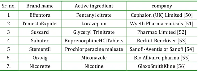
also avoid hepatic first pass elimination. For evaluation of this dosage form both in vivo and in vitro methods have been developed. Examples of some marketed buccal formulation are shown in table 3. Recently researchers facing many more challenges in development of such formulation and it requires a multidisciplinary approach.
Table 3: Marketed formulations
Sr. no. Brand name Active ingredient company
1 Effentora Fentanyl citrate Cephalon (UK) Limited [50]
2 TemestaExpidet Lorazepam Wyeth Pharmaceuticals [51]
3 Suscard Glyceryl Trinitrate Pharmax Limited [52] 4 Subutex BuprenorphineHClTablets Reckitt Benckiser [53]
5 Stementil Prochlorperazine maleate Sanofi-Aventis or Sanofi [54]
6. Oravig Miconazole Bio Alliance pharma [55] 7. Nicorette Nicotine GlaxoSmithKline [56]
REFERENCES
1. Shojaei A.H. Buccal Mucosa as A Route for Systemic
Drug Delivery: A Review. J Pharm Pharmaceut. Sci.
1998; 1(1):15-30.
2. Khanna R., Agraval S.P., Ahuja A. Mucoadhesive
buccal drug delivery a potential alternative to
conventional therapy. Ind. J. pharma. sci. 1998;
60(1):1-11.
3. Kamath K.R., Park K., Swarbrick J. and Boylan J.C.
Eds, Encyclopedia of pharmaceutical technology, vol
10, Marcel Dekker, New York, 1994.
4. Margret C., Mehul D., Chiranjib, Kumudhavalli,
Jayakar. Formulation, design and development of
buccoadhesive tablets of verapamil hydrochloride
Int. J. Pharm.Tech. Res. 2009; 1(4):1663-77.
5. Miller N.S., Chittchang M., Johnston T.P. The use of
mucoadhesive polymers in buccal drug delivery,
Adv. Drug Deliv. Rev. 2005; 57(1):1666–1691.
6. Sampath Kumar KP, Bhowmik D, Dutta A, Paswan S,
Deb L. Critical review on pharmaceutical science.
2012;1:79-93 (www.earthjournals.org)
7. Vyas S.P., Khar R.K. Controlled Drug Delivery and
Advances, 1st ed.; Vallabh prakashan: New Delhi,
2002.
8. Janet A.J., Hoogstraate and Philip W. Drug delivery
via the buccal mucosal. WertzPSTT.1998; 1(7):
306-316.
9. Harris D. and Robinson J.R. Drug delivery via the
mucous membranes of the oral cavity. J. Pharm. Sci.
1992; 81:1-10.
10. Shakya P., Satheesh Madhav N.V., Shakya A.K.,
Kuldeep singh. Palatal mucosa as a route for
systemic drug delivery: review. Journal of
Controlled Release. 2011; 151:2–9.
11. Patel M. et al. Buccal drug delivery system: the
current interest. Int. J. of pharmacy. 2011; 2
(12):4-11.
12. Smart J.D. The role of water movement and polymer
hydration in Mucoadhesion, in: E. Mathiowitz, D.E.
Chickering, CM. Lehr (Eds.), Bioadhesive Drug
Delivery Systems: Fundamentals, Novel Approaches
and Development, Marcel Decker, New York, 1999.
13. Phanindra B., Krishna Moorthy B. and
Muthukumaran M. Recent advances in
mucoadhesive/bioadhesive drug delivery system: A
review. Int. J. Pharm. Med. & Bio. Sc. 2013; 2(1):
68-84.
14. Dharmendra S., Surendra J.K., Sujata M., Ashish P.,
Shweta S. Mucoadhesive Drug Delivery System: A
Review. International Journal of Pharmaceutical &
Biological Archives. 2012; 3(6):1287-1291.
15. Tangri P. et al. Oral mucoadhesive drug delivery
system: A review. Int. J. of biopharm. 2011; 2(1):
16. Huang Y., Leobandung W., Foss A. and Peppas N.A.
Molecular aspects of mucoadhesion and
bioadhesion: tethered structures and site specific
surfaces. J Control Release, 2000; 65:63-71.
17. Patel Mitul et al. Buccal drug delivery system: the
current interest. Int. res. J. of pharm. 2012;
2(12):4-11.
18. Salamat-Miller N., Chittchang M., Johnston T.P. The
use of mucoadhesive polymers in buccal drug
delivery. Adv. Drug. Del. Rev. 2005; 57 (11): 1666–
1691.
19. Hagerstrom H., Paulsson M., Edsman K. Evaluation
of mucoadhesion for two polyelectrolyte gels in
simulated physiological conditions usinga
rheological method. Eur. J. Pharm. Sci. 2000; 9 (3):
301–309.
20. Sudhakar Y.,Kuotsu K.,Bandopadhyay A.K. Buccal
bioadhesive drug delivery- a promising option for
orally less efficient drugs, Jour. of controlled
release.2006; 114:15-40.
21. Saraswathi B., Balaji A. And Umashankar M.S.
Polymers in mucoadhesive drug delivery system-
latest updates. Int. J. Pharm. and Pharmace. Sci.
2013; 5(3):424-430
22. Zaheer A., Sachin, Swamay N.G.N. Mucoadhesive
Polymers: Drug Carriers for Improved Nasal Drug
Delivery, Indian Journal of Novel Drug Delivery.
2012; 4(1):2-16.
23. Yadavet V.K. et al., Mucoadhesive Polymers: Means
of Improving the Mucoadhesive Properties of Drug
Delivery System. J. Chem. Pharm. Res. 2010;
2(5):418-432
24. Andrews G.P. et al. mucoadhesive polymeric
platform for controlled drug delivery. Eur. J.
Biopharm. 2009; 71:505-518.
25. Bhalodia R., Basu B., Garala K., Joshi B. and Mehta K.
Bucoadhesive drug delivery system: A review. Int. J.
of Pharma. And Bio. Sci. 2010; 1(2).
26. Parthasarathy G., Bhaskar K., Jayaveera
K.N.,Prasanth V.V.Buccal Mucosa: a Gifted Choice for
Systemic Drug Delivery. Int. J. of Drug Deli. 2011;
3:586-596.
27. Narasimha R.R., Sindhu R.K., Swapna D., Konasree
S.D., Swathi E. Formulation and evaluation of
rapidly dissolving buccal patches. Int. J. Pharm. Bio
Sci. 2011; 1(3):145-159.
28. Smart J.D. Drug delivery using buccal-adhesive
systems, Advanced Drug Delivery Reviews.ll (1993)
253-270.
29. Kamimori G.H., Karyekar C.S., Otterstetter R., Cox
D.S., Balkin T.J., Belenky G.L., Eddington N.D. The
rate of absorption and relative bioavailability of
caffeine administered in chewing gum versus.
Capsules to normal healthy volunteers. Int. J. Pharm.
2002; 234:159-67.
30. Lee J., Kellaway I.W. Buccal permeation of (D-Ala2,
DLeu5) enkephalin from liquid crystal-line phases
of glycerylmonooleate. Int.J. Pharm. 2000; 195:35–
38.
31. Sudharshini et al. Design and evaluation of Baclofen
mucoadhesive tablets. Int. J. of Biomedical and Adv.
Res. 2010; 1(1): 25-35.
32. Pandey Sonia et al. Formulation and In-vitro
evaluation of bilayered buccal tablets of carvedilol.
Ind J. of Pharma. Edu. And Res. 2010; 44(3):
259-266.
33. Satishbabu B.K., Shriniwasan B.P. Preparation and
evaluation of buccoadhesive film of atenolol. Ind. J.
of phar. sci. 2008; 70 (2): 175-179.
34. Giunchedi et al. Formulation and in vivo evaluation
of Chlorhexidine buccal tablets prepared using
drug-loaded chitosan microspheres. Eur. J. Pharm.
Biopharm. 2002; 53: 233-239
35. Manivannan et al. Formulation and In-Vitro
evaluation of mucoadhesive buccal tablets of
Diltiazem Hydrochloride. Res. J. of Pharm. and Tech.
2008; 1(4):478-80.
36. Perioli et al. Mucoadhesive bilayered tablets for
buccal sustained release of Flurbiprofen, AAPS
Pharm SciTech. 2007; 8(3): E1-E2.
37. Doijad R.C. et al. Buccoadhesive drug delivery
system of Isosorbide dinitrate: Formulation and
evaluation. Ind. J. of phar. sci. 2006, 68(6): 744-746.
38. .Perioli L. Novel mucoadhesive buccal formulation
containing metronidazole for the treatment of
periodontal disease. Journal of Controlled Release.
2004; 95: 521– 533.
39. Park and Munday. Development and evaluation of a
biphasic buccal adhesive tablet for Nicotine
replacement therapy. Int. J. Pharm. 2002; 237:215–
226.
Gite Shital Shridhar et al.: Mucoadhesive buccal drug delivery: An Overview
40. Choi H.G. and Kim C.K. Development of Omeprazole
buccal adhesive with stability enhancement in
human saliva. J. Control Release.2000; 68: 397- 404.
41. Tsutsumi K. et al. Buccal absorption of ergotamine
tartrate using the bioadhesive tablet system in
guinea-pigs. Int. J. of Pharm. 2002; 238:161–170.
42. Samani et al. Formulation and in vitro evaluation of
Prednisolone buccoadhesive
tablets.Ilfarmaco.2005; 60:339-334.
43. Derle et al. Formulation and evaluation of
buccoadhesive bi-layer tablet of Propranolol
Hydrochloride. Int J. of Pharm. and Pharm. Sci.
2009; 1(1):206-212.
44. Arya et al. Development and evaluation of
mucoadhesive buccal tablets of Salbutamol
Sulphate. Int. J. of Pharmacy and Pharmaceutical Sci.
2010; 2(2):40-2.
45. Shivanand et al. Mucoadhesive bilayered buccal
tablets of Tizanidine Hydrochloride. Int. J. of Pharm.
Tech. Res. 2010; 2(3):1861-1869.
46. Shinkar D.M., Dhake A.S., Shetty C.M. Drug delivery
from oral cavity: A focus on mucoadhesive buccal
drug delivery system. PDA journal of
pharmaceutical science and technology.2012;
66(5): 466-500.
47. Santos C.A., Jacob J.S., Hertzog B.A., Carino G.P.,
Mathiowitz E. Methods and Compositions for
Enhancing the Bioadhesive Properties of Polymers
Using Organic Excipients. U.S. Patent 6,156,348,
December 5, 2000.
48. Muraleedhara K.K. et al. Senthil Kumar SK,
Parthiban SA.Mucoadhesive vaginal drug delivery
system: A review on advance status. Int. J.of pharm.
Res.And analysis. 2013; 3(1):33-46.
49. Patel V.F., Liu F., Brown M.B. Modeling the oral
cavity: In vitro and in vivo evaluations of buccal
drug delivery systems. Journal of Controlled
Release. 2012; 161: 746–756.
50. http://www.medicines.org.uk/emcmobile/medicin
e/27819/spc#companyDetails (2 Sept. 2013)
51. Drug Information, iDruginfo.com,
http://www.idruginfo.com/?cat=drug&s=Temesta
%20Expidet&ingredient=Lorazepam (2 Sept. 2013)
52. Netdoctor, suscard tablet,
http://www.netdoctor.co.uk/heart-and-blood/medicines/suscard-tablets.html (2 Sept.
2013)
53. Generic subutex,
http://pain.emedtv.com/subutex/generic-subutex.html (2 Sept. 2013)
54. Pharmabiz.com,http://pharmabiz.com/ArticleDetai
ls.aspx?aid=76475&sid=2 (2 Sept. 2013)
55. Datapharm, www.datapharm.org.uk (2 Sept. 2013)
56. MRP.com,http://www.empr.com/nicorette/drug/2
873/ (2 Sept. 2013)
How to cite this article: Gite Shital Shridhar*, Shinkar Dattatraya Manohar, Saudagar Ravindra Bhanudas1;
Mucoadhesive buccal drug delivery: An Overview; J. Adv. Pharm. Edu. & Res. 2013: 3(4): 319-332.