RESEARCH
Evolution and development of three highly
specialized floral structures of bee-pollinated
Phalaenopsis
species
Dewi Pramanik
1,2,3, Nemi Dorst
4, Niels Meesters
5, Marlies Spaans
4, Erik Smets
1,2,6, Monique Welten
1and Barbara Gravendeel
1,2,7*Abstract
Background: Variation in shape and size of many floral organs is related to pollinators. Evolution of such organs is driven by duplication and modification of MADS-box and MYB transcription factors. We applied a combination of micro-morphological (SEM and micro 3D-CT scanning) and molecular techniques (transcriptome and RT-PCR analysis) to understand the evolution and development of the callus, stelidia and mentum, three highly specialized floral structures of orchids involved in pollination. Early stage and mature tissues were collected from flowers of the bee-pollinated Phalaenopsis equestris and Phalaenopsis pulcherrima, two species that differ in floral morphology: P. equestris has a large callus but short stelidia and no mentum, whereas P. pulcherrima has a small callus, but long stelidia and a pronounced mentum.
Results: Our results show the stelidia develop from early primordial stages, whereas the callus and mentum develop later. In combination, the micro 3D-CT scan analysis and gene expression analyses show that the callus is of mixed petaloid-staminodial origin, the stelidia of staminodial origin, and the mentum of mixed sepaloid-petaloid-stamino-dial origin. SEP clade 1 copies are expressed in the larger callus of P. equestris, whereas AP3 clade 1 and AGL6 clade 1 copies are expressed in the pronounced mentum and long stelidia of P. pulcherrima. AP3 clade 4, PI-, AGL6 clade 2 and PCF clade 1 copies might have a balancing role in callus and gynostemium development. There appears to be a trade-off between DIV clade 2 expression with SEP clade 1 expression in the callus, on the one hand, and with AP3 clade 1 and AGL6 clade 1 expression in the stelidia and mentum on the other.
Conclusions: We detected differential growth and expression of MADS box AP3/PI-like, AGL6-like and SEP-like, and MYB DIV-like gene copies in the callus, stelidia and mentum of two species of Phalaenopsis, of which these floral structures are very differently shaped and sized. Our study provides a first glimpse of the evolutionary developmental mechanisms driving adaptation of Phalaenopsis flowers to different pollinators by providing combined micro-mor-phological and molecular evidence for a possible sepaloid–petaloid–staminodial origin of the orchid mentum. Keywords: Callus, Mentum, Stelidia, RT-PCR, Transcriptomics
© The Author(s) 2020. This article is licensed under a Creative Commons Attribution 4.0 International License, which permits use, sharing, adaptation, distribution and reproduction in any medium or format, as long as you give appropriate credit to the original author(s) and the source, provide a link to the Creative Commons licence, and indicate if changes were made. The images or other third party material in this article are included in the article’s Creative Commons licence, unless indicated otherwise in a credit line to the material. If material is not included in the article’s Creative Commons licence and your intended use is not permitted by statutory regulation or exceeds the permitted use, you will need to obtain permission directly from the copyright holder. To view a copy of this licence, visit http://creat iveco mmons .org/licen ses/by/4.0/. The Creative Commons Public Domain Dedication waiver (http://creat iveco mmons .org/publi cdoma in/ zero/1.0/) applies to the data made available in this article, unless otherwise stated in a credit line to the data.
Background
One of the key innovations in the evolution of flowering plants is the transfer of pollen by pollinators. This adap-tation enabled plants to reproduce much more efficiently and played a major role in the diversification of the flow-ering plants [1]. In more basal flowering plant lineages, such as Magnoliales, pollinators carry pollen all over
Open Access
*Correspondence: [email protected]
1 Naturalis Biodiversity Center, Endless Forms Group, Darwinweg 2, 2333
CR Leiden, The Netherlands
their body, resulting in a low pollination success. In more derived plant lineages such as orchids, very precise place-ment of pollen on very specific body parts of pollinators evolved, that ensured reproductive isolation and species diversification.
Apart from very precise pollen placement in orchids, deceptive pollination also evolved, in which pollinators are cheated when they visit flowers that offer no reward. Deceptive pollination promotes cross-fertilization by reducing the visitation period of pollinators and discour-aging returns to a single flower or inflorescence, in this way preventing self-pollination. Seven mechanisms of deceptive pollination of orchids have been described so far: generalized food deception, Batesian floral mimicry, brood-site imitation, shelter imitation, pseudo-antago-nism, rendezvous attraction and sexual deception [2]. The generalized food deception syndrome [3] is characterized by a model mimicking general floral signals of reward-ing plants such as a similar inflorescence shape, floral color, scent, nectar guides, spurs and pollen-like papillae [4, 5]. These signals attract recently emerged, immigrant or exploratory pollinators [2]. The Batesian mimicry syndrome copies one particular rewarding model plant species growing nearby [6–8]. In the case of brood-site imitation, orchids deceive insects by mimicking oviposi-tion substrates such as rotting fruit, dung, or fungi [9]. In the case of shelter imitation, flowers are shaped in the form of a tube [10] to provide shelter or a warm up place for insects in need of thermoregulation [11, 12]. The pseudo-antagonism syndrome relates to foraging behav-ior of kleptoparasitic pollinators by employing small flo-ral hairs easily vibrating in a small breeze that are thought to mimic insect prey captured in spider webs [13, 14]. The first step in evolution towards pseudocopulation might be related with the rendezvous syndrome [15]. In the lat-ter syndrome, flowers emit signals that attract pollina-tors of both sexes such as aggregation pheromones [16]. In the first syndrome, flowers mimic the shape and scent of female insects; this elicits male insect sexual behavior with the mimicking flower [17–19]. From the seven strat-egies described above, the most common one in orchids is general food deception. Around one-third of all pol-lination cases in orchids are categorized as general food deceptive [20]. Deceptive flowers look, feel and smell like flowers of nearby rewarding plants and only after visiting a cheating flower several times, inexperienced pollina-tors learn to distinguish a cheater from the model. This is sufficient to transfer pollen from one orchid to another and pass on this trait to future generations. Essential for the betrayal is that the deceptive flower manages to posi-tion the pollinator very accurately in front or below the stamen to remove pollen, and in a subsequent visit the stigma, to deposit pollen for cross-fertilization. Several
floral organs and structures in different floral whorls play an important role in this process.
Orchid flowers consist of five whorls. The two outer whorls contain three sepals and three petals, of which the median petal is transformed into a labellum. There are two staminal whorls. The outer whorl consists of two staminodes and one functional stamen. The inner whorl is assumed to contain three staminodes but this is a long debated issue. In the center of the flower there is a fifth whorl that contains a gynoecium that is formed by three fused carpels and at least one stamen. The median petal of orchid flowers, the labellum, is often enlarged and ornamented with a wart-like structure, the callus. Dur-ing landDur-ing on the orchid flower, the callus on the label-lum provides a holdfast for the insect to grasp with its front legs [21] (Fig. 1a). The reproductive organs (style, stigma, stamen) have become fused and incorporated in a gynostemium [22, 23]. To prevent the insect from walking off the labellum, wing-shaped structures on both sides of the gynostemium, so-called stelidia [24], are vital as they keep the insect trapped and also posi-tion the head or other body parts of the animal correctly in front of the reproductive organs (Fig. 1b). Lastly, the so-called mentum, an outgrowth formed by the bases of the gynostemium and labellum and the lateral sides of the lateral sepals [25] can act as a hinge, slamming an insect that walks over the labellum against the upper part of the gynostemium, where the anther and stigma are positioned, to ensure that removal and/or deposi-tion of the pollinia takes place at a very precise posideposi-tion on the pollinator and orchid flower (Fig. 1c). The size and shape of these three highly specialized floral structures became adapted to the bodies of specific pollinators dur-ing orchid evolution. This resulted in diversification of the shape and size of these structures. Such a diversifica-tion can for example be found in different species of the orchid genus Phalaenopsis. Examples of a large callus can be found in Phalaenopsis amabilis (Fig. 2a), P. celebensis
(Fig. 2c), and P. equestris (Fig. 2d), pronounced stelidia are present in P. bellina (Fig. 2b), P. celebensis (Fig. 2c) and P. pulcherrima (Fig. 2f), whereas a large mentum is present in P. pulcherrima (Figs. 1d and 2f).
Models of floral organ development in orchids
Multiple molecular studies show that the floral develop-ment of orchids involves interaction of different MADS box and other genes that duplicated early during orchid evolution [26–31]. The genes that are involved in floral development are interpreted with different models. The first model developed was the ABCDE model and this includes the following genes: MADS box A-class gene
APETALA1 (AP1), B-class genes APETALA3 (AP3) and
gene SEEDSTICK (STK) and E-class gene SEPALLATA
(SEP) [32]. Combinations of multiple MADS box genes are required to form different floral organs in the peta-loid monocots to which orchids belong: class A + B + E genes specify sepals and petals, B + C + E determine sta-mens, C + E specify carpels, and D + E are necessary for ovule development [33, 34]. Since this model could not entirely explain the shape of orchid flowers, it had to be expanded. First of all, petaloid monocots do not possess AP1 orthologues and FRUITFULL (FUL)-like genes are the closest homologues available [35]. The development of the orchid labellum cannot not be explained with the ABCDE model, so the Homeotic Orchid Tepal (HOT) model was developed [36], followed by the Perianth code [29] and the Oncidiinae model [31]. According to the HOT model, four AP3 gene copies (AP3-1, AP3-2, AP3
-3, and AP3-4) and PI, in combination with other classes MADS-box genes, regulate the flower identity program, resulting in distinct sepals, petals and a labellum (median petal) in orchids [36]. The Perianth code model explains
the ratio of two complexes of the MADS-box proteins
AP3/AGL6/PI, called SP and L, respectively, that play a
pivotal role in sepal-petal development and also deter-mine the identity of the labellum. When the complexes are skewed towards the L-complex, the labellum is large; when they are skewed towards the SP complex, the labellum is small or absent [29]. The Oncidiinae model summarizes differential expression of selected MADS-box genes in the perianth of Oncidium Gower Ramsey
and Erycina pusilla. Clade 1 AP3-like OMADS5 and
EpMADS15, and clade 1 AGL6-like genes OMADS7 and
EpMADS3 are expressed in the sepals and petals of both
species. Clade 2 AP3- like OMADS3 is expressed in the entire perianth of O. Gower Ramsey, while its ortholog,
EpMADS14, is only expressed in the lateral sepals of E.
pusilla. Moreover, clade 3 AGL6-like gene EpMADS4 is
solely expressed in the lateral sepals of E. pusilla, causing the median sepal to look different from the lateral ones in this species [31].
Other transcription factors that play an important role in shaping zygomorphy of flowers are RADIALIS (RAD), DIVARICATA (DIV) and DRIF (Divaricata Radialis interacting factor) that belong to the MYB gene family. They interact with each other and with TCP family genes to regulate floral dorsiventral asymmetry.
In Antirrhinum majus, DIV promotes the ventral
iden-tity of petals [37], and RAD promotes the dorsal iden-tity by binding to DRIF and preventing the formation of a DIV/DRIF complex. RAD and DIV interact with
DRIF in the regulation of floral symmetry. In the dorsal region, CYC/DICH genes interact with the promotor and intron of the RAD gene to activate its expression [38]. RAD proteins bind to DRIF I, preventing the bind-ing of DIV with DRIF, and ensure that a DIV/DRIF
complex cannot be formed. In the ventral region, the absence of RAD allows DIV to interact with DRIF, and this allows the formation of a DIV/DRIF complex that promotes the expression of genes that are specific to the ventral region of flowers [39]. Recently, it was dis-covered that MYB transcription factors play a role in shaping zygomorphy of the orchid flower as well [40, 41]. It was detected that the MYB transcription factors
DIV, RAD and DRIF can form a regulatory module that enriches the orchid developmental code [42, 43].
MADS‑box genes drive the development of the orchid callus, stelidia and mentum
So far, the expression of genes related to the development of a callus on the orchid labellum was only reported for
E. pusilla. Dirks-Mulder et al. [31] found a mixed
peta-loid-staminodial origin for this particular organ based on combined micro-CT evidence and expression of MADS box A (FUL-like), B (AP3, PI), E (SEP) and AGL6-2
genes. These results are in line with the ABCDE model, that predicts MADS box B-class genes in organs derived from petals, and the Perianth code model, which predicts expression of AGL6-2 genes in a labellum-derived organ.
expression of MADS box B and C-class genes in organs derived from stamens. To the best of our knowledge, no gene expression analyses of the orchid mentum have been reported yet.
To understand more about the evolution and develop-ment of the orchid callus, stelidia and develop-mentum, and the MADS-box and MYB transcription factors expressed in these structures, two different Phalaenopsis species, P.
equestris, and P. pulcherrima were studied with a
com-bination of micro-morphological and molecular tech-niques. A phylogenetic analysis of Phalaenopsis based on the nuclear internal transcribed spacers (nrITS) and plastid DNA showed that both species are a member of subgenus Phalaenopsis. Phalaenopsis equestris is part of section Stauroglottis, whereas P. pulcherrima is a member of section Esmeralda [50]. These species differ in terms of floral morphology as the first has a large callus, short stelidia and no mentum, whereas the second has a small callus, long stelidia and a pronounced mentum. Species of Phalaenopsis with a large callus, short stelidia and an inconspicuous mentum are pollinated by large pollina-tors such as Xylocopa bees [51, 52] whereas species with a small callus, long stelidia and a conspicuous mentum, like
P. pulcherrima, are pollinated by smaller pollinators such as Amegilla bees (Fig. 1d) [53]. The expectation was that the differences in floral morphology of P. equestris and P.
pulcherrima are correlated with differential expression
of a selection of MADS box genes earlier found to be expressed in the callus and stelidia of flower of E. pusilla
and other transcription factors. Phalaenopsis and Erycina
belong to the Aeridinae and Oncidiinae tribes, respec-tively, that are both members of the so-called ‘advanced’ Epidendroideae according to a phylogenetic analyses based on the nuclear internal transcribed spacers (nrITS), nuclear xanthine de-hydrogenase (XDH), mitochondrial
nad1 and plastid DNA [54]. Questions that we aimed to answer were (i) is the callus in Phalaenopsis flowers of mixed petaloid-staminodial origin, (ii) are the stelidia of
Phalaenopsis flowers derived from staminodes, (iii) what
is the evolutionary origin of the mentum in Phalaenopsis,
and (iv) can differences in the size and shape of the callus, mentum and stelidia of P. equestris and P. pulcherrima be explained by differential expression of MADS box and/or MYB transcription factors?
Materials and methods
Plant material
A total of three P. equestris and three P. pulcherrima
plants, obtained from commercial growers, were reared in the tropical greenhouses of the Hortus botanicus of Leiden University. Based on the size of the floral buds, five different developmental stages were defined (Fig. 3). Floral samples were freshly collected and stored at
− 80 °C for molecular analysis or in standard formalin-aceto-alcohol (FAA: absolute ethanol, 90%; glacial ace-tic acid, 5%; formalin aceace-tic acid, 5%) for imaging. Floral organs for SEM imaging and fully open flowers for micro 3-D CT scanning were immersed in FAA solution for 1 h under vacuum pressure at room temperature, then left for a minimum of 7 days under room temperature.
Scanning electronic microscopy (SEM)
Spirit samples of floral organs and structures preserved in a FAA solution were first transferred to 70% ethanol solutions and then further dissected under light micro-scope (Leitz). Subsequently the samples were washed in 96% ethanol (two times, 20 min) and in 96% acetone (two times, 30 min). To dry the samples and remove any pres-ence of water, a critical point dryer (Leica EM CPD300) was used. The samples were mounted on aluminum stubs of 12.2 × 10 mm diameter (Ted Pella, Inc) using a conductive carbon cement (Structure Probe Inc, United States) or sticker glue (Carbon adhesive tabs, EMS). To inhibit charging, reduce thermal damage, and improve secondary electron emission, the samples were coated with a Platina-Palladium layer of 20 nm, by using a sput-ter coasput-ter (Quorum Q150TS). Sample observations were obtained using a JSM-7600F SEM field emission scanning electron microscope (JEOL).
Micro‑3‑D computed tomography scanning
Spirit samples of mature flowers stored in FAA were treated with 0.1% Phosphotungstic acid (PTA) in 70% ethanol over a timespan of five days and daily refreshed. Prior to scanning, the samples were washed in 70%
ethanol two times for 30 min each, then embedded in 1.5% low melting point agarose (Promega). The scans were performed on a Zeiss Xradia 510 Versa 3D X-ray with a Sealed transmission of 30–160 kV, with a maxi-mum of 10 W X-ray source. Scanning of the flower was performed using the following settings: lens magnitude 0.5x; acceleration voltage/power 80 kV/7 W; exposure time 21 s; projections 1400. The total exposure time was approximately 9 h and 17 min.
Vascular bundles reconstruction and serial layer sectioning
Image stacking and vascular bundle reconstruction was performed in Amira-Avizo 9.5 (Thermo Fisher Scientific). With this software, 3-D structures were made through voxels (3-D pixels). Vascular bundle coloration was pro-duced by creating a script pipeline on the project panel and coloring the bundles in the segmentation editor. Through 2-D segmentation of the images, and electronic volume removal of the lobes, the vascular bundles in the callus, mentum and stelidia could be studied. To better understand organ development, serial layer sectioning was produced from processed 3-D CT scan images. The layer sectioning was made using the script of the bound-ing box and color wash.
RNA extraction
Floral buds were collected from stage 1, whereas the cal-lus and gynostemium (including mentum and stelidia, but excluding the anther cap with pollinia) were dis-sected and collected from stage 5 (Fig. 3). A maximum of 100 mg of each sample was transferred into sterile 2.2-mL micro-centrifuge tubes, together with a 7-mm glass bead (Assistent), and stored at − 80 °C. The frozen samples were grinded by using a TissueLyser II (QIAGEN). RNA was extracted by using a RNeasy Plant Mini Kit (QIA-GEN), under RNase-free conditions. DNase I Amp Grade (Invitrogen 1U/μl) was applied to the extracted RNA to digest single- and double-stranded DNA. To assess the quantity and quality of the samples for cDNA synthesis, they were measured with a nanodrop (NanoDrop 2000c, ThermoFisher), whereas for sequencing, they were meas-ured by Agilent 2100 Bioanalyzer Systems (Agilent Tech-nologies). The quantity of the RNA samples analyzed was at least 50 ng/μL RNA in 50 μL volume, the quality as assessed by the RNA integrity Number (RIN) was at least 7. All RNA samples with a RIN < 7 were discarded. Samples used for further downstream experiments were stored at − 80 °C.
Transcriptomic and bioinformatic analyses
RNA samples isolated from three biological replicates of early floral buds, mature callus and gynostemium of P.
equestris and P. pulcherrima were used for sequencing,
and they were processed separately per organ. These samples were sequenced by the Beijing Genome Insti-tute (BGI) using de novo sequencing with an Illumina HiSeq PE150. BGI cleaned the generated reads and uploaded these to their cloud service. The cleaned reads were downloaded and further analyzed with an in-house designed bioinformatics pipeline [55] using the Natura-lis OpenStack server, which was accessed via PuTTY, a free SSH and telnet client (https ://www.putty .org/). An in-house designed bioinformatics pipeline was used for quality control, assembly, annotation and differential expression analysis. First, the reads were checked for quality with FastQC v0.10.1 [56]. Reads with low quality were trimmed or removed with Trimmomatic v0.32 [57]. With the cleaned reads a de novo assembly was made using Trinity v2.5.1 [58]. Trinity used Bowtie2 v2-2.3.3.1 [59] for aligning the reads. The output of Trinity was clustered to remove redundant data and reduce the size of the transcriptomes using CDHIT-EST [60]. Longer consensus transcripts called ‘unigenes’ resulting from the assembly step were subsequently annotated with gene names using a dedicated local orchid gene database that was made by filtering orchid sequences from the NCBI GenBank Nucleotide (nt) and Non-Redundant (nr) pro-tein databases. The reads were aligned to these unigenes, which enabled the next step: creating a count table with expression values. This count table was based on Trinity identifiers, which were used to link the unigenes to the annotations.
Differential gene expression analysis
expression levels shown in the heatmaps depicted in Figs. 7 and 8.
cDNA synthesis
cDNA was reverse-transcribed through reverse tran-scriptase (RT) by using the iScript cDNA Synthesis Kit (Bio-RAD). The reaction mix (20 µL) consisted of 5 × iscript reaction mix (4 µL), iscript reverse tran-scriptase (1 μL), 1 μg RNA template and water for the rest of the volume. The cDNA was synthesized under the fol-lowing conditions: priming of the RT at 25 °C for 5 min; reverse transcription at 46 °C for 20 min; RT inactivation at 95 °C for 1 min.
Primer design
Nucleotide sequences of MADS-box A, B, C, D and E class genes of Phalaenopsis and control genes PeACT
and PpACT (Actin) were generated from reads obtained
from the transcriptomes. Specific primers were designed in Geneious version 8.1.8 (http://www.genei ous.com) (Additional file 1: Table S1). For screening of bind-ing specificity, all primer pairs were blasted against the Orchidstra database and further tested using PCR and gel electrophoresis.
Semi‑quantitative reverse transcriptase PCR (RT‑PCR)
A semi-quantitative RT-PCR was used to quantify the expression of selected MADS box genes. Total cDNA from three biological replications of early floral buds, mature callus and gynostemium of P. equestris and early floral buds, mature callus and mix of gynostemium, stel-idia and mentum of P. pulcherrima was analyzed in triplo for each primer set. PeACT and PpACT were used as positive control, while the non-template control (reac-tion without cDNA template) was assigned as nega-tive control. Each amplicon group contained the tested samples, a positive control, and a non-template control. The amplification reaction was prepared by using 90 ng cDNA template. The reaction mix contained 10 × Coral-Load Buffer (Qiagen), 25 mM MgCl2 (Qiagen), 100 mM Bovine Serum Albumin, Acetylated-BSA (Promega), 1.25x DMSO (Qiagen), 5x Q-Solution (Qiagen), 0.2 μM of forward and reverse primer (IDT), 2.5 mM dNTPs (Qiagen), 1.25 units/50 µl DNA Taq Polymerase (Qia-gen), and milliQ water (Ultrapure) was used to reach the final volume of 25 µl. The amplification protocol started with a polymerase activation and initial denaturation at 94 °C for 5 min; continued with 35 cycles of denatura-tion at 94 °C for 30 s; then an annealing step at 56 °C for 30 s; an extension step at 72 °C for 1 min 40 s; and a final extension at 72 °C for 7 min. PCR products were run on a 1.0% agarose gel (DNAase/RNAase-free, Bio-line) in 0.5× Ultrapure Tris–Borate–EDTA (TBE) buffer
(ThermoScientific) for 30 min at 100 V. The gels were stained with ethidium bromide (Gibco-BRL) for 30 min and digitally photographed using an Ultima 10si gel doc system (ISOGEN, Life Science).
Phylogenetic analysis
A multiple sequence alignment was performed by using the ClustalW alignment tool within Geneious version 8.1.8 based on translated nucleotides. The nucleotide sequences of MADS box genes of P. equestris and P.
pul-cherrima from transcriptome analysis were first
trans-lated to amino acids in the correct translation frame (Additional file 1: Table S2). The reference protein sequences of floral development genes were downloaded from databases (Additional file 1: Table S3). Additional references of Orchis italica and Apostasia shenzenica
MADS box genes sequences were obtained from Val-oroso et al. [40] and Zhang et al. [62], respectively. The created alignment was trimmed down to the most con-served regions (protein domains and amino acid motifs) to make sure that all sequences had the same length. Regions that did not align were removed prior to further analysis and the presence/absence of shared indels was coded with an extra 1 or 0. Phylogenetic trees were gen-erated with the Geneious Tree Builder plug-in using the Maximum Likelihood (ML) method with gymnosperm gene lineages as outgroup based on Pabón-Mora et al. [63]. A bootstrapping of 100 times was carried out, of which the resulting support values are shown as numbers above the branches.
Results
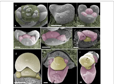
Floral ontogeny and micromorphology
the gynostemium at the beginning of developmental stage 2, from which they further elongate. This part of the floral ontogeny was largely congruent between P. equestris (Fig. 4) and P. pulcherrima (data not shown). Compared to P. equestris, the callus in P. pulcherrima
remained smaller, but the stelidia elongated much more during developmental phases 3-5 (Fig. 5a–e). The men-tum of P. pulcherrima flowers starts to form in develop-mental stage 4 (Fig. 3b).
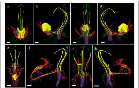
To study the anatomy of mature flowers of P. eques-tris and P. pulcherrima, and verify the results from the SEM analyses, micro 3-D CT scans were made from mature stage 5 flowers of P. equestris (Fig. 6a–d; Addi-tional file 2: Movie S1) and P. pulcherrima (Fig. 6e–h; Additional file 2: Movie S2). Six vascular bundles could
be visualized in the inferior ovary, indicated in purple (Fig. 6b–h). Three of these bundles, indicated in green, run to the median and abaxial sepals (Fig. 6b–h). Three main groups of bundles, indicated in red, were found to feed the petals, including the labellum, in which they further split up (Fig. 6a–h). Four vascular bundles, indi-cated in yellow, could be detected, running towards the fertile stamen and staminodes. One of these bundles runs to the fertile stamen (Fig. 6b–d, f–h); two other bundles, originating from two pairs each, run up into the stelidia (Fig. 6a–h) and the remaining bundle runs all the way up into the callus on the labellum (Fig. 6a– h). The mentum is vascularized by bundles indicated in green (Fig. 6 g–h), red (Fig. 6e–h), and yellow (Fig. 6e– h). When following all vascular bundles downwards, Fig. 4 Floral ontogeny of Phalaenopsis equestris. a Undifferentiated oval-shaped early stage primordia covered by bracts, ×50 magnitude; b
they connect in a plexus situated on top of the inferior ovary, where they connect with the remainder of the vascular system of the flower.
Duplications and expression of selected developmental genes in early floral buds and mature floral structures
Phylogenetic analyses recovered two clades of MADS box FUL-like genes, four clades of AP3 genes, one clade of PI genes, three clades of AG genes, three clades of STK
genes, four clades of SEP genes and three clades of ALG6 -genes. Of the MYB transcription factors, we found three clades of DIV genes, one clade of RAD genes and three clades of DRIF genes. Of the TCP transcription factors, we found two clades of PCF genes, two clades of CIN
genes and three clades of CYC/TB1 genes (Additional file 3: Figs. S1–S11).
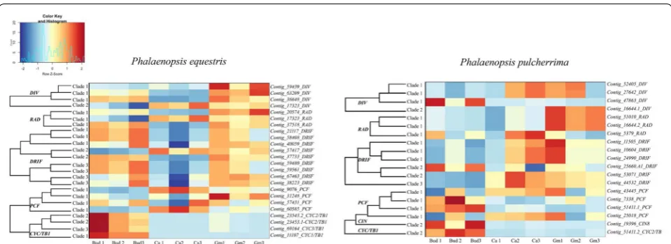
The transcriptome analysis showed that the differ-ent gene copies investigated were not expressed in all floral structures studied and the level of expression also varied. Below, we summarize the patterns found for the MADS box, MYB and TCP transcription factors. Of the MADS-box genes, the A-class genes were found to have a higher number of counts in early than late floral bud
development. B-class genes were mostly expressed in the mature callus. C and D-class genes were highly expressed in the mature gynostemium. The AGL6-like and E class genes had variable differential expression patterns. In P.
equestris these genes were predominantly expressed in
the early floral bud and mature gynostemium, while in
P. pulcherrima, these genes were mostly expressed in the
mature callus and gynostemium (Fig. 7).
DIV and RAD genes in both Phalaenopsis species had less expression in the early floral bud but more in the mature callus and gynostemium. The expression of DIV
clade 2 was further analyzed using semi-quantitative RT-PCR. The DIV clade 2 gene was found to be highly expressed in the gynostemium of P. equestris and in the callus of P. pulcherrima. Similar to the AGL6 and E-class genes, DRIF genes were highly expressed in the early flo-ral bud and mature gynostemium of P. equestris. In P.
pul-cherrima, most DRIF copies were highly expressed in the
mature callus and gynostemium. Most TCP genes were mainly expressed in the early floral bud, except for two
PCF-like gene copies, that were expressed in the mature callus and gynostemium of both species of Phalaenopsis
Fig. 6 Reconstruction of vascular bundle patterns in a mature Phalaenopsis flower based on μCT scanning. a–d P. equestris; e–h. P. pulcherrima. a Frontal view; b successive clockwise turn of 45 degrees; c successive clockwise turn of 90 degrees; d successive clockwise turn of 135 degrees; e frontal view; f successive clockwise turn of 45 degrees; g successive clockwise turn of 90 degrees; h. successive clockwise turn of 135 degrees. Color codes: purple = vascular bundles in ovary; yellow = vascular bundles in stamen and staminodes; green = vascular bundles in sepals; red = vascular bundles in labellum and petals. Scale bar: 100 μm
1 lineage was highly expressed in all tissue types analyzed in both Phalaenopsis species (Fig. 9). To further under-stand the molecular basis of the development of the cal-lus, stelidia, and mentum in Phalaenopsis, we also further analyzed MADS-box gene expression profiles by using semi-quantitative RT-PCR (Fig. 9). These results are described in more detail below.
MADS box A‑class genes
Two copies of FUL-like genes could be detected in the transcriptomes of P. equestris and P. pulcherrima. These were identified as ORAP13 and ORAP11, which repre-sent FUL clade 1 and FUL clade 2 lineages (Additional file 3: Fig. S1). No representative of FUL clade 3 could be detected, which is consistent with previous findings [64]. Both ORAP13 and ORAP11 were highly expressed in early stage floral buds, and their expression decreased in later developmental stages (Fig. 7).
MADS box B‑class genes
One copy of PI and four copies of AP3 gene lineages were detected in the transcriptomes of P. equestris and P. pul-cherrima (Additional file 3: Figs. S2 and S3). There was no expression of AP3 clade 3 (PeMADS3 and PpMADS3) in the early floral bud, mature callus and gynostemium of P. equestris and P. pulcherrima. The AP3 clade 1 gene
line-age PeMADS2 was only expressed in the mature callus of
P. equestris, while the ortholog PpMADS2 was expressed
in the early floral buds, mature callus and gynoste-mium of P. pulcherrima. AP3 clade 2 gene lineages were expressed in early floral buds and mature gynostemium of both Phalaenopsis species. AP3 clade 4 gene line-ages PeMADS4 and PpMADS4 were highly expressed in
all floral structures of both Phalaenopsis species and a similar expression pattern was found for the PI lineages
PeMADS6 and PpMADS6 (Fig. 9).
MADS box C and D‑class genes
Three copies of AG and two copies of STK could be detected in the transcriptomes of P. equestris and
P. pulcherrima. PeMADS 1, LOC110032491 and
LOC110029636 were found to belong to AG clades 1,
2 and 3, whereas PeMADS7 and LOC110021571 were found to belong to STK clades 1 and 2, respectively (Additional file 3: Figs. S4 and S5). AG-like and STK gene lineages were mostly expressed in the mature gynoste-mium of P. equestris. In P. pulcherrima, the expression of these gene lineages was more varied. Low expression of AG and STK genes was found in early floral buds and the mature callus of both P. equestris and P. pulcherrima, except for AG clade 3 and STK clade 2 gene lineages, that were found to have a higher expression in early stage flo-ral buds of both species (Fig. 7).
MADS box E‑class genes
Four copies of SEP-like genes could be detected in the transcriptomes of P. equestris and P. pulcherrima. In P.
equestris, PeSEP2 was found to be a member of clade
1 and PeSEP1 a member of clade 2, while PeSEP3 and
PeSEP4 were found to be members of clade 3 and 4,
respectively. PpSEP1, PpSEP3, PpSEP2 and PpSEP4 could be assigned to clades 1–4 (Additional file 3: Fig. S7).
buds, mature callus and gynostemium of P. equestris.
However, there was no expression of its ortholog PpSEP1
in the early floral buds, mature callus and gynostemium
of P. pulcherrima. The SEP-like gene lineages showed
similar expression patterns: SEP clade 2, 3, and 4 cop-ies were expressed in the early floral buds and mature gynostemium of P. equestris, and in the early floral bud, mature callus and gynostemium of P. pulcherrima (Figs. 7 and 9).
MADS box A–E‑class genes
AGL6-like genes, categorized as AE-class genes [65], were detected with three different copies in both transcrip-tomes analyzed, in agreement with Cai et al. [66]. The first clade of AGL6 was found to contain PeMADS16 and
PpAGL6-1, the second clade existed of PeMADS14 and
PpAGL6-2, and LOC110023965/PeAGL6-3 were found to
be associated with AGL6 clade 3 (Additional file 3: Fig. S6). The AGL6-like copy PpAGL6-1 was expressed in early floral buds, mature callus, and gynostemium. No expression of its ortholog PeAGL6-2 was found in early floral buds, mature callus and gynostemium of P.
eques-tris. PeAGL6-3 was highly expressed in early floral buds
and mature gynostemium of both species. Its ortholog in
P. pulcherrima was highly expressed in the callus (Figs. 7 and 9).
Discussion
Homology of the callus
petaloid vascular bundles. The staminodial vascular bun-dle, connected to the base of the callus, apparently fed the callus to develop and grow in size since no bundles were detected inside the callus. Its first appearance occurs dur-ing the second stage of floral development. A small callus is located on the base of the labellum, eventually guarded by the lateral lobes of the labellum that are still develop-ing durdevelop-ing this stage. The callus grew significantly durdevelop-ing all developmental stages, reaching an approximate size of 4 mm in diameter in mature flowers. The development of the callus of E. pusilla and P. equestris was highly similar. A different pattern was found for the development of the callus of P. pulcherrima: in early development, the size ratio between the labellum and callus was 2:1, whereas in later stages, the ratio between labellum and callus became 3:1, indicating more expansion of the labellum as compared to the callus (Figs. 10, 11).
Because of the mixed petaloid-staminodial origin of the callus found in our micro-CT analyses, we expected a combined expression of MADS-box A, B, and E-class genes in this particular floral organ. Expression of MADS box AE-class AGL6-clade 2, and B-class PI-like and AP3 -like clade 4 copies was indeed found in the mature cal-lus of both species of Phalaenopsis investigated (Fig. 9a). The observed expression of PI-like genes is in agreement with other studies that report this gene to be involved in the regulation of sepals, petals, labellum and gynoste-mium in Phalaenopsis and Dendrobium [27, 45, 67]. Dif-ferences in expression of certain gene copies among the two species of Phalaenopsis investigated were found as well. The SEPALLATA E-class MADS-box gene PeSEP2
was expressed in the large callus of P. equestris, but its ortholog PpSEP1 was not expressed in the small callus of
P. pulcherrima. Expression of the AE-class AGL6 clade 1
and AGL6 clade 3, B-class AP3 clade 1 and AP3 clade 2, and E-class SEP clade 2, 3 and 4 copies was only observed in the small callus of P. pulcherrima but not in the large callus of P. equestris. These results agree with a previ-ous study that found that the Phalaenopsis B-class AP3
and PI-like genes and AE-class AGL6 genes may form higher protein complexes with several PeSEP gene copies to determine floral organ identity [28]. We found higher expression of DIV 2 and lower expression of DIV 1 copies in the larger callus of P. equestris than in the smaller cal-lus of P. pulcherrima.
Homology of the stelidia and mentum
The stelidia develop differently in comparison to the cal-lus in both P. equestris and P. pulcherrima. Four vascular bundles can be found inside the stelidia that are derived from the merged staminodes a1 plus A3 and a2 plus A2 [68]. In later developmental stages, the gynostemium develops by fusion of the fertile stamen A1, median
carpel, and staminodes, which were still separate floral structures in early developmental stages. In some spe-cies, such as P. equestris, staminodial tissue fuses with the gynostemium in an earlier stage than in other species. In
P. pulcherrima, the staminodial tissue remains separate
and increases in size during later developmental stages. Our findings support those of Kurzweil and Kocyan [24], who found that staminodes a1 and a2 are almost always present during early ontogeny of monandrous orchids, but become incorporated in various degrees in the gynostemium in later stages. The staminodes either remain visible as stelidia or become incorporated in the gynostemium (Fig. 11).
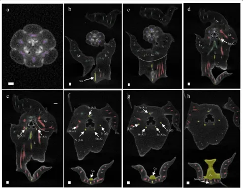
Our ontogenetic study showed that a mentum devel-ops quite late in P. pulcherrima as it started to develop only in phase 4 (Fig. 11). This result is congruent with observations obtained for other orchids with a mentum. For example, in Bletia purpurea, where the mentum was described as an organ originating from a basal extension of the gynostemium and fusion with staminodes and lat-eral sepals [24, 69]. Our micro-CT results provide further support for this hypothesis as both staminodial (a3, A2 and A3) and sepaloid vascular bundles were found to be feeding the mentum, next to vascular bundles also feed-ing the labellum and petals.
Because of the staminodial origin of the stelidia, we expected expression of MADS-box C-, D- and E-class genes in these floral structures as previously found in the stelidia of Erycina pusilla [31]. Expression of MADS box AE-class AGL6 clade 2 and 3 genes was indeed detected in these structures together with E-class SEP clade 2,3 and 4 lineages (Fig. 9b). Because of the mixed petaloid, sepaloid, staminodial origin of the mentum, we expected expression of all five classes of MADS-box genes in this organ. As expected, B-class AP3 clade 3 and 4 and PI-like genes were expressed in the gynostemium of both spe-cies of Phalaenopsis investigated. Expression of MADS box B-class AP3 clade 1 and AE-class AGL6 clade 1 cop-ies was detected in the gynostemium with pronounced stelidia and mentum of P. pulcherrima, whereas these gene copies were not expressed in the gynostemium of P.
equestris, lacking a pronounced mentum and stelidia. In
the gynostemium of the latter species, expression of the E-class SEP clade 1 gene lineage was higher than in the gynostemium of P. pulcherrima. In both species of
Phal-aenopsis investigated, SEP clade 1 copies were found to
expression could be detected in the stelidia and mentum of the two species of Phalaenopsis analyzed.
Implications for current orchid floral models
We found that most of the MADS-box AE-class AGL6 -like, B-class AP3-like and AE-class SEP-like gene line-ages investigated were expressed in early floral buds of
both Phalaenopsis species investigated. These results are in line with previous findings of AP3 gene copies being involved in orchid floral primordia development [36]. Similarly, AGL6-like OSMADS6 was reported to have a function in regulating early meristem identity and floral development in rice [73]. During later devel-opment, these genes showed differential expression in separate floral organs.
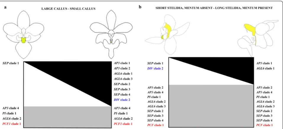
In our study, we discovered possible new roles of B-class and AGL6-like gene lineages in the formation of the callus, mentum and stelidia in orchid flowers. We found that the SP complex PpAP3-1/PpAGL6-1/PpPI,
that promotes sepal and petal development in the peri-anth, is also expressed in the callus, mentum and stelidia (Fig. 12). AGL6 proteins can interact with several MADS proteins during floral development [29, 47, 72, 74]. Our results suggest that duplication of AGL6 might have led to novel functions of these genes in the flowers of
Phal-aenopsis, especially regarding the development of the
stelidia and mentum. Combined expression of the SP complex genes together with SEP clade 2, 3 and 4 line-ages, but lack of expression of the SEP clade 1 lineage, results in a small callus, pronounced mentum and long stelidia in P. pulcherrima. The reverse, namely the expres-sion of the SEP clade 1 gene lineage but lack of expression of the SP complex and SEP clade 2, 3 and 4 gene lineages, results in a large callus in P. equestris. The expression of all four SEP lineages but lack of expression of the SP complex results in the lack of a mentum and short ste-lidia in P. equestris. No expression of AP3 clade 3 line-ages was detected in either the callus or gynostemium of both species of Phalaenopsis investigated. We therefore hypothesize that the role of the AP3 clade 3 lineage, one of the three gene lineages active in the L-complex, may be replaced by the AP3 clade 4 lineage in the callus and by AP3 clade 2 and/or 4 in the gynostemium.
We detected similar expression patterns of E-class SEP
clade 2, 3, and 4 lineages in the callus and gynostemium of both species of Phalaenopsis investigated (Fig. 10). In
Arabidopsis thaliana these lineages have highly
redun-dant functions [75, 76] but the role of the individual SEP
genes in the floral organs and structures of Phalaeno-psis seems very different. Pan et al. [28] discovered that downregulation of the SEP clade 1 lineage in P. eques-tris did not produce any morphological effect, in con-trast with downregulation of the SEP clade 3 lineage,
which resulted in a change in floral morphology. Pelaz et al. [75] reported that downregulation of the activity of
SEP1, 2, and 3 results in all organs developing into sepals in Arabidopsis thaliana. This implies that SEP genes are required for the development of petals in the second, sta-mens in the third and carpels in the fourth floral whorl. Our results suggest that these genes are also involved in the regulation of the first, second and third whorl men-tum, second whorl callus and third whorl stelidia in the flowers of Phalaenopsis.
Zygomorphy of flowers improves the efficiency of their pollination. Bilateral symmetry ensures that pollina-tors can only make contact with the stamen and stigma from one particular direction for pollen removal and deposition. This makes the placement and removal of pollen more precise [77]. In orchid flowers, zygomorphy involves multiple structures, both in the outer two floral whorls and the inner three floral whorls. Part of these structures are of mixed origin such as the mentum, made up of fused sepal, petal and staminodial parts, and the remainder of the gynostemium, made up of fused stami-nodes, stamens and the stigma. MYB and TCP transcrip-tion factors code for floral zygomorphy. The involvement of MYB and TCP genes in orchid floral symmetry has previously been examined in Orchis italica, Dendrobium
catenatum, Cattleya triana and Phalaenopsis equestris
[40, 41, 78, 79] but none of these studies investigated dif-ferential expression among the callus, mentum and stel-idia. Expression of PCF clade 1 was detected in the early floral buds, and mature callus and gynostemium of both
Phalaenopsis species. This finding is similar to that of Li et al. [80], who detected expression of PePCF8 gene, a
PCF clade 1 lineage, in floral organs and vegetative tis-sue of P. equestris. To our knowledge, we are the first to report the expression of DIV 2 in the small labellar cal-lus of P. pulcherrima and short stelidia and gynostemium lacking a mentum of P. equestris. Interestingly, there appears to be a trade-off between DIV clade 2 and SEP
Fig. 11 Reconstruction of vascular bundle patterns in a mature flower of P. pulcherrima based on μCT scanning. Figures a–i shown in sequence from base to apex. a. Section through the base of the ovary, showing six vascular bundles indicated in purple; b. base of the receptacle, also showing the vascular bundles in the base of the mentum (Me) derived from the lateral sepals indicated in green, lateral petals indicated in red and staminode a3 indicated in yellow; c. position where the labellum connects to the receptacle, showing the vascular bundles that are feeding the fertile stamen (Fs/A1) and stelidia (St/A2-A3) indicated in yellow, and two red vascular bundles feeding the lateral petals (Lp) and labellum (L); d. further development of fertile stamen (Fs) and staminodal (Sm/a3) vascular bundles; e–f. position where dorsal sepal (Ds), lateral petals (Lp) and labellum (L) are merging with the receptacle. g. Position where the first vascular bundles that feed the stelidia (St/A2-a2 and St/A3) become visible in the gynostemium; h. transversal section through the gynostemium showing the vascular bundles running towards the dorsal sepal (Ds), fertile stamen (Fs), and the stelidia (St/A2-a2 and St/A3-a1); i. section through the labellum showing that the callus is supported by the vascular bundle running towards staminode a3 (Sm/a3). Ds dorsal sepal, Ls lateral sepal, Lp lateral petal, L labellum, Me mentum, Ca callus, Fs fertile stamen, Ss sterile stamen, St stelidia, A1–A3 staminodes in outer floral whorl, a1–a3 staminodes in inner floral whorl. Color codes: green = vascular bundles in sepals; red = vascular bundles in labellum and petals; yellow = vascular bundles in androecium; purple = vascular bundles in gynoecium. Scale bars: a = 0.5 mm; b–i = 100 μm
clade 1 expression in the callus of Phalaenopsis. Similarly, a trade-off seems present between the expression of DIV
clade 2 and AP3 clade 1 and AGL6 clade 1 in the stelidia
Conclusions
Evolution and development of [1] the callus on the labellum, [2] the stelidia on the lateral sides of the gynostemium, and [3] the mentum at the base of the gynostemium, labellum and adaxial sepals were stud-ied in two species of Phalaenopsis with very differently shaped flowers. In both P. equestris and P. pulcherrima, the callus first emerges in developmental stage 1, and it stops growing in stage 3, with a more pronounced growth in P. equestris as compared with P. pulcherrima. In both species, vascular bundles were found feeding the cal-lus at the position of the staminode in the outer whorl. Considering the MADS-box genes, expression of SEP
clade 1 gene copies was related to the larger sized cal-lus of P. equestris. Expression of AP3-4/PI/AGL6-2 gene copies found in the callus of both species further sup-ports a staminodial origin of this particular tissue, as also found earlier for the emergent orchid model system
Ery-cina pusilla. Of the MYB and TCP transcription factors
analyzed, DIV clade 2 lineages were found to be highly expressed in the small callus of P. pulcherrima but not in the large callus of P. equestris. PCF clade 1 lineages were
found to be highly expressed in the callus of both Phalae-nopsis species investigated.
In both species, the stelidia emerge as separate struc-tures in stage 2. In P. equestris, they stop elongating in stage 3 whereas in P. pulcherrima the stelidia continue to elongate up to stage 5. In both species, vascular bun-dles were found feeding the stelidia at the position of the outer and inner whorl stamens. Expression of AP3 clade 2 and 4, PI, AGL6 clade 2 and 3, and SEP clade 2,3 and 4 gene lineages found in the stelidia of both species further supports a staminodial origin of these particular struc-tures, as also found earlier for the emergent orchid model system E. pusilla. DIV clade 2 lineages were found to be highly expressed in short stelidia P. equestris but not in the long stelidia of P. pulcherrima.
In P. pulcherrima, the mentum first emerges as
genes in this floral organ. To our knowledge, our study is the first to find combined micro-morphological and molecular evidence for a possible sepaloid-petaloid-staminodial origin of the orchid mentum. Transfor-mation studies should be carried out to find proof for this hypothesis. DIV clade 2 lineages were found to be highly expressed in the gynostemium without a men-tum of P. equestris but not in the pronounced mentum
of P. pulcherrima. PCF clade 1 lineages were found to
be highly expressed in the gynostemium of both
Phal-aenopsis species investigated.
In summary, we detected differential growth and expression of MADS box AP3/PI-like, AGL6-like and
SEP-like, and MYB DIV-like gene copies in the callus, stelidia and mentum of two species of Phalaenopsis, of which these floral structures are very differently shaped and sized. By unraveling the first developmental genetic factors driving the floral diversity of this genus, we have come a step closer to understanding how different fits between flowers and small and larger pollinators evolved that ultimately gave rise to reproductive isolation and eventually diversification in these orchids.
Supplementary information
Supplementary information accompanies this paper at https ://doi. org/10.1186/s1322 7-020-00160 -z.
Additional file 1: Table S1. List of primers used for semi quantitative RT PCR analysis. Table S2. List of Phalaenopsis sequences generated from transcriptome analysis. Table S3. List of sequences used for phylogenetic analyses. Table S4. Results of differential gene expression of MADS-box gene analyses using DESeq in R. Table S5. Results of differential gene expression of MYB and TCP genes analyses using DESeq in R.
Additional file 2: Movie S1. 3D visualization of vascular bundle patterns in a mature flower of P. equestris based on μCT scanning. Movie S2. 3D visualization of vascular bundle patterns in a mature flower of P. pulcher-rima based on μCT scanning.
Additional file 3: Figure S1. Maximum likelihood tree of the FUL subfam-ily. Figure S2. Maximum likelihood tree of the AP3 subfamily. Figure S3. Maximum likelihood tree of the PI subfamily. Figure S4. Maximum likelihood tree of the AG subfamily. Figure S5. Maximum likelihood tree of the STK subfamily. Figure S6. Maximum likelihood tree of the AGL6 sub-family. Figure S7. Maximum likelihood tree of the SEP subfamily. Figure S8. Maximum likelihood tree of the DIV subfamily. Figure S9. Maximum likelihood tree of the RAD subfamily. Figure S10. Maximum likelihood tree of the DRIF subfamily. Figure S11. Maximum likelihood tree of the TCP family.
Acknowledgements
We thank Stephen K. Donovan for his considered and incisive comments on an earlier version of this manuscript. Bertie-Joan van Heuven, Elza Duijm and Marcel Eurlings (Naturalis Biodiversity Center) are thanked for their assistance with lab work. Jaco Kruizinga (Hortus botanicus of Leiden University) took care of living plants. Suskandari Kartikaningrum provided spirit samples of P. pul-cherrima. Anne Fleur von Barnau Sythoff, Lieke Helderman, Shadee Albronda, and Winny Phan assisted with bioinformatic analyses.
Authors’ contributions
DP and BG designed the experiments and DP, MP, MS and NM carried out the labwork; DP, MW and NM carried out the imaging; ND carried out the
transcriptomic analysis; DP wrote the manuscript. All authors read and approved the final manuscript.
Funding
This study was financially supported by a personal grant from SMARTD-IAARD to Dewi Pramanik.
Availability of supporting data
All supporting data are available in Additional files.
Ethics approval and consent to participate
The transfer of Phalaenopsis flowers from the Indonesian Ornamental Crops Research Institute, Indonesia, to Naturalis Biodiversity Center, The Netherlands, is specified in a material transfer agreement (MTA). This transfer was approved by CITES and Indonesian law.
Consent for publication
All authors have provided consent for publication.
Competing interests
The authors declare that they have no competing interests.
Author details
1 Naturalis Biodiversity Center, Endless Forms Group, Darwinweg 2, 2333
CR Leiden, The Netherlands. 2 Intitute of Biology Leiden, Leiden University,
Syl-viusweg 72, 2333 BE Leiden, The Netherlands. 3 Indonesian Ornamental Crops
Research Institute (IOCRI), Jl. Raya Ciherang, Pacet-Cianjur 43253, West Java, Indonesia. 4 Faculty of Science and Technology, University of Applied Sciences
Leiden, Zernikedreef 11, 2333 CK Leiden, The Netherlands. 5 Life Sciences, HAN
University of Applied Sciences, Ruitenbergerlaan 31, 6826 CC Arnhem, The Netherlands. 6 Ecology, Evolution and Biodiversity Conservation, KU Leuven,
Kasteelpark Arenberg 31, P.O. Box 2435, 3001 Heverlee, Belgium. 7 IWWR,
Rad-boud University, Heyendaalseweg 135, 6500 GL Nijmegen, The Netherlands.
Received: 14 March 2020 Accepted: 29 June 2020
References
1. Magallón S, Sánchez-Reyes LL, Gómez-Acevedo SL. Thirty clues to the exceptional diversification of flowering plants. Ann Bot. 2018;123(3):491–503.
2. Jersáková J, Johnson SD, Kindlmann P. Mechanisms and evolu-tion of deceptive pollinaevolu-tion in orchids. Biol Rev Camb Philos Soc. 2006;81(2):219–35.
3. Der Pijl V, Ev- S. The evolution of Beetle pollination in a South African orchid. Am J Bot. 1998;85(9):1180–93.
4. Berlin FU, Ag B, Str K. Colour similarity to rewarding model plants affects pollination in a food deceptive orchid. Biol J Lin Soc. 2001;72(3):419–33. 5. Galizia CG, Kunze J, Gumbert A, Borg-karlson A, Sachse S, Markl C, et al.
Relationship of visual and olfactory signal parameters in a food-deceptive flower mimicry system. Behav Ecol. 2005;16(1):159–68.
6. Dafni A, Ivri Y. Floral mimicry between Orchis israelitica Baumann and Dafni (Orchidaceae) and Bellevalia flexuosa Boiss (Liliaceae). Oecologia. 1981;49(2):229–32. https ://doi.org/10.1007/BF003 49193 .
7. Johnson SD. Evidence for Batesian mimicry in a butterfly-pollinated orchid. Biol J Linn Soc. 1994;53(1):91–104.
8. Johnson SD, Alexandersson R, Linder HP. Experimental and phylogenetic evidence for floral mimicry in a guild of fly-pollinated plants. Biol J Linn Soc. 2003;80(2):289–304.
9. Urru I, Stensmyr MC, Hansson BS. Pollination by brood-site deception. Phytochemistry. 2011;72(13):1655–66. https ://doi.org/10.1016/j.phyto chem.2011.02.014.
10. Vereecken NJ, Dorchin A, Dafni A, Hötling S, Schulz S, Watts S. A pollina-tors’ eye view of a shelter mimicry system. Ann Bot. 2013;111(6):1155–65. 11. Dafni A, Ivri Y, Brantjes NBM. Pollination of Serapias Vomeracea Briq
12. Felicioli A, Strumia F, Filippi L, Pinzauti M. Observations on the relation between orchids of the genus Serapias and their pollinators in an area of central Tuscany. Frustula Entomol. 1998;21:103–8.
13. Dodson CH, Frymire GP. Natural pollination of orchids. Missouri. Bot Gard Bull. 1961;49:133–9.
14. Neirenberg L. The mechanism for the maintenance of species integrity in sympatrically occurring equitant onci- diums in the Caribbean. Am Orchid Soc Bull. 1972;41:873–82.
15. BogarÍn D, FernÁndez M, Borkent A, Heemskerk A, Pupulin F, RamÍrez S, et al. Pollination of Trichosalpinx (Orchidaceae: Pleurothal-lidinae) by biting midges (Diptera: Ceratopogonidae). Bot J Linn Soc. 2018;186(4):510–43.
16. Karremans AP, Pupulin F, Grimaldi D, Beentjes KK, Butôt R, Fazzi GE, et al. Pollination of Specklinia by nectar-feeding Drosophila: the first reported case of a deceptive syndrome employing aggregation pheromones in Orchidaceae. Ann Bot. 2015;116(3):437–55.
17. Bergstrom G. Role of volatile chemicals in Ophrys- pollinator interac-tions. In: Harborne JB, editor. Biochemical aspects of plant and animal co-evolution. London: Academic Press; 1978. p. 207–32.
18. Roy BA, Widmer A. Floral mimicry: a fascinating yet poorly understood phenomenon. Trends Plant Sci. 1999;4(8):325–30.
19. Schiestl FP. On the success of a swindle: pollination by deception in orchids. Naturwissenschaften. 2005;92(6):255–64.
20. Dafni A, Bernhardt P. Pollination of terrestrial orchids of Southern Australia and the Mediterranean region. Syst Ecol Evol Implic Evolutiona. 1990;24:193–252.
21. Carmona-Díaz G, García-Franco JG. Reproductive success in the Mexican rewardless Oncidium cosymbephorum (Orchidaceae) facilitated by the oil-rewarding Malpighia glabra (Malpighiaceae). Plant Ecol. 2009;203(2):253–61.
22. Dressler R. Orchids – natural history and classification. 1st ed. Cambridge: Hardvard University Press; 1981.
23. Rudall PJ, Bateman RM. Roles of synorganisation, zygomorphy and heter-otopy in floral evolution: the gynostemium and labellum of orchids and other lilioid monocots. Biol Rev Camb Philos Soc. 2002;77(3):403–41. 24. Kurzweil H, Kocyan A. Ontogeny of Orchid Flowers. In: Kull T, Arditti J,
editors. Orchid biology: reviews and perspective VIII. Dordrecht: Kluwer Academic Publishers; 2002. p. 581.
25. Dressler R. Orchids – natural history and classification. 2nd ed. Cam-bridge: Hardvard University Press; 1990.
26. Tsai W, Kuoh C, Chuang M, Chen W. MADS box genes displayed distinct floral morphogenetic roles in. Plant Cell Physiol. 2004;45(7):831–44. 27. Tsai WC, Pan ZJ, Hsiao YY, Jeng MF, Wu TF, Chen WH, et al. Interactions of
B-class complex proteins involved in tepal development in Phalaenopsis orchid. Plant Cell Physiol. 2008;49(5):814–24.
28. Pan ZJ, Chen YY, Du JS, Chen YY, Chung MC, Tsai WC, et al. Flower development of Phalaenopsis orchid involves functionally divergent SEPALLATA-like genes. New Phytol. 2014;202(3):1024–42.
29. Hsu H-F, Hsu W-H, Lee Y-I, Mao W-T, Yang J-Y, Li J-Y, et al. Model for peri-anth formation in orchids. Nat Plants. 2015;1(5):15046. http://www.natur e.com/artic les/nplan ts201 546.
30. Huang J-Z, Lin C-P, Cheng T-C, Huang Y-W, Tsai Y-J, Cheng S-Y, et al. The genome and transcriptome of Phalaenopsis yield insights into floral organ development and flowering regulation. PeerJ. 2016;4(2016):e2017.
https ://peerj .com/artic les/2017.
31. Dirks-Mulder A, Butôt R, van Schaik P, Wijnands JWPM, van den Berg R, Krol L, et al. Exploring the evolutionary origin of floral organs of Erycina pusilla, an emerging orchid model system. BMC Evol Biol. 2017;17(1):89. 32. Theißen G. Development of floral organ identity: stories from the MADS
house. Curr Opin Plant Biol. 2001;4(1):75–85.
33. Coen E, Meyerowit E. The war of the whorls: genetic interactions control-ling flower development. Nature. 1991;353(September):31.
34. Theißen G, Saedler H. Floral quartets. Nature. 2001;409:469–71. https :// doi.org/10.1038/35054 172.
35. Irish VF, Litt A. Flower development and evolution: gene duplica-tion, diversification and redeployment. Curr Opin Genet Dev. 2005;15(4):454–60.
36. Pan ZJ, Cheng CC, Tsai WC, Chung MC, Chen WH, Hu JM, et al. The dupli-cated B-class MADS-box genes display dualistic characters in orchid floral organ identity and growth. Plant Cell Physiol. 2011;52(9):1515–31.
37. Galego L, Almeida J. Role of DIVARICATA in the control of dorsoventral asymmetry in Antirrhinum flowers. Genes Dev. 2002 Apr 1;16(7):880–91.
http://genes dev.cshlp .org/conte nt/16/7/880.abstr act. 38. Costa MMR, Fox S, Hanna AI, Baxter C, Coen E. Evolution of
regu-latory interactions controlling floral asymmetry. Development. 2005;132(22):5093 LP – 5101. http://dev.biolo gists .org/conte nt/132/22/5093.abstr act.
39. Raimundo J, Sobral R, Bailey P, Azevedo H, Galego L, Almeida J, et al. A subcellular tug of war involving three MYB-like proteins underlies a molecular antagonism in Antirrhinum flower asymmetry. Plant J. 2013;75(4):527–38.
40. Valoroso MC, Sobral R, Saccone G, Salvemini M, Costa MMR, Aceto S. Evolutionary Conservation of the Orchid MYB Transcription Factors DIV, RAD, and DRIF. Front Plant Sci. 2019;15:1359.
41. Madrigal Y, Alzate JF, González F, Pabón-Mora N. Evolution of RADIALIS and DIVARICATA gene lineages in flowering plants with an expanded sampling in non-core eudicots. Am J Bot. 2019;106(3):334–51. https ://doi. org/10.1002/ajb2.1243.
42. De Paolo S, Salvemini M, Gaudio L, Aceto S. De Novo Transcriptome Assembly from Inflorescence of Orchis italica: analysis of Coding and Non-Coding Transcripts. PLoS ONE. 2014;9(7):e102155. https ://doi. org/10.1371/journ al.pone.01021 55.
43. Deaolo S, Gaudio L, Aceto S. Analysis of the TCP genes expressed in the inflorescence of the orchid Orchis italica. Sci Rep. 2015;5:1–11. https ://doi. org/10.1038/srep1 6265.
44. Song I-J, Nakamura T, Fukuda T, Yokoyama J, Ito T, Ichikawa H, et al. Spati-otemporal expression of duplicate AGAMOUS orthologues during floral development in Phalaenopsis. Dev Genes Evol. 2006;216(6):301–13. 45. Xu Y, Teo LL, Zhou J, Kumar PP, Yu H. Floral organ identity genes in the
orchid Dendrobium crumenatum. Plant J. 2006;46(1):54–68. 46. Hsu HF, Hsieh WP, Chen MK, Chang YY, Yang CH. C/D class MADS box
genes from two monocots, orchid (oncidium gower ramsey) and lily (lilium longiflorum), exhibit different effects on floral transition and formation in Arabidopsis thaliana. Plant Cell Physiol. 2010;51(6):1029–45. 47. Wang SY, Lee PF, Lee YI, Hsiao YY, Chen YY, Pan ZJ, et al. Duplicated
C-Class MADS-Box genes reveal distinct roles in gynostemium devel-opment in cymbidium ensifolium (Orchidaceae). Plant Cell Physiol. 2011;52(3):563–77.
48. Chen Y-Y, Lee P-F, Hsiao Y-Y, Wu W-L, Pan Z-J, Lee Y-I, et al. C- and D-class MADS-Box Genes from Phalaenopsis equestris (Orchidaceae) display functions in gynostemium and ovule development. Plant Cell Physiol. 2012;53(6):1053–67.
49. Salemme M, Sica M, Gaudio L, Aceto S. The OitaAG and OitaSTK genes of the orchid Orchis italica: a comparative analysis with other C- and D-class MADS-box genes. Mol Biol Rep. 2013;40(5):3523–35.
50. Tsai CC, Chiang YC, Huang SC, Chen CH, Chou CH. Molecular phylogeny of Phalaenopsis Blume (Orchidaceae) on the basis of plastid and nuclear DNA. Plant Syst Evol. 2010;288(1):77–98. https ://doi.org/10.1007/s0060 6-010-0314-1.
51. Christenson EA. Phalaenopsis: A Monograph. Portland: Timber Press; 2001. p. 330.
52. Alec M. Pridgeon, Phillip J. Cribb FNR, editor. Genera Orchidacearum Volume 6: Epidendroideae. Oxford: Oxford University Press; 2014. 544 p. 53. Xiaohua J, Dezhu L, Zongxin R, Xiaoguo X. A generalized deceptive
pollination system of Doritis pulcherrima (Aeridinae: Orchidaceae) with non-reconfigured pollinaria. BMC Plant Biol. 2012;12:1–8.
54. Freudenstein JV, Chase MW. Phylogenetic relationships in Epidendroideae (Orchidaceae), one of the great flowering plant radiations: progressive specialization and diversification. Ann Bot. 2015;115(4):665–81. 55. Dirks-Mulder A. Evolution and development of orchid flowers and fruits.
Leiden University; 2020. Available from: http://hdl.handl e.net/1887/84583
.
56. Andrews S. FastQC [Internet]. 2010. Available from: 30 October 2018. 57. Bolger AM, Lohse M, Usadel B. Trimmomatic: a flexible trimmer for
Illu-mina sequence data. Bioinformatics. 2014;30(15):2114–20.
58. Grabherr MG, Haas BJ, Yassour M, Levin JZ, Thompson DA, Amit I, et al. Full-length transcriptome assembly from RNA-Seq data without a refer-ence genome. Nat Biotechnol. 2011;29(7):644–52.
•fast, convenient online submission
•
thorough peer review by experienced researchers in your field
• rapid publication on acceptance
• support for research data, including large and complex data types
•
gold Open Access which fosters wider collaboration and increased citations maximum visibility for your research: over 100M website views per year
•
At BMC, research is always in progress.
Learn more biomedcentral.com/submissions
Ready to submit your research? Choose BMC and benefit from: 60. Li W, Godzik A. Cd-hit: a fast program for clustering and
compar-ing large sets of protein or nucleotide sequences. Bioinformatics. 2006;22(13):1658–9. https ://doi.org/10.1093/bioin forma tics/btl15 8. 61. Anders S, Huber W. Differential expression analysis for sequence
count data. Genome Biol. 2010;11(10):R106. https ://doi.org/10.1186/ gb-2010-11-10-r106.
62. Zhang GQ, Liu KW, Li Z, Lohaus R, Hsiao YY, Niu SC, et al. The Apostasia genome and the evolution of orchids. Nature. 2017;549(7672):379–83.
https ://doi.org/10.1038/natur e2389 7.
63. Pabón-Mora N, Suárez-Baron H, Ambrose BA, González F. Flower develop-ment and perianth identity candidate genes in the basal angiosperm Aristolochia fimbriata (Piperales: aristolochiaceae). Front Plant Sci. 2015;6:1–20.
64. Acri-Nunes-Miranda R, Palomino M. Expression of paralogous SEP-, FUL-, AG- and STK-like MADS-box genes in wild-type and peloric Phalaenopsis flowers. Front Plant Sci. 2014;5:76. https ://doi.org/10.3389/fpls.2014.00076
.
65. Su CL, Chen WC, Lee AY, Chen CY, Chang YCA, Chao YT, et al. A modified ABCDE model of flowering in orchids based on gene expression profiling studies of the moth orchid Phalaenopsis aphrodite. PLoS ONE. 2013;8:11. 66. Cai J, Liu X, Vanneste K, Proost S, Tsai W-C, Liu K-W, et al. The genome
sequence of the orchid Phalaenopsis equestris. Nat Genet. 2015;47(1):65– 72. https ://doi.org/10.1038/ng.3149.
67. Tsai WC, Kuoh CS, Chuang MH, Chen WH, Chen HH. Four DEF-like MADS box genes displayed distinct floral morphogenetic roles in Phalaenopsis orchid. Plant Cell Physiol. 2004;45(7):831–44.
68. Kurzweil H. Developmental studies in orchid flowers I: epidendroid and vandoid species. Nord J Bot. 1987;7(4):427–42. https ://doi. org/10.1111/j.1756-1051.1987.tb009 64.x.
69. Endress PK. Floral structure and evolution of primitive angiosperms: recent advances. Plant Syst Evol. 1994;192(1/2):79–97. http://www.jstor .org/stabl e/23674 608.
70. Ohmori S, Kimizu M, Sugita M, Miyao A, Hirochika H, Uchida E, et al. MOSAIC FLORAL ORGANS1, an AGL6-Like MADS Box Gene, Regulates Flo-ral Organ Identity and Meristem Fate in Rice. Plant Cell. 2009;21(10):3008– 25. https ://doi.org/10.1105/tpc.109.06874 2.
71. Thompson BE, Bartling L, Whipple C, Hall DH, Sakai H, Schmidt R, et al. bearded-ear Encodes a MADS Box Transcription Factor Critical for Maize
Floral Development. Plant Cell. 2009;21(9):2578 LP–2590. http://www. plant cell.org/conte nt/21/9/2578.abstr act.
72. Koo SC, Bracko O, Park MS, Schwab R, Chun HJ, Park KM, et al. Control of lateral organ development and flowering time by the Arabidopsis thali-ana MADS-box Gene AGAMOUS-LIKE6. Plant J. 2010;62(5):807–16. https :// doi.org/10.1111/j.1365-313X.2010.04192 .x.
73. Li H, Liang W, Jia R, Yin C, Zong J, Kong H, et al. The AGL6-like gene OsMADS6 regulates floral organ and meristem identities in rice. Cell Res. 2010;20(3):299–313. https ://doi.org/10.1038/cr.2009.143.
74. Gravendeel B, Dirks-Mulder A. Floral development: lip formation in orchids unravelled. Nat Plants. 2015;1(5):15056. https ://doi.org/10.1038/ nplan ts.2015.56.
75. Pelaz S, Ditta GS, Baumann E, Wisman E, Yanofsky MF. B and C floral organ identity functions require SEPALLATTA MADS-box genes. Nature. 2000;405(6783):200–3.
76. Ditta G, Pinyopich A, Robles P, Pelaz S, Yanofsky MF. The SEP4 Gene of Arabidopsis thaliana Functions in Floral Organ and Meristem Identity. Curr Biol. 2004;14(21):1935–40. https ://doi.org/10.1016/j.cub.2004.10.028. 77. Hileman LC, Cubas P. An expanded evolutionary role for flower symmetry
genes. J Biol. 2009;8(10):8–11.
78. Valoroso MC, De Paolo S, Iazzetti G, Aceto S. Transcriptome-wide identification and expression analysis of DIVARICATA-and RADIALIS-like genes of the mediterranean orchid Orchis Italica. Genome Biol Evol. 2017;9(6):1418–31.
79. Madrigal Y, Alzate JF, Pabón-Mora N. Evolution and Expression Patterns of TCP Genes in Asparagales. Front Plant Sci. 2017;8:1–17. https ://doi. org/10.3389/fpls.2017.00009 /full.
80. Lin Y-F, Chen Y-Y, Hsiao Y-Y, Shen C-Y, Hsu J-L, Yeh C-M, et al. Genome-wide identification and characterization of TCP genes involved in ovule development of Phalaenopsis equestris. J Exp Bot. 2016;67(17):5051–66.
https ://doi.org/10.1093/jxb/erw27 3.
Publisher’s Note