_____________________________________________________________________________________________________ *Corresponding author: E-mail: [email protected];
Technology
1(1): 1-10, 2017; Article no.AJB2T.34914
Rapid Biodegradation of Phenanthrene by a Novel
Strain
Pseudomonas denitrificans
Fdl
A. Astanin
1*, S. Zagrebelny
1, N. Naumova
2, A. Alekseev
3, L. Adamenko
3,
V. Morozova
4, I. Andreeva
1and M. Selivanova
51Novosibirsk State University, Pirogova 2, Novosibirsk 630090, Russia.
2Institute of Soil Science and Agrochemistry SBRAS, Lavrentieva 8/2, Novosibirsk 630090, Russia.
3Research Centre for Clinical and Experimental Medicine SBRAS, Timakova 2, Novosibirsk 630060,
Russia.
4Institute of Chemical Biology and Fundamental Medicine SBRAS, Lavrentieva 8, Novosibirsk
630090, Russia.
5State Research Centre for Virology and Biotechnology “Vector”, Koltsovo 630559, Russia.
Authors’ contributions
This work was carried out in collaboration between all authors. Author SZ designed the study and wrote the protocol. Author A. Astanin performed laboratory experiments and wrote the first draft of the manuscript. Author A. Alekseev sampled soil. Authors LA, MS and IA performed antibiotic and biochemical testing. Author VM carried out DNA sequencing and strain identification, while author NN managed literature searches and writing the manuscript. All authors read and approved the final manuscript.
Article Information
DOI: 10.9734/AJB2T/2017/34914 Editor(s): (1) Suntud Sirianuntapiboon, Department of Environmental Technology, School of Energy Environment and Materials, King Mongkut’s University of Technology Thonburi, Thung-kru, Thailand. Reviewers: (1) E. S. Hanan Ali, Egyptian Petroleum Research Institute, Egypt. (2) Eliton da Silva Vasconcelos, Federal University of São Carlos, Brazil. Complete Peer review History: http://prh.sdiarticle3.com/review-history/19776
Received 19thJune 2017
Accepted 28thJune 2017
Published 30thJune 2017
ABSTRACT
Aims:The aim of the study was to find, isolate, identify and characterize a phenanthrene degrader strain, and examine its ability to degrade phenanthrene.
Place and Duration of Study:Faculty of Natural Sciences, Novosibirsk State University, between September 2013 and June 2014.
Nenetsk Autonomous region in Russia (63.00729 NL, 76.89418 EL), and the enriched culture of oil-degraders was spread on plates with polyaromatic hydrocarbons (PAH) to isolate PAH degraders. The isolated strain was characterized morphologically, biochemically, physiologically and genetically (16S rRNA gene nucleotide sequence).
Results: By using crude oil-contaminated soil to obtain a culture enriched with oil-degraders followed by plate cultivation on phenanthrene-amended agar a bacterial strain denoted Fdl was isolated. Its cells were Gram-negative motile rods 0.4-0.5 µm wide and 1.5-2.5 µm long with phenotypic traits common for thePseudomonas genus. The 16S rRNA gene fragment nucleotide sequence showed 99% similarity with Pseudomonas denitrificans. The isolated Pseudomonas
denitrificans Fdl strain was deposited into the GenBank under access number KM 436103.
Incubation of the Fdl strain cells in the medium with phenanthrene as a sole carbon source resulted in phenanthrene concentration decrease, accumulation of corresponding metabolites and bacterial proliferation, which confirmed the strain’s ability to utilize phenanthrene. Over 40 hours of incubation the phenanthrene concentration decreased from 100 to 1 ppm, proving the novel strain to be an effective phenanthrene degrader. Addition of Tween-20 non-ionic surfactant into the incubation medium accelerated phenanthrene degradation and cell proliferation as compared to phenanthrene degradation without a surfactant.
Conclusion: The isolated strain Pseudomonas denitrificans Fdl is capable of efficient phenanthrene degradation, especially in the presence of detergent, and hence can be a good candidate for biological preparations to be tested for bioremediation and sewage sludge treatment.
Keywords: Oil-contaminated soil; phenanthrene; phenanthrene degrading bacteria; Pseudomonas denitrificans; novel strain.
1. INTRODUCTION
Polycyclic aromatic hydrocarbons (PAH) are very common organic pollutants that are toxic and often carcinogenic [1]. Sixteen PAH, including phenanthrene, are included into the priority list of pollutants by the US Environmental Protection Agency [2]. The search for effective and cheap ways to clean PAH polluted territories has been increasingly actual issue. Bioremediation, i.e. using microbes, capable to destruct effectively various organic pollutants and preferably isolated from the contaminated site [3], is one of the most effective methods to remove PAH from environment.
Biodegradation of PAH has been quite intensively studied over the last years, and various bacterial strains, capable to degrade PAH in a broad range of environmental conditions, e.g. acidic and alkaline pH, low and high salinity, low and high temperatures, were isolated and characterized in detail [4-6]. Biodegradation of PAH is limited by their extremely low water solubility and hence low availability for bacterial degradation. To increase PAH bioavailability and intensify their biodegradation the addition of surfactants has been investigated [7]. However, surfactants are also known to negatively affect PAH biodegradation, as they can be more preferable carbon substrates for microbial utilization [8],
exert toxic effect on microbial cells [9], inhibit biodegradation by blocking substrate access into the enzyme active center by forming micelle [10], etc. Thus it is important to seek microorganisms, capable to degrade PAH effectively in the presence of surfactants. Phenanthrene, a polycyclic aromatic hydrocarbon with three benzene rings, is a dangerous organic pollutant, and due to its wide environmental occurrence and low water solubility (1.29 g/l) it is often used as a model substrate in microbial degradation studies, including isolation of novel PAH-degrading strains and estimating their potential for PAH biodegradation. The metabolic pathways of low molecular weight PAH degradation, and in particular, phenanthrene, were studied by many
researchers, the established common
intermediate being 1-hydroxy-2-naphthoic acid, which is further metabolized via one of the two pathways [11].
The aim of our study was to find, isolate, identify and characterize a novel phenanthrene degrader strain, and examine its ability to degrade phenanthrene in a liquid medium in the presence of non-ionic detergent.
2. MATERIALS AND METHODS
2.1 Chemicals and Media Composition
basal salts medium (MBS) contained the following (g/L): (NH4)2SO4, 1.0; g KH2PO4, 5.0; g
MgSO4∙ 7H2O, 0.1; Fe(NH4)2(SO4)2, 0.005; and 1
ml of micronutrients’ solution, containing 23 mg MnCl2·2H2O, 31 mg H3BO3, 36 mg CoCl2∙6H2O,
10 mg CuCl2∙2H2O, 20 mg NiCl2∙6H2O, 50 mg
ZnCl2, 30 mg Na2MoO4∙2H2O. The medium рH
was adjusted to 7.0. The solid MBS was prepared by adding 30 g of bacteriological agar into 1 liter of liquid MBS. The standard agarised Luria broth (LB) medium was used for colony-forming units (CFU) counting.
2.2 Isolation of a Phenanthrene Degrader Strain and Enrichment Culture Conditions
Soil was sampled from the long-term oil contaminated area the Purov district of the Yamal-Nenetsk Autonomous region in Russia (63.00729 NL, 76.89418 EL). The enrichment culture approach was employed to isolate the oil-degrading bacterial strains. Initially the enriched culture of oil-degraders was obtained. Then the culture was spread on plates with PAH to isolate PAH degraders. To obtain a culture enriched with oil-degraders 1 g of soil was put into the 250 ml Erlenmeyer flask with 100 ml of sterile liquid MBS medium. The sterilized crude oil at the rate of 2.5% (v/v) was added into the medium as a sole carbon source. Flask cultivations were carried out in a rotary shaker at 150 rpm for 1 week at 35°C. After that a 1 ml aliquot of the enriched medium was transferred into the new flask with similar medium for the next cultivation step. The process was repeated 3 times. After that a 100 μl aliquot of the enriched medium was spread on MBS agar plates. The phenanthrene solution in diethyl ester (0.1% w/v) was pulverized over plates in a sterile box, the solvent being allowed to evaporate under regular laminar flux. The plates were cultivated for 1 week at 35°C. The presence of a ring around a colony was interpreted as positive phenanthrene degradation. The colonies were sampled with a sterile loop and transferred onto LB agar plates to obtain a pure culture.
2.3 Morphological and Biochemical Characterization of a Strain
Cell morphology was studied by phase-contrasting microscopy using Axiscop 40 microscope (Carl Zeiss, Germany). Biochemical and physiological traits, as well as pathogenicity, were determined by standard techniques [12,13]. The antibiotic testing was studied using the antibiotics-impregnated paper discs produced by
the Research Centre of Pharmacotherapy (Saint-Petersburg, Russia), the amount of antibiotics per disc being as following (μg): Rifampicin, 5; penicillin, oxacillin, ampicillin and gentamicin, 10 each; oleandomycin, erythromycin and lincomycin, 15 each; streptomycin, neomycin,
kanamycin, monomycin, tetracycline,
levomycetin and ristomycin, 30 each; karbenicyllin, 100; and polymyxin,100 unites.
2.4 Molecular Identification of a Bacterial Strain
The isolated strain was identified by analyzing its 16S rRNA gene nucleotide sequence. A 1350 bp gene fragment was amplified using the lyzate of the strain colonies. The oligonucleotides 8-f-b 5'-AGRGTTTGATCCTGGCTCA-3' and
16S-1350-r-B 5'–ACGGGCGGTGTGTACAAG-3'
were used as primers to identify bacteria by their 16S rRNA gene [14].
Sequencing of the obtained PCR-amplicons was performed with the same oligonucleotides and BigDye v.3.1 reagent under standard conditions. The reaction products were analysed electrophoretically using ABI Sequencing Analyzer 3500. The obtained nucleotide sequences were analysed using ABI Sequence Scanner and Sequencher v.4.1.4 software and compared to the 16S rRNA gene sequences deposited in GenBank using BLASTN software.
2.5 Phenanthrene Degradation
2.6 Measurement of Phenanthrene and 1-hydroxy-2-naphthoic Acid Concentration
Concentrations of phenanthrene and 1-hydroxy-2-naphthoic acid in the obtained acetonitrile solutions were analysed by reversed-phase HPLC. HPLC was performed on a Milichrome А-02 chromatograph (Econova, Ltd., Russia), equipped with a ProntoSIL 120-5C18 AQ column (2 x 75 mm, B&W Separation Technologies Pvt. Ltd.) under the following conditions: solvent A, 0,2 M LiClO4 - 0.05 M HClO4; solvent B,
acetonitrile; linear gradient of B in A from 0 to 100% in 40 min, flow rate of 100 l/min, column temperature of 400 C, detection at 210, 220, 230, 240, 250, 260, 280 and 300 nm; the sample volume 3 μl. The presence of 1-hydroxy-2-naphthoic acid (HNA) was determined by comparing the retention time and spectrum of the
analysed sample with the respective
characteristics of the reference standard. The HNA and phenanthrene concentrations were determined by measuring the areas of the respective peaks, using the calibration curve with reference compounds.
2.7 Cell Growth during Biodegradation
Bacterial cell proliferation in course of biodegradation was estimated by counting the strain colonies grown over 48 hours on the LB agar. When no detergent was added, the aliquots were sampled after 8, 16, 24, 32, 40, 48 hours of cultivation. When the detergent was added, the aliquots were sampled after 2, 4, 6, 8, 16, 24, 48 hours of cultivation. The aliquots thus sampled were titrated in 0.9% solution of NaCl in water and transferred onto LB agar plates to count CFU after 48 hours incubation at 35°C.
3. RESULTS AND DISCUSSION
3.1 Isolation and Characterization of a Novel Strain of the Phenanthrene-degrading Bacterium
By using crude oil-contaminated soil to obtain a culture enriched with oil-degraders followed by plate cultivation on phenanthrene-amended agar we isolated a bacterial strain denoted Fdl. To identify it we studied its morphological, physiological and biochemical properties and performed 16S rRNA genes sequencing.
3.2 Morphological and Physiological Characteristics of the Isolated Strain
The cells of the isolated strain were found to be Gram-negative motile rods, mostly solitary, but sometimes paired cells 0.4-0.5 µm wide and 1.5-2.5 µm long cells without endospores. On the LB agar the strain produced transparent yellowish glance round-shaped colonies with an even or slightly curving edge; on the solid medium containing fish peptone the strain produces yellow-orange pigment. The strain was found to be aerobic with the optimum growth temperature of 28-30ºС, the moderate and weak growth rate being observed at 37ºС and 1ºС, respectively.
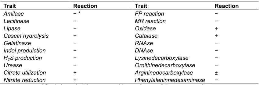
3.3 Biochemical Characteristics of the Strain
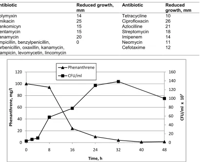
Standard biochemical characterization of the strain showed its negative reaction to indol, hydrogen sulphide, casein hydrolysis, as well as in Voges-Proskauer test reaction and in incubation with the methyl red dye. The strain was found to be able to reduce nitrates into nitrites and to utilize citrate as a carbon source. Under laboratory conditions the strain was not shown to secrete such enzymes as amylase, lipase, gelatinase, licitinase, urease, DNAse, RNAse, while positive reaction was displayed in oxidase and catalase tests (Table 1). The isolated strain was found to be represented by a bacterium with a respiratory, rather than fermentation, metabolism as it does not hydrolyze sucrose, glucose, lactose, arabinose, ramnose, displaying weak acid-formation on media containing maltose and xylose (Table 2). Notably, the strain showed negative results as related to all four pathogenicity tests, i.e. hemolythic, plasmocoagulating, fibrinolythic and gelatinolythic activities. The absence of pathogenic properties in the isolated strain intended for bioremediation is very important as it shows its ecological safety. The strain was also found to be resistant to ampicillin, benzylpeicillin, carbenicillin, oxaxillin, rifampicin, levomycetin and lincomycin, (Table 3).
The isolated strain showed phenotypic traits common for the representatives of the
Pseudomonasgenus. Further identification of the
isolated Fd1 strain revealed its close relationship
with P. denitrifiсans species (Table 4).
stored in the international data base Gen Bank showed that the novel sequence had 99% similarity with the sequences of the following bacteria: Pseudomonas nitroreducens (NR 113601), P. denitrificans (NR 102805) and P.
multiresinivorans (NR 119225). These
information, together with the results of the phenotypic and genotypic analysis of the isolated strain Fdl led us to identify it as representing
Pseudomonas denitrificans species. The 16S
rRNA gene fragment nucleotide sequence of the
Pseudomonas denitrificans Fdl strain was
deposited into the GenBank under access number KM 436103.
3.4 Phenanthrene Degradation by the Isolated Strain
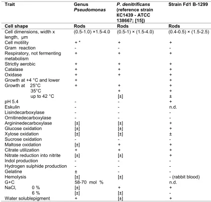
Incubation of the Fdl strain cells in the medium with phenanthrene as a sole carbon source was found to result in phenanthrene concentration decrease, accumulation of corresponding metabolites in the medium and bacterial cell proliferation, all together confirming the ability of the strain to utilize phenanthrene as a carbon source. The kinetic curve of phenanthrene biodegradation was characterized by three portions, coinciding with the main phases of cell proliferation and growth (Fig. 1). After 4 hours of incubation the lag-phase ended, and the exponential growth started and continued up to 24 hours of incubation with the maximal degradation rate being observed between 8 and 16 hours. After 32 hours of incubation, when almost all phenanthrene in the medium was utilized, the number of living cells started to decrease correspondingly. Over 40 hours of incubation the phenanthrene concentration decreased from 100 ppm down to 1 ppm, proving
the novel Fdl strain to be an effective phenanthrene degrader.
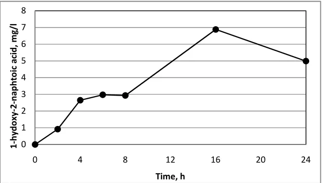
3.5 Identification and Accumulation of 1-hydroxy-2-naphthoic Acid
The HPLC analysis of phenanthrene degradation products in the incubation medium was found to contain significant amount of HNA known to be the key metabolite in phenanthrene degradation. Kinetic studies of the HNA accumulation (Fig. 2) showed increase in HNA concentration during the first 24 hours of incubation. After 32 hours of incubation when phenanthrene concentration decreased below the HNA concentration, the latter started to decrease as well. The further degradation of HNA, as was reported earlier, goes via salicylic or phthalic acid production. However, HPLC analysis found no such compounds in the incubation medium, which may result from their extremely short life time in the medium. The fact that the strain was found to be able to proliferate on/in the medium containing salicylic acid as the sole carbon source and not on/in the medium with the phthalic acid as the sole carbon source (the data are not shown) can serve as indirect evidence in favour of the salicylic acid production pathway.
3.6 The Effect of Tween-20 Surfactant on Phenanthrene Degradation
Addition of Tween-20 non-ionic surfactant into
the incubation medium accelerated
phenanthrene degradation and cell proliferation as compared to phenanthrene degradation without a surfactant (Fig. 3). When Tween 20 was added, the phenanthrene concentration was found to decrease already after 2 hours of
Table 1. Biochemical characteristics of the isolated strain Fdl В-1299
Trait Reaction Trait Reaction
Amilase − * FP reaction −
Lecitinase − MR reaction −
Lipase − Oxidase +
Casein hydrolysis − Catalase +
Gelatinase − RNAse −
Indol produiction − DNAse −
H2S production − Lysinedecarboxylase −
Urease − Ornithinedecarboxylase −
Citrate utilization + Argininedecarboxylase ±
Nitrate reduction + Phenylalaninnedesaminase −
incubation, decreasing to 1 ppm over 24 hours, with the maximal degradation rate being recorded at 2-8 hours interval. Since Tween 20 is known as a good carbon source to be utilized by bacterial cells, its effect on phenanthrene degradation may be due both to a facilitated assimilation/uptake of phenanthrene by bacterial cells and/or increased cell biomass. Judging by the CFU counts, we may conclude that after 4 hours of incubation cell proliferation is supported mostly by Tween 20 as a growth substrate, with phenanthrene contributing not more than 10 %. However, as phenanthrene degradation was observed to start already after 2 hours of incubation, i.e. before the end of the lag-phase, the increased phenanthrene uptake by cells may be suggested to contribute into the accelerated degradation as well.
The presence of Tween 20 in the medium resulted in the accumulation of large amount of HNA, with a pattern similar to the one without
Tween 20 (Fig. 4). The maximal HNA concentration was registered after 16 hours of incubation. Despite the fact that it is a less preferable substrate for bacterial utilization, as compared with Tween 20, HNA did not accumulate in the medium in course of incubation.
Table 2. Carbohydrates utilization by the isolated strain on the of medium
Carbohydrate Gas
emission Acidproduction
Sucrose -
-Mannitol -
-Glucose - +
Lactose -
-Arabinose -
-Ramnose -
-Maltose - +
Xylose - ±
Table 3. Antibiotic activity (zones of the reduced growth of the isolated strain around the antibiotic containing discs, mm)
Antibiotic Reduced growth,
mm Antibiotic Reducedgrowth, mm
Polymyxin 14 Tetracycline 10
Amikacin 25 Ciprofloxacin 26
Vankomicyn 15 Azlocilline 21
Gentamycin 15 Streptomycin 18
Kanamycin 20 Imipenem 14
Ampicillin, benzylpenicillin, carbenicillin, oxaxillin, kanamycin, rifampicin, levomycetin, lincomycin
0 Neomycin 11
Cefotaxime 12
Fig. 1. Phenanthrene degradation and cell proliferation
0 20 40 60 80 100 120 140 160
0 20 40 60 80 100 120
0 8 16 24 32 40 48
CF
U
/m
l
x
10
7
Ph
en
an
th
re
ne
,m
g/
l
Table 4. Morphological, physiological and biochemical characteristics of the bacterial genus
Pseudomonas, its typical representative strainP. denitrifiсansand the novel strain
Fd1В-1299
Trait Genus
Pseudomonas P. denitrifiсans(reference strain КС1439 - АТСС 138667; [15])
Strain Fd1 В-1299
Cell shape Rods Rods Rods
Cell dimensions, width x
length, μm (0.5-1.0) ×1.5-4.0 (0.5-1) × (1.5-4.0) (0.4-0.5) × (1.5-2.5)
Cell motility + * + +
Gram reaction - -
-Respiratory, not fermenting
metabolism + + +
Strictly aerobic + + +
Catalase + + +
Oxidase + + +
Growth at +4 °C and lower + +
Growth at 25°С + +
+ [±]
+ + ± 35°С
up to 42 °С
рН 5.4 - - +
Eskulin - - n.d.
Lisindecarboxylase - -
-Ornitinedecarboxylase - -
-Argininedecarboxylase [±] [±] +
Glucose oxidation [±] [±] +
Xylose oxidation [±] [±] ±
Sucrose oxidation - -
-Maltose oxidation [±] + +
Citrate utilization + + +
Nitrate reduction into nitrite [±] [±] +
Indol production - -
-Hydrogen sulphide production - -
-Gelatine ± -
-Hemolysis [±] [±] - (rabbit blood)
G+C 58-70 mol % n.d.
NaCl, 0 % [±] + +
6 % [±] [±]
-Water solublepigment + [±] +
* Symbols used: “+” denotes a positive reaction, “-” denotes a negative reaction; [±] means variable trait, while “±” means weak trait and “n.d.” Means no datum.
Many bacteria, representing diverse genera such
as Bacillus [16], Nocardia [17], Pseudomonas
[18-21], Sphingomonas [22], Sphingobium [23],
Mycobacterium [24], Sinorhizobium [25],
Rhizobium [26], Novosphignobium [27] and
others [28,29] were reported to degrade phenanthrene. However, their degradation rate in liquid medium with 100 ppm phenanthrene ranged 96-99% over 77-360 hours, whereas the
Pseudomonas denitrificans Fdl strain isolated in
this study was shown to degrade phenanthrene over 48 hours without any additions present, and
twice as rapidly if phenanthrene
assimilation/uptake was facilitated by the detergent present in the medium. The closest to our strain was aPseudomonasstrain, reported to degrade 100% of 60 mg/L phenanthrene within 60 hours [30].
Some phenanthrene-degrading Pseudomonas
Fig. 2. Accumulation of 1-hydroxy-2-naphtoic acid in course of phenanthrene degradation
Fig. 3. Phenanthrene degradation and bacterial cell proliferation in the medium with Tween 20 added
Fig. 4. The accumulation of 1-hydroxy-2-naphtoic acid in course of phenanthrene degradation in the medium with Tween 20 added
0 1 2 3 4 5
0 8 16 24 32 40 48
1-hy
do
xy
-2
-n
ap
ht
oi
c
ac
id
, m
g/
l
Time, h
0 500 1000 1500 2000 2500 3000
0 20 40 60 80 100 120
0 4 8 12 16 20 24
CF
U
/m
l
x
10
7
Ph
en
an
th
re
ne
, m
g/
l
Time, h Phenanthren e
0 1 2 3 4 5 6 7 8
0 4 8 12 16 20 24
1-hy
do
xy
-2
-n
ap
ht
oi
c
ac
id
, m
g/
l
4. CONCLUSION
The isolated strain Pseudomonas denitrificans
Fdl is capable of efficient phenanthrene degradation, especially in the presence of detergent, and hence can be a good candidate for biological preparations to be tested for industrial bioremediation and sewage sludge treatment.
COMPETING INTERESTS
Authors have declared that no competing interests exist.
REFERENCES
1. Mastrangela G, Fadda E, Marzia V. Polycyclic aromatic hydrocarbons and cancer in man. Environ Health Perspect. 1996;104:1166–1170.
PMCID PMC1469515
2. US Environmental Protection Agency. Priority pollutants. (Accessed 13 June 2017).
Available:http://water.epa.gov/scitech/meth ods/cwa/pollutants.cfm
3. Wu M, Chen L, Tian Y, Ding Y, Dick WA. Degradation of polycyclic aromatic hydrocarbons by microbial consortia enriched from three soils using two different culture media. Environ Pollut. 2013;78:152-158.
DOI: 10.1016/j.envpol.2013.03.004
4. Gerbeth A, Krausse S, Gemende B, Muller RH. Search of microorganism that degrade PAHs under alkaline conditions. Eng Life Sci. 2004;311-318.
DOI: 10.1002/elsc.200420034
5. Okere UV, Cabrerizo A, Dachs J, Jones KC, Semple KT. Biodegradation of
phenanthrene by indigenous
microorganisms in soils from Livingstone Island, Antarctica. FEMS Microbiol Lett. 2004;329:69-77.
DOI: 10.1111/j.1574-6968.2012.02501.x 6. Viamajala S, Peyton BM, Richards LA,
Petersen JN. Solubilization, solution equilibria, and biodegradation of PAH’s
under thermophilic conditions.
Chemosphere. 2007;66:1094-1106. DOI: 10.1016/j.chemosphere.2006.06.059 7. Li J, Chen BH. Surfactant-mediated
biodegradation of polycyclic aromatic hydrocarbons. Materials. 2009;2:76-94. DOI: 10.3390/ma2010076
8. Jin DY, Jiang X, Jing X, Ou ZQ. Effects of concentration, head group, and structure of
surfactants on the degradation of phenanthrene. J Hazard Mater. 2007;144: 215-221.
DOI: 10.1016/j.jhazmat.2006.10.012 9. Tiehm A. Degradation of polycyclic
aromatic-hydrocarbons in the presence of synthetic surfactants. Appl Environ Microbiol. 1994;60:258-263.
PMCID PMC201297
10. Efroymson RA, Alexander M.
Biodegradation by an Arthrobacter species of hydrocarbons partitioned into an organic-solvent. Appl Environ Microbiol. 1991;57:1441-1447.
Available:http://aem.asm.org/content/57/5/ 1441
11. Gao S, Seo JS, Wang J, Keuma YS, Li J, Li QX. Multiple degradation pathways of
phenanthrene by Stenotrophomonas
maltophilia C6. Int Biodeterior
Biodegradation. 2013;79:98-104. DOI: 10.1016/j.ibiod.2013.01.012
12. Holt JG. Bergey's manual of systematic bacteriology, V.2. Baltimore-London: Williams and Wilkins; 1986.
13. Gerhardt P. Methods for General and molecular bacteriology. Washington: ASM; 1994.
14. Wang Y, Qian P. Conservative Fragments in Bacterial 16S rRNA Genes and Primer Design for 16S Ribosomal DNA Amplicons in Metagenomic Studies. PLoS One. 2009;4:e7401.
DOI: 10.1371/journal.pone.0007401 15. Weyant R, Moss CW, Weaver RE, Hollis
DG, Jordan JJ, Cook EC, et al. Identification of Unusual Pathogenic Gram-Negative Aerobic and Facultative Anaerobic Bacteria. Baltimore: Williams & Wilkins; 1994.
16. Doddamani HP, Ninnekar HZ.
Biodegradation of phenanthrene by a
Bacillussp. Curr Microbiol. 2000;41:11-14.
DOI: 10.1007/s002840010083
17. Zeinali M, Vossoughi M, Ardestani SK. Degradation of phenanthrene and anthracene by Nocardia otitidiscaviarum
strain TSH1, a moderately thermophilic bacterium. J Appl Microbiol. 2008;105:398-406.
DOI: 10.1111/j.1365-2672.2008.03753.x 18. Phale PS. Biodegradation of phenanthrene
by Pseudomonas sp. strain PP2:
Novel metabolic pathway, role of
biosurfactant and cell surface
assimilation. Appl Microbiol Biotechnol. 2003;61:342-351.
DOI: 10.1099/mic.0.030460-0
19. Prabhu Y, Zeinali M, Vossoughi M,
Ardestani SK. Degradation of
phenanthrene and anthracene byNocardia
otitidiscaviarumstrain TSH1, a moderately
thermophilic bacterium. J Appl Microbiol. 2008;105:398-406.
DOI: 10.1111/j.1365-2672.2008.03753.x 20. Deveryshetty J, Phale PS. Biodegradation
of phenanthrene by Pseudomonas sp.
strain PPD: purification and
characterization of 1-hydroxy-2-naphthoic acid dioxygenase. Microbiology. 2009;155: 3083-3091.
DOI: 10.1099/mic.0.030460-0
21. Sun K, Liu J, Gao Y, Jin L, Gu Y, Wang W. Isolation, plant colonization potential and phenanthrene degradation performance of the endophytic bacterium Pseudomonas
sp. Ph6-gfp. Scientific Reports. 2014; 26:5462.
DOI: 10.1038/srep05462
22. Hesham Ael-L, Mawad AM, Mostafa YM, Shoreit A. Biodegradation ability and catabolic genes of petroleum-degrading
Sphingomonas koreensis strain ASU-06
isolated from Egyptian oily soil. Biomed Res Int. 2014;127674.
DOI: 10.1155/2014/127674
23. Wang C, Wang F, Hong Q, Zhang Y, Kengara FO, Li Z, et al. Isolation and characterization of a toxic metal-tolerant Phenanthrene-degrader Sphingobium sp. in a two-liquid-phase partitioning bioreactor (TPPB). Environ Earth Sci. 2013;70:1765-1773.
DOI: 10.1007/s12665-013-2264-8
24. Moody JD, Freeman JP, Doerge DR,
Cerniglia CE. Degradation of
Phenanthrene and Anthracene by Cell Suspensions of Mycobacterium sp. Strain PYR-1. Appl Environ Microbiol. 2001; 67:1476–1483.
DOI: 10.1128/AEM.67.4.1476-1483.2001 25. Keum YS, Se JS, Hu Y, Li QX.
Degradation pathways of phenanthrene by
Sinorhizobium sp. C4. Applied
Microbiology & Biotechnology. 2006;1: 935–941.
DOI: 10.1007/s00253-005-0219-z
26. González-Paredes Y, Alarcón A, Ferrera-Cerrato R, Almaraz JJ, Martínez-Romero E, Cruz-Sánchez JS, et al. Tolerance, growth and degradation of phenanthrene and benzo[a]pyrene by Rhizobium tropici
CIAT 899 in liquid culture medium. Appl Soil Ecol. 2013;63:105-111.
DOI: 10.1016/j.apsoil.2012.09.010
27. Lyu Y, Zheng W, Zheng T, Tian Y. Biodegradation of polycyclic aromatic
hydrocarbons by Novosphingobium
pentaromativorans US6-1. PLoS One.
2014;9:e101438.
DOI: 10.1371/journal.pone.0101438 Available:http://journals.plos.org/plosone/a rticle?id=10.1371/journal.pone.0101438 28. Roy AS, Baruah R, Borah M, Singh AK,
Boruah HPD, Saikia N, et al. Bioremediation potential of native hydrocarbon degrading bacterial strains in crude oil contaminated soil under microcosm study. Int Biodeterior Biodegradation. 2014;94:79-89.
DOI: 10.1016/j.ibiod.2014.03.024
29. Wu M, Chen L, Tian Y, Ding Y, Dick WA. Degradation of polycyclic aromatic hydrocarbons by microbial consortia enriched from three soils using two different culture media. Environ Pollut. 2013;178:152-158.
DOI: 10.1007/s13205-017-0704-y
30. Feng T, Lin H, Tang J, Feng Y. Characterization of polycyclic aromatic hydrocarbons degradation and arsenate reduction by a versatile Pseudomonas
isolate. Int Biodeterior Biodegradation. 2014;90:79-87.
DOI: 10.1016/j.ibiod.2014.01.015
31. Xia W, Du Z, Cui Q, Dong H, Wang F, He P, Tang YC. Biosurfactant produced by
novel Pseudomonas sp. WJ6 with
biodegradation of n-alkanes and polycyclic aromatic hydrocarbons. J Hazard Mater. 2014;276:489-498.
DOI: 10.1016/j.jhazmat.2014.05.062 _________________________________________________________________________________
© 2017 Astanin et al.; This is an Open Access article distributed under the terms of the Creative Commons Attribution License (http://creativecommons.org/licenses/by/4.0), which permits unrestricted use, distribution, and reproduction in any medium, provided the original work is properly cited.
Peer-review history: