Ultrasound-assisted extraction of bioactive compounds from lemon balm
and peppermint leaves
Jana Šic Žlabur, Sandra Voća*, Nadica Dobričević, Stjepan Pliestić, Ante Galić, Ana Boričević, and Nataša Borić
Department of Agricultural Technology, Storage and Transport, University of Zagreb, Svetošimunska cesta 25, 10000 Zagreb, Croatia
Received June 18, 2015; accepted November16, 2015
*Corresponding author e-mail: [email protected]
A b s t r a c t. The aim of this study was to investigate the influence of conventional and ultrasound-assisted extraction (fre-quency, time, temperature) on the content of bioactive compounds as well as on the antioxidant activity of aqueous extracts from fresh lemon balm and peppermint leaves. Total phenols, flavonoids, non-flavonoids, total chlorophylls, total carotenoids, and radi- cal scavenging capacity were determined. Moreover, the relation-ship between bioactive compounds and antioxidant capacity was studied by linear regression. A significant increase in all studied bioactive compounds during ultrasonic extraction for 5 to 20 min was found. With the classical extraction method, the highest amounts of total phenols, flavonoids, and antioxidant activity were determined, and the maximum amounts of total chlorophylls and carotenoids were determined during 20 min ultrasonic extraction. The correlation analysis revealed a strong, positive relationship between antioxidant activity and total phenolic compounds.
K e y w o r d s: total phenol content, chlorophylls, antioxidant activity, conventional extraction, high intensity ultrasound
INTRODUCTION
Phenolic compounds exhibit a high degree of degrada -tion in terms of technological processes and show distinct thermolability, sensitivity to light, the impact of pathogens,
mechanical damage to plant tissue cells (Ross et al., 2009).
Lately modern, non-invasive extraction techniques of plant metabolites with the main objective of increased extracted compound yield and a shortened period of extraction have
been increasingly popularized. One of the mentioned tech-niques is high-intensity ultrasound, which shows a wide range of application in the technological processes eg in-
activation of microorganisms, crystallization, filtration, drying, extraction, homogenization, stimulation of oxida
-tion, emulsifying (Barba et al., 2015a; Deng et al., 2015; Herceg et al., 2009; Koubaa et al., 2015). High intensity ultrasound treatment does not show any degradation rates on the content of bioactive compounds in the treated food
products (Koubaa et al., 2015; Šic Žabur et al., 2015; Zinoviadou et al., 2015). In addition, high intensity
ultra-sound is applicable for extraction of compounds with
various chemical structures and is characterized by
non-in-vasive temperatures (Cvjetko Bubalo et al., 2013; Koubaa
et al., 2015; Šic Žlabur et al., 2015). The ultrasound
tech-nique is extremely simple in application, and usually two
types of equipment are used, including:
– a system with a directly immersed ultrasonic probe
(transducer)
– an ultrasonic bath.
Phenolic acids, flavonoids, and tannins are the most commonly found polyphenolic compounds in plant extracts (Naik et al., 2006). Phenolic compounds from plant mate-rial have recently gained great popularity because of their
significant positive effects on human health (Auclair et al.,
2008; Barba et al., 2014). Phenolic compounds along with
plant pigments (carotenoids, chlorophylls) have proven significant antioxidant activity, which is directly corelat
-ed with removal of free radicals and superoxide (Hsu et al., 2013; Sinkovič et al., 2015). Therefore, it is import-ant to emphasize the need to increase the consumption of foodstuffs rich in phenolic compounds aimed at preventive action on human health from cancer disease prevention to occurrence and prevention of cardiovascular diseases
(Barba et al., 2014).
Peppermint (Mentha piperita L.) and lemon balm
(Melissa officinalis L.) are mainly used as a spice in many
food products (De Vincenzi et al., 1991) and traditionally
used for medical purposes (Foster and Tyler, 1993).
Pepper-mint and lemon balm are plant species characterized by
a high content of phenolic compounds, mainly flavonoids (Guédon and Pasquier, 1994) but they are also rich in chlo -rophylls. A number of positive effects of peppermint on
human health are known, such as antibacterial (Hajlaoui et al., 2008), antifungal (Bouchra et al., 2003), and
insec-ticidal activity (Saljoqi et al., 2006). Lemon balm is one of the most popular medicinal plants with proven sedative,
antispasmodic, antibacterial, antiviral, anti-inflammatory, and antioxidative effect (Yamasaki et al., 1998).
MATERIALS AND METHODS
Peppermint and lemon balm were cultivated on the
experimental field of the Department of Vegetable Crops, Faculty of Agriculture University of Zagreb. The experi -ments were performed in triplicate. During plant cultivation, no fertilization treatment was applied. Also, the availability
of water to plants was exclusively dependent on climatic conditions; irrigation was not applied on the experimental field. Peppermint and lemon balm plants were harvested with the whole stem in mid-June 2014. Before analysis,
only green, healthy leaves with no visible signs of damage were torn off from the plant stems, while the stem and leaf petiole were removed. The leaves were washed under cold running water and dried. All chemical analyses were made
on leaves and leaf water extracts in triplicate.
The water extracts of plant leaves for each plant material,
peppermint and lemon balm, were prepared with a uni-
que procedure: 2 g (±0.01) of plant leaves were cut into smaller pieces and weighed in an Erlenmeyer flask with
a volume of 100 ml, onto which 50 ml of distilled water at
room temperature (22°C) were added. Prepared leaf solu -tions were allowed to stand for 24 h at room temperature
with intermitted shaking. After 24 h, the solutions were
filtered through Whatman filter paper and used for the
designed chemical analysis.
An ultrasonic bath (Bandelin, RK 103 H, Germany)
with frequency 35 kHz and nominal power 140 W was
used for ultrasound extraction. Peppermint and lemon
balm water solutions were prepared with a unique
proce-dure: 2 g (±0.01) of fresh plant leaves were weighed in an Erlenmeyer flask with a volume of 100 ml, onto which 50 ml of distilled water at room temperature (22°C) were added. During the ultrasonic treatment, the time of extraction
was 5, 10, 15, 20, 25, and 30 min. After each ultrasonic
extraction, solutions were filtered through Whatman filter
paper and were used for the designed chemical analysis. During the sonication of the samples, temperature change was measured after 5, 10, 15, 20, 25, and 30 min with an
infrared thermometer (Batronix, IC UT-300B, Germany) (Table 1).
The total phenol contents in peppermint and lemon balm
leaves (raw material) were determined with the Ough and Amerine (1998) method. In turn, the total phenolic content of peppermint and lemon balm extracts (aqueous solutions)
were obtained according to Shukla et al. (2012) with some modification during the analysis. Measurement was carried out as follows: 1 ml of the prepared aqueous extract and 1 ml of the Folin-Ciocalteu reagent diluted with distilled water (1:2) were added in the volumetric flask with a
volu-me of 50 ml and allowed to stand for 3 min. Additionally, 3 ml of a saturated sodium carbonate solution was added, the
flask was filled to the mark with distilled water and allowed
to stand for 3 h at room temperature with intermittent shak-ing. The absorbance of the blue colour that developed was
measured spectrophotometrically (Shimadzu UV 1650 PC) at 750 nm with distilled water as a blank. The flavonoid content of the peppermint and lemon balm extracts were determined with the Abou-Arab and Abou-Salem (2010) method with some modification during the analysis: 1 ml of T a b l e 1. Experimental conditions
Extraction
technique Solvent Solvent volume (ml) Time Temperature (°C) Ultrasonic bath
Conventional Distilled water 50 24 h 22.1 –
UAE Distilled water 50
5 min 28.2
35 kHz/140 W
10 min 40.3
15 min 46.0
20 min 54.2
25 min 54.7
30 min 56.4
the prepared aqueous extracts, 1 ml of 20% (v/v) HCl, and 0.5 ml formaldehyde were added in the volumetric flask
with a volume of 25 ml. The prepared samples were purged
with nitrogen (N2) and allowed to stand for 24 h at room
temperature. After 24 h, the same Folin-Cioacalteu reaction
as for total phenols was performed. The absorbance of the blue colour that developed was measured
spectrophotomet-rically (Shimadzu UV 1650 PC) at 750 nm with distilled water as a blank. The non-flavonoid content was expressed
mathematically as the difference between total phenols and
flavonoids. Gallic acid was used as an external standard and the concentration of total phenols, flavonoids, and non-fla
-vonoids was expressed as mg g-1 of the aqueous extract.
The content of total chlorophylls (chlorophyll a and
chlorophyll b) and total carotenoids were determined
by Holm (1954) and Wettstein (1957). The sample was
weighed into a test tube and 10 ml of acetone was ad- ded.The solution prepared in this way was homogeni-
zed with a laboratory homogenizer (IKA, UltraTurax T-18, Germany). The solution was filtered and transferred into a volumetric flask with a known volume. The absorbance was measured spectrophotometrically (Shimadzu UV 1650
PC) at 662, 644, and 440 nm with acetone as a blank probe. The absorbance values were recalculated according to the Holm-Wettstein equations in the pigment concentration
(mg l-1), and the final result of the pigment content was
expressed in mg g-1:
chlorophyll a = 9.784 A662- 0.990 A644, chlorophyll b = 21.426 A644- 4.65 A662, chlorophyll a+b = 5.134 A662+ 20.436 A644, carotenoids = 4.695 A440-0.268 (chlorophyll a+b).
The antioxidant activity of leaves and aqueous extracts
of peppermint and lemon balm leaves was determined
with the ABTS method. Trolox (6-hydroxy-2,5,7,8-tetra-methylchroman-2-carboxylic acid; SigmaAldrich) was used as an antioxidant standard. Trolox (2.5 mM) was pre
-pared in ethanol (80 %) for use as a stock standard. ABTS, 2,2’-azinobis(3-ethylbenzothiazoline-6-sulfonic acid), and
potassium persulfate were obtained from Sigma-Aldrich.
The experiments were performed according to Miller et al. (1993). ABTS (7 mM) and potassium persulfate (140 mM)
were dissolved in distilled water. These two solutions were
mixed (88 µl of prepared potassium persulfate (140 mM) and 5 ml of prepared ABTS solution) and the mixture was
allowed to stand in the dark at room temperature for 16 h
before use in order to produce ABTS radicals (ABTS•+). On the day of the analysis, 1% ABTS•+ solution (1 ml ABTS•+ in a volumetric flask of 100 ml filled with 96% ethanol to the mark) was prepared. For the study, the absor
-bance of the ABTS radical solution was measured at 734 nm and set to absorbance of 0.70±0.02. 160 μl of the extracts obtained were directly injected in the cuvette, mixed with
2 ml 1% ABTS•+, and absorbance was measured at 734 nm. The final results of the antioxidant activity was calculated based on the calibration curve and expressed as mmol TE l-1.
Statistical analysis was performed using the SAS® version 9.3. Data were subjected to the one-way analysis
of variance (ANOVA) according to the treatment structure. Mean values were compared by the t-test (LSD), and they were considered significantly different at p≤0.05.
Regression and correlation analysis were performed in order to investigate the nature and intensity of the relationships
between bioactive compounds and antioxidant activity in vitro. Correlation coefficients with p values ≤0.05 were considered significant.
RESULTS AND DISCUSSION
A high statistically significant difference (p≤0.0001)
was observed for all the analysed chemical compounds
depending on the extraction technique (Table 2). The
amount of total phenolic compounds in fresh lemon balm leaves was 1 977.45 mg l-1. Many literature data indicate a high content of phenolic compounds in lemon balm leaves
(Dastmalchi et al., 2008; Komes et al., 2011; Najafian and
Rowshan, 2013). The aqueous samples of lemon balm
leaves extracted with the classical method for 24 h at room
temperature show a high content of phenolic compounds with the determined total phenol content of 1331.54 mg l-1.
The aqueous samples of lemon balm leaves extracted by
high intensity ultrasound in an ultrasonic bath show a
sig-nificant increase in phenolic compounds depending on the extraction time. The content of all the phenolic compounds
studied show an increasing trend in the time of ultrasonic
extraction from 5 to 25 min. During the period mentioned,
the total phenol content increased by about 6.5 times. The highest value of the total phenolic content in the samples treated with ultrasound was determined in UES5 during
25 min of extraction (Table 2), while sample UES6 (extraction time 30 min) had even a lower content of phe -nolic compounds than sample UES4. The increasing trend of the total phenol content during the ultrasonic treatment
for 30 min is expected, since the extraction time plays a key role in the separation of chemical compounds (Koubaa et al., 2015; Šic Žlabur et al., 2015). The results of total
phe-nols extracted with the classical method show higher yields
compared to the phenol results obtained in samples treated with ultrasound. In aqueous samples of lemon balm leaves
extracted classically, only 1.5-fold higher total phenol con -tent was determined compared to sample UES5. During
the classical extraction, even 24 h was required to extract
the high content of phenolic compounds. High-intensity
ultrasound shows significantly better results compared to conventional extraction because already during 25 min
of application thereof a very high content of phenolic
(İnce et al., 2013). The antioxidant activity is in correla -tion with the total phenolic content, vitamins, and generally
bioactive compounds (Barba et al., 2010, 2013; Carbonell-Capella et al., 2014; Criado et al., 2014). According to the results obtained in this study, a strong relationship between
antioxidant activity and the content of total phenolic com
-pounds, flavonoids, and non-flavonoids in the aqueous samples of fresh lemon balm leaves extracted with the
clas-sical and ultrasonic methods was determined (Table 3). The
highest antioxidant activity was determined in a sample of fresh lemon balm leaves (2.42 mmol TE l-1) that also contains the highest amount of all the studied phenolic
compounds. The antioxidant activity was the lowest in the aqueous extract of lemon balm leaves treated with ultra
-sound for 5 min (UES1), in which the total phenol content was the lowest. The increasing trend of antioxidant
acti-vity was determined for samples UES2, UES3, UES4, and
UES5, while, as expected, sample UES6 showed the lowest antioxidant activity. The aqueous samples of lemon balm leaves extracted classically showed high antioxidant acti-vity. Generally, lemon balm is exceptionally rich in pheno -lic compounds. The results of this research indicate a high content of phenolic compounds in all the studied aqueous
extracts of lemon balm leaves as well as strong antioxidant activity and are in line with other literature data (Caniova and Brandsteterova, 2001; İnce et al., 2013; Karasová and Lehotay, 2006).
The linear regression between the antioxidant activ -ity and the total phenol content of lemon balm aqueous
extracts is presented in Fig. 1a. The total phenol content of the lemon balm aqueous extracts was found to correlate significantly with the antioxidant activity (ABTS). In this research, the relationship between the antioxidant activity
T a b l e 2. Total phenol, non-flavonoid, flavonoid content and antioxidant capacity of leaves and aqueous samples of lemon balm leaves extracted with utrasound and classical methods
Extraction technique Total phenols Non-flavonoids Flavonoids Antioxidant capacity
(mg l-1 extract) (mmol TE l-1)
Leaves 1977.45±4.53 a 1739.46±1.335 a 237.99±0.45 d 2.42±12.77 a
Classical extraction 1331.54±0.575 b 750.90±1.82 b 580.64±1.25 a 2.29±16.5 b
UES1 138.11±0.765 h 80.31±1.45 h 57.79±1.36 h 1.91±5.8 e
UES2 225.92±0.45 g 144.82±0.45 g 81.10±0.02 g 1.99±3.0 ed
UES3 310.86±0.07 f 210.40±0.065 f 100.46±0.45 f 2.09±15.3 cd
UES4 720.93±1.92 d 500.16±1.92 d 213.02±0.6 e 2.10±16.86 cd
UES5 891.76±0.825 c 507.92±0.065 c 391.61±1.255 b 2.17±4.15 c
UES6 637.97±0.45 e 290.36±0.125 e 347.61±0.355 c 2.05±3.2 d
UES1, UES2, UES3, UES4, UES5, UES6 – ultrasound extracted samples: 5, 10, 15, 20, 25, and 30 min, respectively. Different letters indicate significant differences between means at p<0.0001.
T a b l e 3. Correlation coefficients (r) between the selected che- mical compounds and antioxidant activity (mmol TE l-1) in differ-ent aqueous extracts of lemon balm and peppermint leaf samples
Lemon balm aqueous extracts Antioxidant activity
Total phenols 0.932***
Total flavonoids 0.869 **
Non flavonoids 0.938***
Total chlorophylls 0.444 NS
Chlorophyll a 0.380 NS
Chlorophyll b 0.471 NS
Total carotenoids 0.800*
Peppermint aqueous extracts
Total phenols 0.874**
Total flavonoids 0.961***
Non flavonoids 0.836**
Total chlorophylls 0.019 NS
Chlorophyll a -0.078 NS
Chlorophyll b 0.091 NS
Total carotenoids 0.549 NS
and total phenols was linear with a coefficient of deter -mination R2=0.8683, which assumes a strong relationship between the observed parameters. Table 3 shows the results
of the correlation coefficients (r) between the total phenol content (mg l-1) and antioxidant activity (mmol TE l-1) in
the aqueous samples of lemon balm leaves extracted with
the classical method and with ultrasound. A high
correla-tion coefficient (p≤0.0001) was observed between the total phenol content (r=0.932), non-flavonoids (r=0.938), and antioxidant activity of lemon balm aqueous leaf extracts.
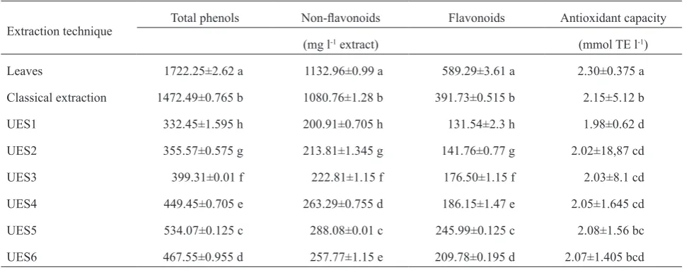
Table 4 presents the results of the total phenolic, flavo
-noid, and non-flavonoid content and antioxidant activity of fresh peppermint leaves and aqueous leaf samples extract
-ed classically and with ultrasound. A high significant statistical difference (p≤0.0001) was observed for all the analysed phenol compounds (total phenols, flavonoids, and non-flavonoids) depending on the extraction technique. In
the samples of fresh peppermint leaves, a high content of
total phenols (1 722.25 mg l-1) was determined, which is
a slightly lower value, compared with the total phenol con-tent of fresh lemon balm leaves. The results obtained are consistent with other literature data, which emphasize a sig-
nificantly high phenol content of peppermint leaves (Lv et al., 2012; Kapp et al., 2013; Riachi and De Maria,
2015). During the 24 h of classical extraction of fresh
peppermint leaves in water, the total phenol content was 1 472.49 mg l-1,which isan about 15% lower valuein
com-parison with the total phenol content identified in fresh leaves. Based on the data above mentioned, it should be emphasized that the applied classical extraction meth
-od significantly contributed to the separation of phenolic compounds. During the ultrasonic extraction of the aque
-ous peppermint samples, an about 60% increase in the
total phenol content in the period from 5 to 25 min was determined. The lower total phenol content was
deter-mined during ultrasonic extraction in a maximum applied time of 30 min. An increasing trend of the total flavonoid and non-flavonoid content during ultrasonic extraction for Fig. 1. Linear regression between total phenol content (mg l-1) and antioxidant activity (mmol TE l-1) in aqueous extracts of: a – lemon balm and b – peppermint leaf samples. The significance of the correlation coefficient (r) is marked with asterisks (** = significance at p≤0.001).
T a b l e 4. Total phenol, non-flavonoid, and flavonoid content and antioxidant capacity of leaves and aqueous samples of peppermint leaves extracted with the ultrasound and classical methods
Extraction technique Total phenols Non-flavonoids Flavonoids Antioxidant capacity
(mg l-1 extract) (mmol TE l-1)
Leaves 1722.25±2.62 a 1132.96±0.99 a 589.29±3.61 a 2.30±0.375 a
Classical extraction 1472.49±0.765 b 1080.76±1.28 b 391.73±0.515 b 2.15±5.12 b
UES1 332.45±1.595 h 200.91±0.705 h 131.54±2.3 h 1.98±0.62 d
UES2 355.57±0.575 g 213.81±1.345 g 141.76±0.77 g 2.02±18,87 cd
UES3 399.31±0.01 f 222.81±1.15 f 176.50±1.15 f 2.03±8.1 cd
UES4 449.45±0.705 e 263.29±0.755 d 186.15±1.47 e 2.05±1.645 cd
UES5 534.07±0.125 c 288.08±0.01 c 245.99±0.125 c 2.08±1.56 bc
UES6 467.55±0.955 d 257.77±1.15 e 209.78±0.195 d 2.07±1.405 bcd
Explanations as in Table 2.
Total phenol (mg l-1)
Total phenol (mg l-1)
ABTS (mmol
TE l
a period from 5 to 25 min was recorded. The antioxidant
activity of fresh peppermint leaves was lower than the
anti-oxidant values determined for fresh lemon balm leaves, which was expected, since fresh lemon balm leaves had
higher total phenol content. Oh et al. (2013) and Riachi and De Maria (2015) emphasize the very high antioxidant
capacity of aqueous peppermint solutions, comparing
dif-ferent methods for testing the antioxidant activity (FRAP, ORAC, ABTS, DPPH).
A strong correlation between total phenolic compounds
(flavonoids and non-flavonoids) of peppermint samples (extracted classically and with ultrasound) and antioxidant
activity was determined. The linear regression between
antioxidant activity measured with the ABTS method and the total phenol content of the peppermint aqueous extracts
are presented in Fig. 1b. The total phenol content of pepper
-mint aqueous extracts was found to correlate significantly with the antioxidant capacity (ABTS). The relationship between antioxidant capacity and total phenolic compounds was linear with a coefficient of determination R2=0.7628, which assumes a strong relationship between the observed
parameters. A high correlation coefficient (p≤0.0001) was observed between the total flavonoids (r=0.961), total phenol content (r=0.874), non-flavonoids (r=0.836 with p≤0.001) and antioxidant activity of peppermint aqueous leaf extracts (Table 3).
The total chlorophyll, chlorophyll a, chlorophyll b, and carotenoid content of fresh lemon balm and
pepper-mint leaves and their aqueous samples extracted classically
and ultrasonic are shown in Tables 5 and 6. Lemon balm
T a b l e 5. Total chlorophylls, chlorophyll a, chlorophyll b, and total carotenoids in leaves and aqueous samples of lemon balm leaves extracted with the ultrasound and classical methods
Extraction technique Total chlorophylls Chlorophyll a Chlorophyll b Total carotenoids (mg g-1)
Leaves 177.21±1.92 a 115.57±1.16 a 61.64±0.76 a 56.09±0.22 a
Classical extraction 2.79±0.94 d 0.83±0.29 cd 1.96±0.64 de 3.83±0.85 b
UES1 0.93±0.01 e 0.31±0.01 d 0.62±0.01 f 0.56±0.004 g
UES2 2.24±0.04 de 0.74±0.015 cd 1.49±0.03 e 0.79±0.004 f
UES3 3.01±0.11 d 0.99±0.04 cd 2.02±0.07 de 1.03±0.02 e
UES4 6.39±0.02 b 2.16±0.007 b 4.23±0.013 b 3.85±0.02 b
UES5 4.64±0.09 c 1.55±0.04 bc 3.09±0.05 c 3.01±0.03 c
UES6 3.24±0.06 cd 1.06±0.01 cd 2.19±0.045 d 2.35±0.004 d
Explanations as in Table 2.
T a b l e 6. Total chlorophylls, chlorophyll a, chlorophyll b, and total carotenoids of leaves and aqueous samples of peppermint leaves extracted with the ultrasound and classical methods
Extraction technique Total chlorophylls Chlorophyll a Chlorophyll b Total carotenoids (mg g-1)
Leaves 703.15±6.67 a 454.48±4.04 a 248.66±2.63 a 163.43±1.53 a
Classical extraction 4.37±0.64 f 1.33±0.23 h 3.04±0.41 f 6.66±0.21 b
UES1 4.10±0.001 g 1.45±0.02 g 2.58±0.04 g 2.72±0.015 f
UES2 5.04±0.04 e 1.83±0.006 e 3.21±0.03 e 3.06±0.017 e
UES3 5.39±0.001 d 1.96±0.001 d 3.43±0.001 d 3.32±0.011 d
UES4 9.84±0.102 b 3.55±0.03 b 6.29±0.07 b 5.49±0.04 c
UES5 6.77±0.123 c 2.40±0.04 c 4.37±0.08 c 3.28±0.04 d
UES6 4.52±0.06 f 1.59±0.02 f 2.93±0.04 f 1.09±0.001 g
and peppermint are characterized by high content of green
pigments, chlorophylls, which is confirmed by numerous scientific investigations (Grzeszczuk and Jadczak, 2009;
Loranty et al., 2010; Komes et al., 2011). According to the result of this research, fresh lemon balm leaves con-tained on average 177.21, 115.57, and 61.64 mg g-1 of total chlorophylls, chlorophyll a, and chlorophyll b; while fresh
peppermint leaves exhibited significantly higher amounts:
703.15, 454.48, and 248.66 mg g-1 of total chlorophylls,
chlorophyll a, and chlorophyll b, respectively. Fresh lemon balm and peppermint leaves also contained significant
amounts of total carotenoids: lemon balm 56.09 mg g-1 and peppermint even three times higher ie 163.43 mg g-1.
The extraction technique significantly affected the total chlorophyll (chlorophyll a and chlorophyll b) and
carote-noid content in lemon balm and peppermint aqueous sam-ples with p value less than 0.0001. During the ultrasonic
extraction of lemon balm leaves for 20 min (UES4), a sig-nificant increase in the total chlorophyll content was determined, even 56% higher compared with the sam
-ples extracted classically. Also, the same increasing trend during ultrasonic extraction for 20 min was determined
for the chlorophyll a and chlorophyll b content. The
chlo-rophyll a content was even 62%, and chlochlo-rophyll b 54% higher compared to the samples extracted classically. In the period from 25 (UES5) to 30 min (UES6) of ultrason
-ic extraction, a signif-icant decrease in the total chlorophyll (chlorophyll a and b) content was determined, but the final
value was still higher than the value obtained in the sample
extracted classically. Numerous literature data emphasize the extreme effectiveness and significant yields in the application of the ultrasound techniques in the extraction
of chlorophylls and its derivatives from various plant
mate-rials (Barba et al., 2015b; Grimi et al., 2014; Roselló-Soto
et al., 2015a, 2015b). In their research, Kong et al. (2014) emphasize a significant yield increase (59%) in total chlo -rophyll content recovered from a Chlorella vulgaris residue by ultrasound application compared to conventional heat
extraction. The total carotenoid content determined in fresh
lemon balm leaves was 56.09 mg g-1, which is a significant -ly lower value than the value determined for the carotenoid
content in fresh peppermint leaves (163.43 mg g-1). Also,
the carotenoid content significantly changed depending on the applied extraction technique in the samples of lemon
balm and peppermint leaves. The highest total carotenoid content was determined for the aqueous samples of lemon
balm leaves extracted with ultrasound (3.83 mg g-1), while the highest content 6.66 mg g-1 in the aqueous samples of peppermint leaves was determined by applying the
clas-sical extraction. During the ultrasonic extraction of the
lemon balm samples in the period from 5 to 20 min, an in-
crease in the total carotenoid content from 0.56 (UES1) to
3.85 mg g-1 (UES4) was determined, which is an increase
by approximately 85%. In the period of 25(UES5) and 30 min (UES6) of ultrasonic extraction, a decrease in the
total carotenoid content was determined. At the ultrasonic
extraction of the aqueous peppermint leaf samples, an
increase in the total carotenoid content in the period from 5 to 20 min was determined, with the highest value of 5.49 mg g-1 (UES4), which is an increase by about 51%. The increasing trend for the total carotenoid content is in
line with other literature data, which emphasize the extreme effectiveness of ultrasound in carotenoid extraction from
various plant materials such as carrots, algae, tomatoes etc.
in comparison with the classical techniques (Kumcuoglu et al., 2014; Roselló-Soto et al., 2015b).
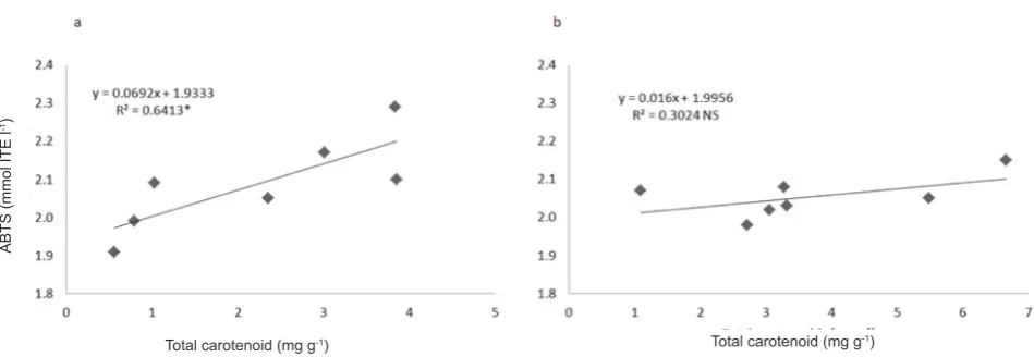
Figures 2 and 3 show linear regression between the to-tal chlorophyll and toto-tal carotenoid contents (mg g-1), and
antioxidant activity (mmol TE l-1) measured in different
aqueous extracts of lemon balm and peppermint leaves.
Linear regression analysis between the total chlorophyll
content and antioxidant activity of aqueous lemon balm and peppermint samples did not show a significant correlation (Fig. 2), which was also determined by analysing the correla-tion coefficients (Table 3). The results obtained differ from
other literature data. Namely, chlorophylls are chemical
Fig. 2. Linear regression between the total chlorophyll content (mg g-1) and antioxidant activity (mmol TE l-1) in the aqueous extracts of: a – lemon balm and b – peppermint leaf samples. Significance of the correlation coefficient (r) is marked with asterisks, ***signifi-cance at p≤0.0001.
Total chlorophyll (mg g-1) Total chlorophyll (mg g-1)
ABTS (mmol
TE l
compounds with a proven significant antioxidant effect,
due to which they show a positive effect on human health
(Roselló-Soto et al., 2015b). The total carotenoid content
of the lemon balm aqueous extracts significantly correlate (0.01<p<0.05) with the antioxidant activity (ABTS) at a coefficient of determination R2=0.6413 (Fig. 3a). Linear regression analysis between the total carotenoid content
and antioxidant activity of the aqueous peppermint samples did not show a significant correlation with the determined coefficient R2=0.3024 (Fig. 3b) and correlation coefficient
r=0.549 (Table 3). Carotenoids and their derivatives are compounds that exhibit strong antioxidant activity (Mueller and Boehm, 2011; Roselló-Soto et al., 2015b). In this
research, the extraction method is the main reason why no significant correlation between the total chlorophyll, carot
-enoid content, and antioxidant activity was determined.
CONCLUSIONS
1. The extraction technique significantly affected the
content of all the investigated bioactive compounds of the
lemon balm and peppermint aqueous extracts.
2. With the conventional extraction method, higher con
-tents of total phenolic compounds and antioxidant activity were determined, in comparison with ultrasonic extraction. 3. Ultrasonic extraction for 25 min yielded a significant total phenol content and antioxidant activity.
4. By ultrasonic extraction for 20 min, higher values of total chlorophylls (chlorophyll a and b) and carote -noids were obtained, in comparison with the conventional method.
5. Application of ultrasound significantly shortens the extraction period compared with the conventional method in which the extract time amounts to 24 h.
6. During ultrasonic extraction for 20 and 25 min, high
values of total phenolic compounds, total chlorophylls,
carotenoids, and antioxidant activity were obtained.
7. A significant, positive correlation between the total phenol content and antioxidant activity of the aqueous lemon balm and peppermint extracts was determined.
REFERENCES
Abou-Arab A.E. and Abu-Salem M.F., 2010. Evaluation of bio-active compounds of Stevia rebaudiana leaves and callus. Afr. J. Food Sci., 4, 627-634.
Auclair S., Silberberg M., Gueux E., Morand C., Mazur A., Milenkovic D., and Scalbert A., 2008. Apple polyphenols and fibers attenuate atherosclerosis in apolipoprotein E-deficient mice. J. Agric. Food Chem., 56, 5558-5563. Barba F.J., Esteve M.J., and Frígola A., 2010. Ascorbic acid is
the only bioactive that is better preserved by high hydro-static pressure than by thermal treatment of a vegetable beverage. J. Agric. Food Chem., 58, 10070-10075.
Barba F.J., Esteve M.J., Tedeschi P., Brandolini V., and Frígola A., 2013. A comparative study of the analysis of antioxidant activities of liquid foods employing spectrophotometric, fluorometric, and chemiluminescent methods. Food Anal. Methods, 6, 317-327.
Barba F.J., Esteve M.J., and Frígola A., 2014. Bioactive com-ponents from leaf vegetable products. Stud. Nat. Prod. Chem., 41, 321-346.
Barba F.J., Grimi N., and Vorobiev E., 2015a. Evaluating the potential of cell disruption technologies for green selective extraction of antioxidant compounds from Stevia rebau- diana Bertoni leaves. J. Food Eng., 149, 222-228. Barba F.J., Grimi N., and Vorobiev E., 2015b. New approaches
for the use of non-conventional cell disruption technologies to extract potential food additives and nutraceuticals from microalgae. Food Eng. Rev., 7, 45-62.
Bouchra C., Achouri M., Idrissi H.L.M., and Hmamouchi M., 2003. Chemical composition and antifungal activity of essential oils of seven Moroccan Labiatae against Botrytis cinerea Pers. J. Ethnopharmacol., 89, 165-169.
Caniova A. and Brandsteterova E., 2001. HPLC analysis of phenolic acids in Melissa officinalis. J. Liq. Chrom. Rel. Technol., 24, 2647-2659.
Fig. 3. Linear regression between the total carotenoid content (mg g-1) and antioxidant activity (mmol TE l-1) in the aqueous extracts of: a – lemon balm and b – peppermint leaf samples. Explanations as in Fig. 2.
Total carotenoid (mg g-1)
ABTS (mmol ITE l
-1)
Carbonell-Capella J.M., Barba F.J., Esteve M.J., and Frígola A., 2014. Quality parameters, bioactive compounds and their correlation with antioxidant capacity of commercial fruit-based baby foods. Food Sci. Technol. Int., 20, 479-487. Criado N., Barba F.J., Frígola A., and Rodrigo D., 2014. Effect
of Stevia rebaudiana on oxidative enzyme activity and its correlation with antioxidant capacity and bioactive com-pounds. Food Bioprocess Technol., 7, 1518-1525.
Cvjetko Bubalo M., Sabotin I., Radoš I., Valentinčič J., Bosiljkov T., Brnčić M., and Žnidaršič-Plazl P., 2013. A comparative study of ultrasound, microwave and micro-reactor-assisted imidazolium-based ionic liquid synthesis. Green Proc. Synth., 2, 579-590.
Dastmalchi K., Damien Dorman H.J., Oinonen P.P., Darwis Y., Laakso I., and Hiltunen R., 2008. Chemical composition and in vitro antioxidative activity of a lemon balm (Melissa officinalis L.) extract. Food Sci. Technol. –LWT, 41, 391-400.
De Vincenzi M., Maialetti F., Di Pascquale M., and Dessi M.R., 1991. Monographs on botanical flavouring substances used in foods. Fitoterapia, 62, 47-63.
Deng Q., Zinoviadou K.G., Galanakis C.M., Orlien V., Grimi N., Vorobiev E., Lebovka N., and Barba F.J., 2015. The effects of conventional and non-conventional processing on glu-cosinolates and its derived forms, isothiocyanates: extraction, degradation, and applications. Food Eng. Rev., 7, 357-381.
Foster S. and Tyler V.E., 1993. Tyler’s Honest Herbal: A Sensible Guide to the Use of Herbs and Related Remedies. The Haworth Herbal Press, Binghamton, NY, USA.
Grimi N., Dubois A., Marchal L., Jubeau S., Lebovka N.I., and Vorobiev E., 2014. Selective extraction from microal-gae Nannochloropsis sp. using different methods of cell disruption. Bioresource Technol., 153, 254-259.
Grzeszczuk M. and Jadczak D., 2009. Estimation of biological value of some species of mint (Mentha L.). Herba Polonica, 55, 193-199.
Guédon D.J. and Pasquier B.P., 1994. Analysis and distribution of flavonoid gycosides and rosmarinic acid in 40 Mentha x piperita clones. J. Agric. Food Chem. 42, 679-684.
Hajlaoui H., Snoussi M., BenJannet H., Mighri Z., and Bakhrouf A., 2008. Comparison of chemical composition and antimicrobial activities of Mentha longifolia L. sp. longifolia essential oil from two Tunisian localities (Gabes and Sidi Bouzid). Ann. Microbiol., 58, 103-110.
Herceg Z., Brnčić M., Jambrak Režek A., Rimac Brnčić S., Badanjak M., and Sokolić I., 2009. Possibility of applica-tion high intensity ultrasound in milk industry. Mljekarstvo, 59, 65-69.
Holm G., 1954. Chlorophyll mutations in barley. Acta Agr. Scand., 4, 457-471.
Hsu C.Y., Chao P.Y., Hu S.P., and Yang C.M., 2013. The Antioxidant and free radical scavenging activities of chlo-rophylls and pheophytins. Food Nutr. Sci., 4, 1-8.
İnce A.E., Şahin S., and Şümnü S.G., 2013. Extraction of phe-nolic compounds from melissa using microwave and ultrasound. Turk. J. Agric. For., 37, 69-75.
Kapp K., Hakala A.E., Anne Orav A., Pohjala L., Vuorela P., Püssa T., Vuorela H., and Raal A., 2013. Commercial peppermint (Mentha piperita L.) teas: Antichlamydial effect and polyphenolic composition. Food Res. Int., 53, 758-766.
Karasová G. and Lehotay J., 2006. Comparison of several extraction methods for the isolation of benzoic acid deriva-tives from Melissa officinalis. J. Liq. Chromatogr. Rel. Technol., 29, 1633-1644.
Komes D., Belščak-Cvitanović A., Horžić D., Marković K., and Kovačević Ganić K., 2011. Characterisation of pig-ments and antioxidant properties of three medicinal plants dried under different drying conditions. Proc. 11th Int. Congress Engineering and Food, May 22-26, Athens, Greece.
Kong W., Liu N., Zhang J., Yang Q., Hua S., Song H., and Xia C., 2014. Optimization of ultrasound-assisted extraction parameters of chlorophyll from Chlorella vulgaris residue after lipid separation using response surface methodology. J. Food Sci. Technol., 51, 2006-2013.
Koubaa M., Roselló-Soto E., Šic Žlabur J., Režek Jambrak A., Brnčić M., Grimi N., Boussetta N., and Barba F.J., 2015. Current and new insights in the sustainable and green recovery of nutritionally valuable compounds from Stevia rebaudiana Bertoni. J. Agric. Food Chem., 63, 6835-6846. Kumcuoglu S., Yilmaz T., and Tavman S., 2014. Ultrasound
assisted extraction of lycopene from tomato processing wastes. J. Food Sci. Technol., 51, 4102-4107.
Loranty A., Rembiałkowska E., Rosa E.A.S., and Bennett R.N., 2010. Identification, quantification and availability of carotenoids and chlorophylls in fruit, herb and medicinal teas. J. Food Compos. Anal. 23, 432-441.
Lv J., Huang H., Yua L., Whent M., Niu Y., Shi H., Wang T.T.Y., Luthria D., Charles D., and Yu L.L., 2012. Phenolic com- position and nutraceutical properties of organic and con-ventional cinnamon and peppermint. Food Chem., 132, 1442-1450.
Miller N.J., Diplock A.T., Rice-Evans C., Davies M.J., Gopinathan V., and Milner A., 1993. A novel method for measuring antioxidant capacity and its application to moni-toring the antioxidant status in premature neonates. Clin. Sci., 84, 407-412.
Mueller L. and Boehm V., 2011. Antioxidant activity of β-caro- tene compounds in different in vitro assays. Molecules, 16, 1055-1069.
Naik G.H., Priyadarsini K.I., and Mohan H., 2006. Free radical scavenging reactions and photochemical analysis of tripha-la, an ayurvedic formulation. Curr. Sci., 90, 1100-1105. Najafian S. and Rowshan V., 2013. Polyphenolic compounds of
Mentha longifolia and Lemon Balm (Melissa officinalis L.) in Iran. Int. Res. J. Appl. Basic Sci., 4, 608-612.
Oh J., Jo H., Cho A.R., Kim S.J., and Han J., 2013. Antioxidant and antimicrobial activities of various leafy herbal teas. Food Control, 31, 403-409.
Ough C.S. and Amerine M.A., 1998. Methods for Analysis of Musts and Wines. Wiley Press, Washington, DC, USA. Riachi L.G. and De Maria C.A.B., 2015. Peppermint
antioxi-dants revisited. Food Chem., 176, 72-81.
Roselló-Soto E., Koubaa M., Moubarik A., Lopes R.P., Saraiva J.A., Boussetta N., Grimi N., and Barba F.J., 2015b. Emerging opportunities for the effective valorization of wastes and by-products generated during olive oil produc-tion process: Non-convenproduc-tional methods for the recovery of high-added value compounds. Trends Food Sci. Tech., 2, 296-310.
Ross K.A., Beta T., and Arntfield S.D., 2009. A comparative study on the phenolic acids identified and quantified in dry beans using HPLC as affected by different extraction and hydrolysis methods. Food Chem., 113, 336-344.
Saljoqi A.U.R., Afridi M.K., Khan S.A., and Rehman S., 2006. Effects of six plant extracts on rice weevil Sitophilus oryzae L. in the stored wheat grains. J. Agric. Biol. Sci., 1, 1-5. SAS/STAT, 2010. Ver. 9.3., SAS Institute, Cary, NC.
Shukla S., Mehta A., Mehta P., and Bajpai V.K., 2012. Anti- oxidant ability and total phenolic content of aqueous leaf extract of Stevia rebaudiana Bert. Exp. Toxicol. Pathol., 64, 807-811.
Sinkovič L., Demšar L., Žnidarčič D., Vidrih R., Hribar J., and Treutter D., 2015. Phenolic profiles in leaves of chi- cory cultivars (Cichorium intybus L.) as influenced by organic and mineral fertilizers. Food Chem., 166, 507-513. Šic Žlabur J., Voća S., Dobričević N., Rimac Brnčić S., Dujmić F.,
and Brnčić M., 2015. Optimization of ultrasound assisted extraction of functional ingredients from Stevia rebaudiana Bertoni leaves. Int. Agrophys., 29, 231-237.
Wettstein D., 1957. Chlorophyll letale und der submikrosko- pische Formwechsel der Plastiden. Exp. Cell. Res., 12, 427-434.
Yamasaki K., Nakano M., Kawahata T., Mori H., Otake T., Ueba N., Oishi I., Inami R., Yamane M., Nakamura M., Murata H., and Nakanishi T., 1998. Anti-HIV-1 activity of herbs in Labiatae. Biol. Pharm. Bull., 21, 829-833. Zinoviadou K.G., Galanakis C.M, Brnčić M., Grimi N.,