DETERMINATION OF THE VAGAL THRESHOLD AND CHANGES OF IT’S USING
Michal Botek, Pavel Stejskal, Jakub Krejčí, Aleš Jakubec, Aleš Gába
Faculty of Physical Culture, Palacký University, Olomouc, Czech Republic
Submitted in August, 2008
Exercise intensity causes changes in the activity of both branches of the autonomic nervous system (ANS) as involved in cardiovascular system regulation. Reduction in vagal activity and an increase in sympatho-adrenal activity is associated with an increase in death risk from both cardiac and arrhythmic causes during exercise. The main aim of this work was to develop a simple mathematic algorithm for determination of critical exercise intensity, at which, if exceeded, the cardiovascular system starts to be influenced dominantly by rising sympathetic activity including catecholamine and a significant withdrawal in cardiac vagal activity (vagal threshold – TVA) occurs. The testing group consisted of 10 volunteers (men). Their mean age was 27.24 ± 3.23 years and the mean value of their maximal oxygen
uptake (VO2max) was 50.24 ± 4.63 ml·kg–1·min–1. ANS activity was monitored by the microprocessor diagnostics
sys-tem VarCor PF 7 and assessed by a non invasive spectral analysis (SA) of heart rate variability (HRV) method. The power of the high frequency component (PHF) was calculated by integrating the area under the power spectral density curve in the frequency range from 0.15 to 0.5 Hz. Changes in autonomic cardiac regulation were assessed during walking in the steady state with exercise intensities ranging from 20 to 70% of maximal heart rate reserve (MHRR) on the treadmill. Each exercise intensity increases of about the 10% MHRR in a range from 20 to 70% MHRR led to a significant decrease in vagal activity. A designed mathematic algorithm for detecting the deflection point of the vagal activity during incremental exercise intensity revealed TVA at 43.63 ± 4.66% MHRR. We can state that the designed
algorithm for detection of TVA enables an estimation of such a “safe” intensity, when the vagal activity is still preserved and sympathetic activity does not markedly rise up during exercise. The estimation of TVA could be recommended especially for the exercise prescription for patients with both reduction HRV, and at risk for sudden cardiac death.
Keywords:Spectral analysis of heart rate variability, exercise intensity, vagal threshold, prescription of the training exer-cise.
INTRODUCTION
A sedentary lifestyle or physical inactivity is gener-ally associated with the appearance of the most relevant chronic diseases. On the other hand, regular endur-ance exercise represents an effective preventive and therapeutic tool (Hilberg, 2008) against such diseases. However, the patients with cardiovascular diseases who practice exercise are exposed to certain risks resulting from insufficient heart function, an increased tendency to myocardial arrhythmias and also from changes in the bloodstream (ACSM, 2007). The level of intensity seems to be a crucial risk factor of sudden cardiac death or myocardial arrhythmias; therefore, its optimizing is a basic requirement for safe exercise.
The cardiovascular system is mostly controlled by au-tonomic activity through the activity of the sympathetic and parasympathetic pathways of the ANS, and their activity depends mainly on the intensity of the exercise (Arai et al., 1989; Casedei et al., 1995; Parekh & Lee, 2005; Stejskal et al., 2001; Yamamoto, Hughson, & Pe-terson, 1991). At low intensity, tachycardia occurred
mainly due to a withdrawal of efferent vagal activity (Perini et al., 1990). Rising sympathetic activity together with the level of circulating catecholamine plays a major role in heart regulation at higher intensities (Breuer et al., 1993; Kluess, Wood, & Welsch, 2000). With the dominant increase in sympatho-adrenal activity, higher demands on compression heart work are associated therewith (Ganong, 1999), and thereby, increase in the risk of both heart and circulation failure are related. From the presented data it is clear that patients with a higher risk of sudden cardiac failure should exercise only at such an intensity as does not lead to a signifi-cant increase in sympatho-adrenal activity, but which enables changes in the vagal activity regulation of the cardiovascular system.
Izdeb-ska et al., 2004). So far, published studies have brought us different results describing exercise intensity, from which the efferent cardiac vagal modulation disappears, and the activity of the heart will be mainly mediated by the sympatho-adrenal system. For example, Perini et al. (1990) and Orizio et al. (1989) presented an intensity of about 30 and 33% VO2max. Hautala et al. (2003) described 40% VO2max as the dividing intensity in vagal and sympathetic cardiac modulation. According to the studies of Achten and Jeukendrup (2003) and Nakamu-ra, Yamamoto and Muraoka (1993), the target intensity is found at 50–60% VO2max.
Heart rate variability (HRV) is generally accepted as a feedback marker of cardiac vagal and sympathetic activity (Akselrod et al., 1981; Task Force, 1996). Due to obesity, diabetes mellitus, hypertension or several cardiovascular diseases, there is significantly decreased HRV (Kuch et al., 2004; Matsunaga et al., 2004; Nolan et al., 1998; Shibata et al., 2002) which has been asso-ciated with higher risk of sudden death or myocardial arrhythmias (Schwarz, La Rovere, & Vanoli, 1992). On the contrary, regular endurance exercise leads to an in-crease in vagal cardiac activity and a dein-crease in sym-pathetic activity (increase in HRV) in healthy people as well as in patients who exercised during their reha-bilitation process (Cornelissen & Fagard, 2005; Dixon, Kamath, McKartney, & Fallen, 1992; Fujimoto et al., 1999; Goldsmith, Bigger Jr., Steinman, & Fleiss, 1992; Mueller, 2007; Takeyama et al., 2000).
The spectral analysis (SA) of HRV is a non invasive method for the direct assessment of vagal cardiac ac-tivity and for the indirect evaluation of sympathovagal balance. It is known that exercise intensity decreases the vagal parameters of SA HRV (Stejskal et al., 2001). Therefore, the aim of this study was to identify the criti-cal exercise intensity level which is linked with a very mild reduction of vagal cardiac modulation (vagal threshold – TVA). A sophisticated determination of TVA may allow us to prescribe “safe” exercise for patients with a higher risk of sudden cardiac death or myocardial arrhythmias.
METHODS
The testing group consisted of 10 men who were studying or working at the Faculty of Physical Culture (FPC), Palacký University in Olomouc. Each testing protocol and situation was clearly and precisely de-scribed to them. The study protocol was approved by the ethics committee of the Faculty of Physical Culture, and the subjects gave written informed consent. During this study, all measurements were performed in the labo-ratories belonging to the Department of Functional An-thropology and Physiology of FPC PU. The investigated
persons were instructed to keep an optimal regime, and vigorous physical activity was forbidden minimally for 48 hours before the testing. Further, the volunteers were not allowed to eat and drink coffee, tea, nor any substance, which could influence ANS activity, 2 hours prior to the measurement of ANS activity. All subjects were non smokers, and they were asked to come on an empty stomach for all measurements.
Before the testing, the volunteers had been inves-tigated to preclude any medical or health limitations to perform the maximal exertion test. Usually in the morning, each tested person underwent an initial tests battery, which was performed 14 days before the start of the study in the exercise laboratory. Subjects under-went basic anthropometric measurements (height [cm], weight [kg]) and body composition was assessed using the bioimpedance method (In Body 720, South Korea). The measurement of HRV and oxygen consumption fol-lowed anthropometric measurements. The maximal run-ning test was performed on a Lode Valliant treadmill (Netherlands). The test started with a warm up phase: 4 minutes (min.) at 8 km·h–1 and 10 km·h–1, respectively. Immediately after the warm up, the inclination of the treadmill increased from 0 to 5%, and speed remained at 10 km·h–1. Then the speed increased every minute by 1 km·h–1 till exhaustion. During the test, the subject breathed in a mask: ventilation and both O2 and CO2 exchange were analyzed by a gas ventilator ZAN 600 Ergo USB (Germany).
ECG data were collected during a standardized or-tho-clinostatics maneuver of lying–standing–lying by the VarCor PF 7 system (Salinger & Gwozdziewicz, 2008), which requires for HRV analysis 256 artifacts free of subsequent R–R intervals for each position. Frequency domain analyses were performed according to the meth-ods described by Salinger et al. (1998). The amplitude density of the collected signal was estimated using the fast Fourier Transform method with a partly modified Coarse-Graining Spectral Analysis algorithm (Yamamo-to & Hughson, 1991). The power of the mean spectral component with high frequency (PHF) was calculated by integrating the area under the power spectral density curve in the frequency ranges according to Salinger et al. (1998) with a result of: PHF from 0.15 to 0.5 Hz.
both HRV and oxygen consumption lasted 5 minutes. The measurement itself followed a 5 minute warm up aimed at the achievement of HRT. The investigations at 50, 60 and 70% MHRR coursed separately in different days. Only intensities of 20–30–40% MHRR were done by the subject all together during one measurement. The intensities were chosen randomly.
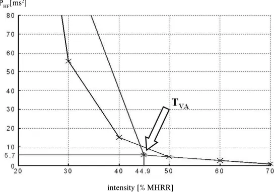
An algorithm of TVA calculation based on load curve is described in this article. The result of a load curve measurement is the set of parameters {xi, yi}, i = 1, 2,…n, where yi is the value of PHF [ms2] measured within load xi [%]. In our ramp test schedule, the load is increased step by step at levels of 20%, 30%,…70%, and so on, in our case bringing a result of n = 6. The mathematics algorithm does not include resting PHF.
The algorithm splits the measured set of parameters into two subsets, calculates a regression line in each subset and calculates the TVA as a point of intersection of two regression lines. The first subset contains the parameters {xi, yi}, i = 1, 2,…k and the second subset contains the parameters {xi, yi}, i = k, k + 1,…n. The pa-rameters with index k belong to both subsets.
The regression line
y = a1 + b1x (1)
with coefficients = = = = = = − − = k i i k i i k i i k i i k i i k i i i x x x k y x y x k b 1 1 1 1 1 1 1 2 , = = − = k i i k i i x k b y k a 1 1 1 1 1 (2)
is calculated from values in the first subset. The regres-sion line
y = a2 + b2x (3)
with coefficients = = = = = = − + − − + − = n k i i n k i i n k i i n k i i n k i i n k i i i x x x k n y x y x k n b 2 ) 1 ( ) 1 ( 2 , = = − + − + − = n k i i n k i i x k n b y k n a ) 1 ( ) 1 ( 1 2 2 (4)
is calculated from values in the second subset. The TVA is calculated as the point of intersection [xP, yP] of lines (1) and (3), the resulting equations are
1 2 2 1 b b a a xP − −
= , yP=a1+b1xP=a2+b2xP. (5)
The algorithm calculates the value of index k, which splits the measured set into two subsets, by means of an iterative procedure. At the beginning of this iterative procedure, the index k is set for a starting value, i. e. k = 2, and the condition
b2 < 0 ∧a2 + b2xn > 0 (6)
is tested. If the condition (6) is fulfilled, the iterative procedure is stopped and the value of index k is taken. If the condition (6) is not fulfilled, the index k is incre-mented, i. e. k = k + 1, the coefficients (4) are calculated again and the condition (6) is tested. The iterative pro-cedure continues until the condition (6) is fulfilled. The meaning of this condition is that the regression line (3) has a decreasing slope and lies above zero level within the interval 〈xk, xn〉. It can be said that the goal of the
iterative procedure is to find such a regression line (3) which does not cross the x axis.
All statistical processes were performed in MS Excel 2003, Statistica 6.0 software and the algorithm for detec-tion of the TVA was created in MatLab 7.5.
RESULTS
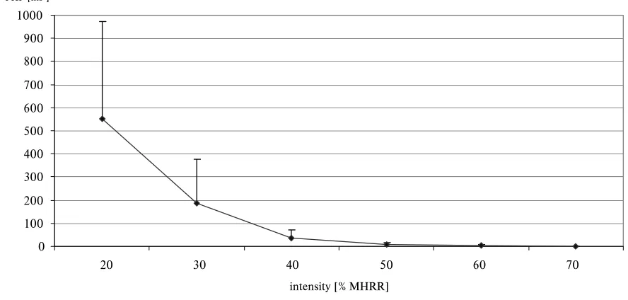
The Wilcoxon test revealed significant differences in PHF values between all assessed exercise intensi-ties. Fig. 2 clearly shows that the highest reduction in PHF values occurred up to 40% MHRR. On the contrary, minimal attenuation of PHF values was detected at over 50% MHRR.
TABLE 1
Basic characteristics of the testing group
Age
[year]
BMI
[kg·m–2]
Body Fat
[%]
HRrest
[beat·min–1]
HRmax
[beat·min–1]
ANTR
[beat·min–1]
VO2max
[ml·kg–1·min–1]
27.24 ± 3.23 24.89 ± 1.81 9.39 ± 3.97 50.30 ± 2.21 193.0 ± 9.32 170.0 ± 8.82 50.24 ± 4.63
Fig. 1
Illustrated example of the vagal threshold assessment
Legend: % MHRR – percent of the maximal heart rate reserve, PHF – high frequency power, TVA – vagal threshold.
TABLE 2
Statistical differences of the PHF values during different exercise intensity
Parameter [units] R 20% MHRR 30% MHRR 40% MHRR 50% MHRR 60% MHRR 70% MHRR HR [beat·min–1]
M SD
83.23 5.90
78.02 4.13
91.05 2.75
106.42 3.67
122.03 4.42
136.86 6.51
151.08 6.80
PHF [ms2]
M SD
288.94 402.36
551.62 421.37
185.80 189.63
37.24 34.18
9.05 5.06
4.25 2.54
0.86 0.52
PHF [ms2]
p
R vs 20* R vs 30* R vs 40** R vs 50** R vs 60** R vs 70**
20 vs 30** 20 vs 40** 20 vs 50** 20 vs 60** 20 vs 70**
30 vs 40** 30 vs 50** 30 vs 60** 30 vs 70**
40 vs 50** 40 vs 60** 40 vs 70**
50 vs 60* 50 vs 70**
60 vs 70**
Legend: R – rest HR during standing position, PHF – high frequency power, M – mean value, SD – standard deviation, % MHRR – percent of the maximal heart rate reserve, HR – heart rate, p – significant values (Wilcoxon test), *p ≤ 0.05, **p ≤ 0.01, vs – versus.
PHF[ms2]
intensity [% MHRR]
DISCUSSION
Cardiovascular adjustment in exercise represents a combination and integration of the following factors: central command, autonomic cardiac regulation, reflex-es originating in the baroreflex, and circulating catecho-lamine (Aubert, Seps, & Beckers, 2003). Mechanisms of cardiac regulation apply their influence to increasing cardiac output during exercise, when demands of work-ing muscles for oxygen and energy substrates delivery in-crease (Åstrand, Rodahl, Dahl, & Strømme, 2003). The increase in cardiac output is caused by heart rate accel-eration and an increase in stroke volume during exercise. It is well accepted that decrease in efferent cardiac vagal
Fig. 2
Dynamics of spectral measure PHF within different exercise intensity
JOUFOTJUZ<.)33>
1)'<NT>
Legend: % MHRR – percent of the maximal heart rate reserve, PHF – high frequency power.
TABLE 3
Level of the vagal threshold expressed by various parameters
Parameters M SD Max Min
INT [% MHRR] 43.63 4.66 49.73 37.10
HR [beat·min–1] 112.06 9.30 132.97 100.18
PHF [ms2] 10.87 5.87 19.53 1.80
Legend: INT – exercise intensity, PHF – high frequency power, % MHRR – percent of the maximal heart rate reserve, HR – heart rate, M – mean value, SD – standard deviation, Max – maximal value, Min – minimal value.
Fig. 3
3D Graphs of the SA HRV during different intensity
and chronotropic effects of increases in sympatho-adre-nal activity (Breuer et al., 1993; Ganong, 1999; Kluess, Wood, & Welsch, 2000).
The conclusions associated with investigation of au-tonomic cardiac behavioral during exercise have used rather misleading data. In a relatively older study, Rob-inson, Epstein, Beiser and Braunwald (1966) described that an initial rise in HR connected with vagal withdraw-al can reach a maximum increase of 30 beats·min–1. This further HR increment has been ascribed to increased cardiac sympathetic activity. On the basis of the dy-namics of the relative value of the very low frequency power during exercise, Perini et al. (1990) states that 30% VO2max represents a threshold in cardiovascular adjustment when increases in the sympathetic activity during exercise occur. It is interesting to note the study by Hautala et al. (2003), who used the fractal method of HRV during exercise for analysis of R–R intervals, and investigated that sympathetic activity starts to domi-nate after vagal activity in the cardiac regulation at an intensity of 40% VO2max. According to Tulppo et al. (1998), the vagal modulation disappears at the level of 50–60% VO2max, whereafter the increase in heart activ-ity is mainly mediated by sympathetic activactiv-ity.
Cottin, Papelier and Escourrou (1999) and Warren Jaffe, Wraa and Stebbins (1997) together concluded that HRV is a valid technique for the non invasive assess-ment of vagal activity during exercise, but its validity as measure of sympathetic activity during exercise is equivocal. Therefore, PHF as a good index of vagal activ-ity (Task Force, 1996) was measured during walking in a steady state at that intensity level, which ranges from 20 to 70% MHRR in this study. Our results showed that each enhancement in the intensity by about 10% MHRR evoked a significant reduction in vagal activity. This study confirmed the previously published statement about a negative relationship between intensity and HRV (Arai et al., 1989; Parekh & Lee, 2005; Stejskal et al., 2001; Yamamoto, Hughson, & Peterson, 1991).
The dynamics of the PHF clearly shows that the most pronounced attenuation in vagal activity during exer-cise occurred between 20–50% MHRR. Otherwise, the higher intensity led to another significant withdrawal in vagal activity, but these changes were not so obvious compared to changes in vagal activity observed at low intensity. According to our opinion, such a residual va-gal activity at above 50% MHRR has neither marginal nor any regulatory effect on the heart action.
Several chronic diseases cause significant withdrawal in vagal cardiac regulation. It has been also revealed that such a decline in vagal activity has been associated with the electrical instability of the myocardium, malig-nant arrhythmias or ventricular fibrillations (Nolan et al., 1998; Schwarz, La Rovere, & Vanoli, 1992; Vanoli
& Schwarz, 1990). Therefore, exercise below TVA may have a protective effect against the mentioned medical complications due to persisting vagal activity. On the other hand, exercise intensity over TVA level induces a marked enhancement in sympato-adrenal system ac-tivity, which causes not only a rising risk of myocardial arrhythmia development, but also increases demands on the compression of the heart’s work, which may lead to its failure in threatened patients.
In 2002, Shibata et al. came up with the idea of a new method to determine exercise intensity for obese women based on cardiac vagal activity. They suggested that exercise at the TVA level represents a safe exercise intensity in the light of cardiac stress. Hence, TVA may be recommended generally for people who might pos-sess a lower cardiac sympatho-vagal balance. They have established the TVA level at HR 114.6 ± 8.5 beats·min–1. The HR value of our TVA was almost equal to the HR value of TVA of the last cited study. However, our tested subjects were about 13 years younger than volunteers in the study of Shibata et al. (2002), and therefore the relative load level in our case (with a mean value of TVA 45% MHRR) was lower than in their study. From this point of view, we state that it is better to express exercise intensity in relative values (% VO2max or % MHRR) than in absolute value of HR.
The relative small age dispersion (27.24 ± 3.23 years)
can be considered as a limit to the applicability of this study because of the fact that an incremental increase in age causes a reduction in HRV, mainly due to a decrease in vagal activity (Finley & Nugent, 1995; Fukusaki, Kawakubo, & Yamamoto, 2000; Šlachta et al., 2002; Vallejo et al., 2004). It will be in the future very interest-ing to examine the relationship between TVA level and age. Tulppo et al. (1998) have already published that exercise intensity at which instantaneous R–R interval variability disappeared and was not related to age.
CONCLUSIONS
ACKNOWLEDGMENT
The study has been supported by the research grant from the Ministry of Education, Youth and Sports of the Czech Republic (No. MSM 6198959221) “Physical Activity and Inactivity of the Inhabitants of the Czech Republic in the Context of Behavioral Changes”.
REFERENCES
Achten, J., & Jeukendrup, A. E. (2003). Heart rate monitoring: Applications and limitations. Sports Medicine, 33(7), 517–538.
Akselrod, S., Gordon, D., Ubel, F. A., Shannon, D. C., Berger, A. C., & Cohen, R. J. (1981). Power spec-trum analysis of heart rate fluctuation: A quantitative probe of beat to beat cardiovacular control. Science, 213, 220–222.
ACSM (1990). The recommended quantity and qual-ity of exercise for developing and maintaining car-diorespiratory and muscular fitness in health adults. Medicine and Science in Sports and Exercise, 22, 265–274.
American College of Sports Medicine; American Heart Association. (2007). Exercise and acute cardiovas-cular events: Placing the risks into perspective. Medicine and Science in Sports and Exercise, 39(5), 886–897.
Arai, Y. et al. (1989). Modulation of cardiac autonomic activity during and immediately after exercise. Ameri-can Journal of Physiology, 256, 132–141.
Åstrand, P. O., Rodahl, K., Dahl, H. A., & Strømme, S. B. (2003). Textbook of work physiology: Physiolog-ical bases of exercises (4th ed.).Windsor, Canada: McGraw-Hill.
Aubert, A. E., Seps, B., & Beckers, F. (2003). Heart rate variability in athletes. Sports Medicine, 33(12), 889–919.
Breuer, H. W., Skyschally, A., Schulz, R., Martin, C., Wehr, M., & Heusch, G. (1993). British Heart Jour-nal, 70(2), 144–149.
Casadei, B., Cochrane, S., Johnston, J., Conway, J., & Sleight, P. (1995). Pitfalls in the interpretation of spectral analysis of the heart rate during exercise in humans. Acta Physiologica Scandinavica,153(2), 125–131.
Cléroux, J., Feldman, R. D., & Petrella, R. J. (1999). Lifestyle modifications to prevent and control hyper-tension: 4. recommendations for physical exercise training – Canadian Hypertension Society, Canadian Coalition for High Blood Pressure Prevention and Control, Laboratory Centre for Disease Control at Health Canada, Heart and Stroke Foundation of Canada. Canadian Medical Association Journal, 160 (9 Suppl), 21–28.
Cornelissen, V. A., & Fagard, R. H. (2005). Effects of endurance training on blood pressure, blood pres-sure regulating mechanisms, and cardiovascular risk factors. Hypertension, 6(4), 667–675.
Cottin, F., Papelier, Y., & Escourrou, P. (1999). Effects of exercise load and breathing frequency on heart rate and blood pressure variability during dynamic exercise. International Journal of Sports Medicine, 20(4), 232–238.
Dixon, M., Kamath, V., McKartney, N., & Fallen, L. (1992). Neural regulation of heart rate variability in endurance athletes and sedentary controls. Cardio-vascular Research, 26(7), 713–719.
Finley, J. P., & Nugent, S. T. (1995). Heart rate variabil-ity in infants, children and young adults. Journal of the Autonomic Nervous System, 51(2), 103–118. Fujimoto, S., Uemura, S., Tomoda, Y., Yamamoto, H.,
Matsukura, Y., Horii, M., Iwamoto, E., Hashimo-to, T., & Dohi, K. (1999). Effects of exercise train-ing on the heart rate variability and QT dispersion of patients with acute myocardial infarction. Japanese Circulation Journal, 63(8), 577–582.
Fukusaki, C., Kawakubo, K., & Yamamoto, Y. (2000). Assessment of the primary effect of aging on heart rate variability in humans. Clinical Autonomic Re-search, 10(3), 123–130.
Goldsmith, R. L., Bigger, J. T., Jr., Steinman, R. C., & Fleiss, J. L. (1992). Comparison of 24 hour para-sympathetic activity in endurance trained and un-trained young men. Journal of the American College of Cardiology, 20(3), 552–558.
Ganong, W. F. (1999). Přehled lékařské fysiologie (1st ed.) (T. Blažek et al., Trans.). Jinočany: H & H. (Original work published 1997).
Hautala, A. J., Mäkikallio, T. H., Seppänen, T., Huiku-ri, H. V., & Tulppo, M. P. (2003). Short term correla-tion properties of R–R interval dynamics at differ-ent exercise intensity levels. Clinical Physiology and Functional Imaging, 23(4), 215–223.
Hilberg, T. (2008). Physical activity in the prevention of cardiovascular diseases. Epidemiology and mecha-nisms. Hamostaseologie, 28(1), 9–15.
Christensen, N. J., & Galbo, H. (1983). Sympathetic nervous activity during exercise. Annual Review of Physiology, 45, 139–153.
Izdebska, E., Cybulska, I., Izdebskir, J., Makowiecka-Ciesla, M., & Trzebski, A. (2004). Effects of mod-erate physical training on blood pressure variability and hemodynamic pattern in mildly hypertensive subjects. Journal of Physiology and Pharmacology, 55(4), 713–724.
Kuch, B., Parvanov, T., Hense, H. W., Axmann, J., & Bolte, H. D. (2008). Short period heart rate variability in the general population as compared to patients with acute myocardial infarction from the same source population. Annals of Noninvasive Electrocardiology, 9(2), 113–120.
Matsunaga, A., Masuda, T., Ogura, M. N., Saitoh, M., Kasahara, Y., Iwamura, T., Yamaoka-Tojo, M., Sato, K., & Izumi, T. (2004). Adaptation to low intensity exer-cise on a cycle ergometer by patients with acute myo-cardial infarction undergoing phase I cardiac rehabili-tation. Circulation Journal, 68(10), 938–945.
Mueller, P. J. (2007). Exercise training and sympathetic nervous system activity: Evidence for physical activ-ity dependent neural plasticactiv-ity. Clinical and Experi-mental Pharmacology & Physiology, 34(4), 377–384. Nakamura, Y., Yamamoto, Y., & Muraoka, I. (1993).
Autonomic control of heart rate during physical ex-ercise and fractal dimension of heart rate variability. Journal of Applied Physiology, 74(2), 875–881. Nolan, J., Batin, P. D., Andrews, R., Lindsay, S. J.,
Brooksby, P., Mullen, M., Baig, W., Flapan, A. D., Cowley, A., Prescott, R. J., Neilson, J. M., & Fox, K. A. (1998). Prospective study of heart rate variabil-ity and mortalvariabil-ity in chronic heart failure: Results of the United Kingdom heart failure evaluation and as-sessment of risk trial (UK heart). Circulation, 98(15), 1510–1516.
Orizio, C., Perini, R., Comande, A., Castellano, M., Beschi, M., & Veicsteinas, A. (1988). Plasma cat-echolamine and heart rate at the beginning of mus-cular exercise in humans. European Journal of Ap-plied Physiology and Occupational Physiology, 57(5), 644–651.
Parekh, A., & Lee, C. M. (2005). Heart rate variability after isocaloric exercise bouts of different intensities. Medicine and Science in Sports and Exercise, 37(4), 599–605.
Perini, R., Orizio, C., Baselli, G., Cerutti, S., & Veicstei-nas, A. (1990). The influence of exercise intensity on the power spectrum of heart rate variability. Europe-an Journal of Applied Physiology, 61(1, 2), 143–148. Perini, R., Orizio, C., Comande, A., Castellano, M.,
Beschi, M., & Veicsteinas, A. (1989). Plasma nore-pinephrine and heart rate dynamics during recovery from submaximal exercise in man. European Journal of Applied Physiology, 58(8), 879–883.
Robinson, B. F., Epstein, S. E., Beiser, G. D., & Braun-wald, E. (1966). Control of heart rate by the auto-nomic nervous system. Studies in human beings on the interrelation between baroreceptor mechanisms and exercise. Circulation Research, 19(2), 400–411. Salinger, J., & Gwozdziewicz, M. (2008). Systémy
po-užívané pro vyšetření krátkodobé variability srdeční frekvence. In K. Javorka (Ed.), Variabilita
frekven-cie srdca: Mechanismy, hodnotenie, klinické využitie. (pp. 57–60). Martin: Osveta.
Salinger, J., Opavský, J., Stejskal, P., Vychodil, R., Olšák, S., & Janura, M. (1998). The evaluation of heart rate variability in physical exercise by using the telemetric variapulse TF3 system. Acta Universitatis Palackianae Olomucensis. Gymnica, 28, 13–23. Shibata, M., Moritani, T., Miyawaki, T., Hayashi, T.,
& Nakao, K. (2002). Exercise prescription based upon cardiac vagal activity for middle aged obese women. International Journal of Obesity and Related Metabolic Disorders, 26(10), 1356–1362.
Schwartz, P. J., La Rovere, M. T., & Vanoli, E. (1992). Autonomic nervous system and sudden cardiac death: Experimental basis and clinical observations for post myocardial infarction risk stratification. Cir-culation, 85(1 Suppl.), 77–91.
Stejskal, P. et al. (2001). Power spectrum of heart rate variability in exercising humans: The effect of exer-cise intensity. Sports Medicine, Training and Rehabili-tation, 10(1), 39–57.
Šlachta, R., Stejskal, P., Elfmark, M., Salinger, J., Kali-na, M., & Řehová, I. (2002). Age and spectral
analy-sis of heart rate variability. Acta Universitatis Palacki-anae Olomucensis. Gymnica, 32(1), 59–67.
Takeyama, J., Itoh, H., Kato, M., Koike, A., Aoki, K., Fu, L. T., Watanabe, H., Nagayama, M., & Kata-giri, T. (2000). Effects of physical training on the recovery of the autonomic nervous activity during ex-ercise after coronary artery bypass grafting: Effects of physical training after CABG. Japanese Circula-tion Journal,64(11), 809–813.
Task Force of the European Society of Cardiology and the North American Society of Pacing and Elec-trophysiology (1996). Heart rate variability: Stand-ards of measurement, physiological interpretation, and clinical use. Special report. Circulation, 93(5), 1043–1065.
Tulppo, P. M., Mäkikallio, H. T., Seppnen, T., Lauk-kanen, T. R., & Huikuri, V. H. (1998). Vagal modu-lation of heart rate during exercise: Effects of age and physical fitness. American Journal of Physiology: Heart and Circulatory Physiology, 274(2), 424–429. Vanoli, E., & Schwartz, P. J. (1990). Sympathetic-para-sympathetic interaction and sudden death. Basic Re-search in Cardiology, 85 (Suppl. 1), 305–321. Vallejo, M., Marquez, M. F., Borja-Aburto, V. H.,
Carde-nas, M., & Hermosillo, A. G. (2004). Age, body mass index, and menstrual cycle influence young women’s heart rate variability – a multivariable analysis. Clini-cal Autonomic Research, 15(4), 292–298.
Yamamoto, Y., & Hughson, R. L. (1991). Coarse – grain-ing spectral analysis: A new method for studygrain-ing heart rate variability. Journal of Applied Physiology, 71(3), 1143–1150.
Yamamoto, Y., Hughson, R. L., & Peterson, J. C. (1991). Autonomic control of heart rate during exercise stud-ied by heart rate variability spectral analysis. Journal of Applied Physiology, 71(3), 1136–1142.
STANOVENÍ VAGOVÉHO PRAHU A MOŽNOSTI JEHO VYUŽITÍ
(Souhrn anglického textu)
Cílem této studie bylo navrhnout jednoduchý ma-tematický postup, podle kterého by bylo možno sta-novit takovou hraniční intenzitu zatížení, nad kterou se redukovaná vagová aktivita dále výrazně nemění a kardiovaskulární systém je dominantně řízen zvyšu-jící se aktivitou sympatoadrenálního systému (vagový práh – TVA). Testovaný soubor tvořilo 10 mužů ve věku 27,24 ± 3,23 let s hodnotou maximální spotřeby kyslíku
50,24 ± 4,63 ml.kg–1.min–1. Aktivita ANS byla
hodnoce-na pomocí neinvazivní metody spektrální ahodnoce-nalýzy (SA) variability srdeční frekvence (HRV). Změny v autonom-ní kardiálautonom-ní regulaci byly posuzovány během chůze na běhátku v setrvalém stavu při intenzitách zatížení od 20 % do 70 % maximální tepové rezervy (MTR). Zvýšení intenzity zatížení o 10 % MTR v rozmezí od 20 % do 70 % MTR vedlo vždy k signifikantnímu snížení aktivity vagu. Navržený postup pro stanovení deflekčního bodu křivky závislosti PHF na intenzitě zatížení, za kterým již PHF výrazně neklesá, umožnil identifikovat TVA na úrovni 43,63 ± 4,66 % MTR. Navržený algoritmus stanovení
TVA dovoluje odhadnout při tělesné práci „bezpečnou“ intenzitu zatížení, při které je ještě zachována aktivita vagu a aktivita sympatiku se ještě výrazně nezvyšuje. Stanovení TVA se může uplatnit zejména při preskripci intenzity zatížení v rámci programu pohybové aktivity u pacientů s redukovanou aktivitou ANS a se zvýšeným rizikem náhlé srdeční příhody.
Klíčová slova: spektrální analýza variability srdeční frek-vence, intenzita zatížení, vagový práh, preskripce programu pohybové aktivity.
Mgr. Michal Botek, Ph.D.
Education and previous work experience
1998–2003 – Faculty of Physical Culture, Palacký University, Olomouc – Physical education and geogra-phy – Mgr.
2003–2007 – Faculty of Physical Culture, Palacký Uni-versity, Olomouc – Kinanthropology – Ph.D.
Scientific orientation
Exercise physiology and sport, monitoring of the auto-nomic nervous system activity by spectral analysis of heart rate variability method and its application into the sport training and the exercise therapy.
First-line publications
Botek, M., Stejskal, P., & Jakubec, A. (2006). Využití metody spektrální analýzy variability srdeční frek-vence při optimalizaci intenzity tréninkového zatí-žení u atletů. Atletika, 5, 1–2.
Botek, M., Stejskal, P., Jakubec, A., Řehová, I., Ci-pryan, L., Cipryanová, H., & Bartáková, O. (2007). Posouzení rozdílů mezi výsledky ranních a odpoled-ních vyšetření aktivity autonomního nervového sys-tému metodou spektrální analýzy variability srdeční frekvence. Česká kinantropologie, 11(4), 56–63. Botek, M., Stejskal, P., & Neuls, F. (2008). Monitoring
of the autonomic nervous system activity during post marathon recovery by spectral analysis of heart rate variability: A case study. Medicina Sportiva, 12(2), 31–35.
Palacký University, Olomouc Faculty of Physical Culture tř. Míru 115