www.fm.viamedica.pl
Address for correspondence: N. Nezami, Drug Applied Research Center, Tabriz University of Medical Sciences, Tabriz, Iran, tel: +98 (411) 33 111 47, fax: +98 (411) 33 632 31, mobile: +98 91 411 305 60, e-mail: [email protected]
Anatomical differences in the right and left
renal arterial patterns
M.K. Tarzamni
1, N. Nezami
2, 3, R.J. Rashid
1, H. Argani
3 ,4, P. Hajealioghli
1, S. Ghorashi
2, 51Department of Radiology, Tabriz University of Medical Sciences, Tabriz, Iran 2Young Researchers’ Club, Tabriz Islamic Azad University, Tabriz, Iran
3Drug Applied Research Center, Tabriz University of Medical Sciences, Tabriz, Iran 4Department of Nephrology, Shahid Beheshti University of Medical Sciences, Tehran, Iran 5Faculty of Medicine, Tabriz Islamic Azad University, Tabriz, Iran
[Received 4 January 2008; Accepted 25 April 2008]
The aim of this study was to determine the pattern and character of the renal arteries in patients referred for preoperative or diagnostic evaluation of the renal or abdominal arteries by multi-detector computed tomography and, by comparing the arterial anatomy of the right and left kidneys, to evaluate the effect of differences in their anatomical position on the characteristics of the arteries.
During a cross-sectional study from August 2005 to October 2007, 117 pa-tients underwent contrast-enhanced 64-slice multi-detector computed tomog-raphy renal angiogtomog-raphy in Tabriz Imam Khomeini Hospital (Parsian Centre). The number of arteries, the number of branches and the presence of accessory arteries and early branching were assessed in the renal arteries on both sides. In all, the data for 117 patients data were analysed, 76 (65%) of whom were male and 41 (35%) female. The mean of age of the patients was 39.26 ± ± 17.03 years. The mean diameters of the aorta and renal artery were 2.62 ± ± 1.55 mm and 0.62 ± 0.11 mm respectively and the distance to branching was 3.39 ± 1.59 mm. There was no significant difference in diameter between the left and right renal arteries or in the distance to branching (0.62 ± 0.11 vs. 0.61 ± 0.12 mm; p = 0.35; 3.24 ± 1.2 vs. 3.56 ± 1.77 mm; p = 0.11). An accessory artery was presented in 58 kidneys and this significantly more often occurred on the right side than on the left side: 38 of 117 (32.47%) right kidneys vs. 20 of 117 (17.09%) left kidneys (p = 0.01). There was early branch-ing in 42 subjects (35.89%). In a comparison of early branchbranch-ing of the arteries of the right and left kidneys, no significant difference was found, despite the higher incidence of branching on the right side.
The diameters of the right and left renal arteries and the distances to branching did not differ. Apart from width, there was no difference in kidney size. An accessory artery occurred more frequently in the right renal artery than in the left. (Folia Morphol 2008; 67: 104–110)
INTRODUCTION
Accurate radiological assessment of the renal vascular anatomy of a kidney donor is of paramount importance in preoperative planning, allowing the surgeon to plan which kidney to remove [3, 18–20]. As we know, the right and left kidneys have differ-ent anatomical positions with differing proximity to other organs and the aorta, which may affect their vascular anatomy [12]. Recent advances in comput-ed tomography (CT) technology now provide better vascular assessment.
In this study we report on the anatomy of the renal arteries and the characteristics of the kidneys of patients referred to the CT angiography unit for renal or abdominal artery evaluation, and the right and left renal arteries are compared anatomically.
MATERIAL AND METHODS
During cross-sectional renal angiography per-formed between August 2005 and October 2007 117 patients were evaluated who had been re-ferred for pretransplantation or abdominal artery study. The CT angiographic examination was per-formed by multi-detector CT (Somatom Sensation 64, Siemens, Germany) in the Imam Khomeini Hospital, Tabriz.
After fasting for at least three hours, each do-nor ingested 700–800 mL of water over 30 min before the scan in an attempt to improve hydra-tion. First the topogram of the abdomen was scanned and then the selected region of interest from the upper margin of the Th12 vertebra to the lower margin of the L5 vertebra. The bolus tracking method was used for timing. This was carried out by injecting 80 mL iopromide 300 mgI/ /mL (Ultravist 300, Schering, Germany) followed by 40 mL normal saline bolus chase with the use of a dual-head pressure injector (Medrad, USA). Slices of 0.6 mm were acquired in a single breath hold using 0.6 mm collimation, 120 KV, 110 mAs, with a beam pitch of 1.2 and a rotation speed of 0.5 s. After CT angiography images were processed by using various techniques, including multipla-nar reconstructions (MPR), maximum intensity pro-jection (MIP) and volume rendering techniques (VRT) on the Advantage Windows 3D workstation. For arterial phase reconstruction the images were reconstructed at 1 mm slice thickness and 50% overlap.
The following parameters were evaluated: — the length of the main renal artery (from the
os-tium to branching);
— the diameter of the main renal artery at emer-gence from the aorta;
— the number of accessory arteries, if any; — the presence of early branching;
— kidney length, width and anterioposterior diam-eter (APD).
Each observation and measurement was performed in MPR, MIP, and VRT mode to compare the findings. Statistical analyses were performed by the SPSS version 13.0 for Windows software package (SPSS, Chicago, USA). Results are presented as mean ± standard deviation. Statistical significance between the groups compared was estimated using an inde-pendent sample t-test, Fisher’s exact test, and Pear-son’s correlation. The results were considered sig-nificant when the p value was < 0.05.
RESULTS
In all, the data for117 patients data were anal-ysed, 76 (65%) of whom were male and 41 (35%) female. The mean of age of the patients was 39.26 ± ± 17.03 years. The mean diameters of the aorta and renal artery were 2.62 ± 1.55 mm and 0.62 ± 0.11 mm respectively and the distance to branching was 3.39 ± 1.59 mm.
The mean values for main renal artery diameter, distance to branching, length, width, and APD of the kidneys according to side are shown in Table 1. There was no significant difference between the di-ameters of the right and left kidney arteries or in the distance to branching (p = 0.35 and p = 0.11, respectively) (Table 1). A comparison of left and right kidney length and APD showed that the length and APD of the left kidney were greater than of the right, but these differences were not significant (p = 0.48 and p = 0.1, respectively). However, the width of the right kidney was significantly greater than that of the left (p = 0.005).
A comparison between males and females of aortic APD showed the diameters of the left and right renal arteries in males to be significantly higher than those in females (Table 2). Furthermore, a compari-son of kidney size between males and females re-vealed that, although the length and APD of both left and right kidneys in males were higher than in females, there was no statistically significant differ-ence between males and females in the widths of the left and right kidneys (Table 2).
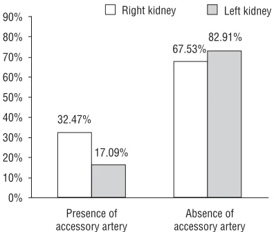
associated with an accessory artery in 47 subjects: in 11 patients on both sides, in 27 cases only on the right side, while in 9 cases only the left side was involved (Fig. 2). A comparison of kidney side and accessory artery development revealed that an
ac-cessory artery occurred significantly more often on the right than on the left side; 38 of 117 (32.47%) and 20 of 117 (17.09%), respectively (p = 0.01).
Early branching of the main renal artery was shown in 42 (35.89%) subjects (Fig. 3), 50 of 234 Table 1. Comparison of right side and left side kidney and artery characteristics
Left side Right side Difference p
Renal artery diameter 0.62 ± 0.11 0.61 ± 0.12 0.014 0.35
Renal artery distance 3.24 ± 1.2 3.56 ± 1.77 0.31 0.11
Kidney length 10.41 ± 1.16 10.31 ± 1.07 0.1 0.48
Kidney width 5.44 ± 0.85 5.71 ± 0.73 0.27 0.01
Kidney anterioposterior 4.97 ± 0.7 4.82 ± 0.69 0.15 0.1
Table 2. Comparison of characteristics of arteries and kidneys in males and females
Male Female p
Aorta diameter 1.61 ± 2.26 1.43 ± 0.18 < 0.001
Left renal artery diameter 0.65 ± 0.10 0.57 ± 0.10 < 0.001
Right renal artery diameter 0.64 ± 0.10 0.56 ± 0.13 0.001
Left kidney length 10.67 ± 1.03 9.95 ± 1.24 0.001
Right kidney length 10.51 ± 0.90 9.92 ± 1.26 0.005
Left kidney width 5.52 ± 0.84 5.29 ± 0.85 NS
Right kidney width 5.79 ± 0.69 5.56 ± 0.79 NS
Left kidney anterior posterior diameter 5.10 ± 0.74 4.74 ± 0.57 0.004
Right kidney anterior posterior diameter 4.92 ± 0.67 4.63 ± 0.70 0.032
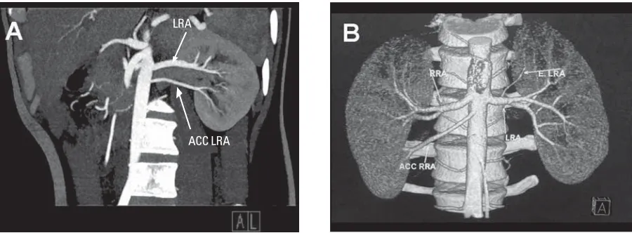
Figure 1. A, B. Multi-slice spiral computerised angiography showing single accessory renal artery on left (MIP) and right (VRT) kidney; LRA — left renal artery, ACC LRA — accessory artery of left renal artery, RRA — right renal artery, ACC RRA — accessory artery of right renal artery, E.LRA — early branching of left renal artery, AL view — anterior left view; AHR view — anterior head right view.
kidneys (21.36%). It occurred bilaterally in 8 cases, in the right kidney in 21 cases and in the left kidney in 13 cases. A comparison of early branching devel-opment in left and right renal arteries showed no significant difference: 21 of 117 (17.94%) vs. 29 of 117 (24.78%), respectively (p = 0.264).
The mean number of segmental arteries on the right and left sides was 2.69 ± 0.782 and 2.73 ± ± 0.738 respectively and there was no difference between right and left sides (p = 0.73). Table 3 shows the in number of segmental arteries accord-ing to the side of renal artery and total.
DISCUSSION
Multi-detector CT angiography provides quick, accurate determination of the anatomical location and course of the renal vessels [6, 10, 11, 13–17]. Angioscopic and MIP views supply additional in-formation about the renal arteries and veins and
complement conventional volume-rendered imag-es (Fig. 4A–D).
Typically, arterial branches of the renal artery can be identified up to the segmental level, but detection of vessels smaller than 2 mm is limited [13]. The sensi-tivity of volume-rendered CT angiography for the dem-onstration and location of the main renal arteries, how-ever, approaches 100% [13, 14, 16]. Surgical and CT findings correlate in over 95% of patients [13] and as CT angiography technology has progressed, its accu-racy has improved. Some researchers have shown that use of 3D volume-rendered CT angiography enables correct identification of renal artery anatomy even up to 100% sensitivity [13, 14, 17].
The ovoid kidneys lie, as is well known, retroperi-toneally on the posterior abdominal wall, one on each side of the vertebral column at the level of the Th12 to that of the L3 vertebrae. The right kidney usually lies slightly inferior to the left kidney because of the Figure 3. Axial volume-rendered image demonstrating early branching in the right renal artery; E.RRA — early branching of right renal artery, R.MRA — right main renal artery, AHR view — anterior head right view.
Figure 2. Comparison of accessory artery development rate be-tween the right and left kidneys.
Table 3. Frequency of segmental arteries
Segmental arteries Left side Right side Total
number Frequency Percentage Frequency Percentage Frequency Percentage
2 55 47% 51 51% 106 45.3%
3 47 40.2% 48 41% 95 40.6%
4 11 9.4% 17 14.5% 28 12%
large size of the right lobe of the liver. Superiorly, the kidneys are associated with the diaphragm and, more inferiorly, the posterior surfaces of the kidney are re-lated to the quadratus lumborum muscle. The liver, duodenum, and ascending colon are anterior to the right kidney and it is separated from the liver by the hepatorenal recess. The left kidney is related to the stomach, spleen, pancreas, jejunum, and descend-ing colon. In view of the differdescend-ing anatomical posi-tions and varying proximities to other organs of the right and left kidneys, we designed the present study to compare the size and vascular anatomy of the right kidney with that of the left and to evaluate the effect of the differences in anatomical position [12].
Although in our study left kidney length and APD were higher than those of the right kidney, these differences were not significant. However, it is in-teresting that the width of the right kidney is signif-icantly greater than that of the left. This may be due to anatomical position and relative proximities. As noted in anatomical references, the right kidney is pushed down and located lower than the left kid-ney because of the pressure of the right lobe of the liver. This pressure on the upper right kidney may result in compression of the kidney in one
dimen-sion, its length, so that this would be less than for the left one. This decrease in length would then be compensated for by an increase in width, so that the right kidney, despite its smaller length, had a greater width.
In most individuals each kidney is supplied by a single renal artery that originates from the abdom-inal aorta [1, 2, 8, 9]. The renal arteries typically arise from the aorta at the level of L2, below the origin of the superior mesenteric artery, with the renal vein being anterior to the renal artery. The renal arteries course anterior to the renal pelvis before they enter the medial aspect of the renal hilum. The right renal artery typically demonstrates a long downward course to the relatively inferior right kidney, travers-ing behind the inferior vena cava. Conversely, the left renal artery, which arises below the right renal artery and has a more horizontal orientation, has a fairly direct upward course to the superiorly posi-tioned left kidney. Both renal arteries usually course in a slightly posterior direction because of the posi-tion of the kidneys [8, 9].
right renal artery traverses a greater distance to reach the kidney hilum than the left. Of course, distance to branching is more important than distance to kid-ney with regard to donation, as the donor renal ar-tery on the side selected is severed before bifurca-tion. We found no difference between the two sides in this respect.
The main renal artery divides into segmental ar-teries near the renal hilum [1, 2, 8, 9]. The first divi-sion is typically the posterior branch, followed by division into four anterior branches as the superior (apical), anterosuperior, anteroinferior, and inferior segmental arteries. Our findings showed there was no difference between right and left sides in the number of segmental arteries. Accessory renal ar-teries constitute the most common, clinically impor-tant renal vascular variant and are seen in up to one third of patients. Multiple renal arteries are unilat-eral in approximately 30% of patients and bilatunilat-eral in approximately 10% [8, 18]. Accessory arteries usu-ally arise from the aorta or iliac arteries anywhere from the level of Th11 to the level of L4. In rare cas-es, they can arise from the lower thoracic aorta or from the lumbar or mesenteric arteries [8]. The ac-cessory artery usually courses into the renal hilum to perfuse the upper or lower renal poles. Accessory vessels to the polar regions are usually smaller than accessory hilar renal arteries, which are typically equal in size to a single renal artery [8].
The prevalence of bilateral and unilateral acces-sory arteries in our study was approximately the same as in previous studies at 9.4% (11 subjects) and 30.76% (36 subjects) respectively. In all cases the accessory arteries arose from the aorta and not from iliac or lower thoracic, lumbar or mesenteric arteries. We also showed that accessory artery de-velopment on the right was twofold that on the left side. This may be due to the greater distance traversed by the right renal artery to reach the kid-ney from the aorta located to the left of vertebral column [12]. On the basis of Poiseuille’s Law, an increase in distance associated with an increase in resistance results in a decrease in flow rate and would result in kidney ischaemia, albeit at a low level [4]. During foetal development, therefore, the kidney probably produces local angiogenetic me-diators and assay to abate this condition by means of an accessory artery, which is associated with supplementary flow.
Early branching, or prehilar arterial branching, is another common variant that must be checked for
in patients being evaluated for donor nephrectomy. Volume-rendered images, MIP images, shaded sur-face display images, and multiplanar reformatted images have all demonstrated a high sensitivity (ap-proaching 100%) in the detection of early branch-ing [6, 7, 13, 14, 17]. Images must be obtained dur-ing the arterial phase of vascular enhancement to obtain such good results [5].
The results of our study demonstrated that al-though early branching occurred in the right renal artery more than in the left, the difference was not significant and early branching may develop on ei-ther side.
Anatomically the arterial patterns of the left and right kidneys do not differ, except in the accessory pattern, which is of importance for donation. In addition the diameter of the aorta, left and right renal arterial diameters and the length and anterio-posterior diameters of the left and right kidneys were higher in males than in females.
REFERENCES
1. Dyer R (1993) Renal arteriography. In: Dyer R ed. Basic vascular and interventional radiology. Churchill Living-stone, New York, pp. 89–95.
2. El-Galley RES, Keane TE (2000) Embryology, anatomy, and surgical applications of the kidney and ureter. Surg Clin North Am, 80: 381–401.
3. Flechner SM, Sandler CM, Houston GK, Van Buren CT, Lorber MI, Kahan BD (1985) 100 living-related kidney donor evaluations using digital subtraction angiogra-phy. Transplantation, 40: 675–678.
4. Guyton AC, Hall JE (2006) Overview of the circulation, medical physics of pressure, flow, and resistance. In: Guyton AC, Hall JE eds. Textbook of medical physiology. Elsevier Saunders, Philadelphia, pp. 161–170. 5. Herts BR, Coll DM, Lieber ML, Streem SB, Novick AC
(1999) Triphasic helical CT of the kidneys: contribution of vascular phase scanning in patients before urologic surgery. Am J Roentgenol, 173: 1273–1277.
6. Johnson PT, Halpern EJ, Kuszyk BS, Heath DG, Wechsler RJ, Nazarian LN, Gardiner GA, Levin DC, Fishman EK (1999) Renal artery stenosis: CT angiography-comparison of real-time volume rendering and maximum intensity projec-tion algorithms. Radiology, 211: 337–343.
7. Kaatee R, Beek FJ, de Lange EE, van Leeuwen MS, Smits HF, van der Ven PJ, Beutler JJ, Mali WP (1997) Renal artery stenosis: detection and quantification with spiral CT angiography and optimized digital subtraction angio-graphy. Radiology, 205: 121–127.
8. Kadir S (1986) Angiography of the kidneys. In: Kadir S ed. Diagnostic angiography. Saunders, Philadelphia, pp. 445–495.
10. Kuszyk BS, Fishman EK (1998) Technical aspects of CT angiography. Semin Ultrasound CT MR, 19: 383–393. 11. Kuszyk BS, Heath DG, Ney DR, Bluemke DA, Urban BA, Chambers TP, Fishman EK (1995). CT angiography with volume rendering: imaging findings. Am J Roentgenol, 165: 445–448.
12. Moore K, Dalley A (1999) Abdomen. In: Moore K, Dalley A eds. Clinically Oriented Anatomy. Lippincott Williams & Wilkins, Philadelphia, pp. 279–289. 13. Platt J, Ellis J, Korobkin M, Reige K (1997) Helical CT
evaluation of potential kidney donors: findings in 154 subjects. Am J Roentgenol, 169: 1325–1330. 14. Rubin GD, Alfrey EJ, Dake MD, Semba CP, Sommer FG,
Kuo PC, Dafoe DC, Waskerwitz JA, Bloch DA, Jeffrey RB (1995) Assessment of living renal donors with spiral CT. Radiology, 195: 457–462.
15. Smith PA, Fishman EK (1998) CT angiography: renal applications. In: Ferris EJ, Waltman AC, Fishman EK, Polak JF, Potchen EJ eds. Syllabus: a categorical course in diagnostic radiology-vascular imaging. Radiological Society of North America, Oak Brook, pp. 35–45.
16. Smith PA, Fishman EK (1998) Three-dimensional CT an-giography: renal applications. Semin Ultrasound CT MR, 19: 413–424.
17. Smith PA, Ratner LE, Lynch FC, Corl FM, Fishman EK (1998) Role of CT angiography in the preoperative eva-luation for laparoscopic nephrectomy. RadioGraphics, 18: 589–601.
18. Spring DB, Salvatierra O Jr, Palubinskas AJ, Amend WJ Jr, Vincenti FG, Feduska NJ (1979) Results and significance of angiography in potential kidney donors. Radiology, 133: 45–47.
19. Walker TG, Geller SC, Delmonico FL, Waltman AC, Athanasoulis CA (1988) Donor renal angiography: its influence on the decision to use the right or left kid-ney. AJR, 15: 1149–1151.