Available online at http://ijpdr.com
Original Research Article
FORMULATION, DEVELOPMENT AND EVALUATION OF
MUCOADHESIVE MICROSPHERE OF STAVUDINE
Raj Karan Pal*1, Girijesh Kumar Pandey1, Amit Joshi1, B.K. Dubey1, Prabhat Jain2 1Technocrats Institute of Technology-Pharmacy Education and Research, Bhopal (M.P.)
2Scan Research Laboratories Bhopal (M.P.)
*Correspondence Info:
Raj Karan Pal,
Department of Pharmaceutics, Technocrats Institute of Technology-Pharmacy Education and Research, Bhopal, M.P
Email:
*Article History:
Received: 20/10/2018 Revised: 26/11/2018 Accepted: 27/11/2018
ABSTRACT
Stavudine (D4T, thymidine) is an FDA-approved drug for clinical use in the treatment of HIV infection, AIDS and AIDS-related conditions either alone or in combination with other antiviral agents. The stavudine has a very short half-life (0.8 to 1.5hr) with rapid absorption. The side effects of stavudine are dose dependent and a reduction of the total administered dose reduces the severity of the toxicity. Stavudine is typically administered orally as a capsule and oral solution. Dosage forms that are retained in the stomach would increase the absorption, improve drug efficiency and decrease dose requirements. The aim of the present study was to develop stavudine loaded chitosan microspheres by the ionic gelation method using sodium tripolyphosphate (Na-TPP) as the crosslinking agent. The use of ionotropic gelation avoids the possibility of the occurrence of the toxic and undesirable effects associated with the use of glutaraldehyde, a chemical crosslinking agent. The prepared microspheres were evaluated for mean particle size and particle size distribution, drug loading, encapsulation efficiency and in-vitro drug release. FT-IR spectroscopic analysis was performed to ascertain drug polymer interaction. The release profiles showed zero-order release behavior up to 12 hours where the highest drug release was 98.78 % of the stavudine loaded in the chitosan microspheres, indicating a strong crosslinking between chitosan and TPP anions. The surface morphology of the prepared microspheres was studied by SEM. With an increase in the crosslinking density the rate of drug release decreased. From the results of the present investigation it may be concluded that drug loaded chitosan microspheres can be prepared by a simple technique which avoids the use of complex apparatus and special precautions.
Key words: Stavudine, HIV infection, Microspheres, Chitosan, Sodium tripolyphosphate.
Introduction:
Oral route is most suitable and preferable route for drug administration to reach systemic circulation due to its low cost and
easy administration. But success of
conventional dosage form is limited due to its
residence time. Hence mucoadhesive
microsphere drug delivery systems are used
to prolong the residence time at the site of
application, maintain therapeutically
Mucoadhesive microspheres become adhesive on hydration and hence used for localizing the drugs to a particular target site of gastrointestinal tract (GIT) for prolonged period of time. Moreover, it is easy for administration, no patient compliances and flexibility in the formulation. One of the most
feasible approaches for achieving a
prolonged and predictable drug delivery in gastrointestinal tract (GIT) is to control gastro retentive drug delivery system which will provide important therapeutic options. Mucoadhesive microspheres delivery system is an attractive due to ability of adherence to the mucosal surface and releases the entrapped drug in a sustained release. Bio adhesion phenomenon is associated with
biological surface and mucoadhesion
associated with i.e. mucin layer of a mucosal tissue. Mucoadhesive microspheres have
advantages like efficient absorption,
enhanced bioavailability of the drugs, maximum utilization of drugs and much more intimate contact with intestinal cells, better patience compliance and targeting to specific absorption site (Anil et al., 2011; Bindu et al., 2010; Parmar et al., 2010; Velmurugana and Ali, 2013). Stavudine
(2',3'-didehydro-3'-deoxythymidine) is a
nucleotide reverse transcriptase inhibitors and primarily used in the treatment of one of the most common chronic disease of the planet, AIDS. It has short biological half-life 0.8–1.5 h and low daily dose of 30 mg is required (Tripathi, 2007; Martin, 1998). The
frequency of dosing is more. To overcome
inherent drawbacks associated with
conventional dosage forms of stavudine, an attempt is being made to develop an alternative drug delivery system in the form of mucoadhesive microspheres.
MATERIALS AND METHODS Materials
Stavudine was obtained as free gift sample from Hetero labs limited, Baddi. Excipients used were Chitosan from Qualigens, Mumbai. Glacial acetic acids were purchased from Merck Specialities pvt. Ltd., Mumbai, Sodium tripolyphosphate was purchased from Loba Chem. Pvt. Ltd (Mumbai, India). All other chemicals and reagent used were of analytical grade. Ultrapure water was used throughout the study.
Preparation of Mucoadhesive Microspheres
Chitosan solution of was prepared in 1% acetic acid using homogenizer (Remi motors, Mumbai) at 5000rpm for about 30 minutes, then drug was added to chitosan solution. Microspheres were formed by dropping the bubble-free dispersion of chitosan –drug solution through a disposable syringe (10 ml) onto a gently stirrer (100 rpm) at room
temperature in 1% TPP (Sodium
tripolyphosphate) solution. Chitosan
microspheres were separated after 2 hours by filtration and rinsed with distilled water and then they were vacuum dried (Kalyankar and Nalanda, 2010). The composition of formulations given in table 1.
Table 1 Formulations of the mucoadhesive microspheres
Sr. No Formulation Code
Stavudine (mg)
Chitosan (mg)
1. F1 10 25
2. F2 10 50
3. F3 10 75
4 F4 10 100
5 F5 10 125
Analytical method development
Determination of absorption maxima
A solution of containing the concentration 10 μg/ ml was prepared in 0.1N HCl. UV spectrum was taken using Double beam
UV/VIS spectrophotometer
(Labindia-3000+). The solution was scanned in the range of 200-400nm.
Preparation calibration curve
Accurately weighed 10 mg of drug was dissolved in 10 ml of 0.1N HCl solution in 10 ml of volumetric flask. The resulted solution 1000µg/ml and from this solution 1 ml pipette out and transfer into 10 ml volumetric flask and volume make up with 0.1N HCl solution. Prepare suitable dilution to make it to a concentration range of 10-50 μg/ml. The spectrum of this solution was run in 200-400 nm range in U.V. spectrophotometer (Labindia-3000+). Linearity of standard curve was assessed from the square of correlation coefficient (r2) which determined by least-square linear regression analysis.
Fourier Transform Infrared (FTIR) spectroscopy
The physical properties of the physical assortment were comparing with those of stavudine pure drug. Samples was assorted comprehensively through 100mg potassium bromide IR powder as well as compacted under vacuum at a pressure of concerning 12 psi for 3 minutes. The ensuing disc was mounted in an appropriate holder in Brukers Alpha IR spectrophotometer and the IR spectrum was recorded from 3500 cm to 500 cm. The resultant spectrum was compared for any spectrum changes.
Evaluation of microspheres Percentage yield
The prepared microspheres with a size range of 200-300nm were collected and weighed from different formulations. The measured weight was divided by the total amount of all non-volatile components which were used for
the preparation of the microspheres
(Venkatesh et al., 2012).
% Yield
= Actual weight of product Total weight of drug and polymer 𝑥 100 Drug entrapment
The various formulations of the
mucoadhesive microspheres were subjected for drug content. 10 mg of mucoadhesive
microspheres from all batches were
accurately weighed and crushed. The powder of microspheres were dissolved in 10 ml 0.1 N HCl and centrifuge at 1000 rpm. This supernatant solution is than filtered through whatmann filter paper No. 44. After filtration, from this solution 0.1 ml was taken out and diluted up to 10 ml with 0.1 N HCl. The percentage drug entrapment was calculated using calibration curve method. Measurement of mean particle size
The mean size of the microspheres was
determined by Photo Correlation
Spectroscopy (PCS) on a submicron particle size analyzer (Horiba Instruments) at a scattering angle of 90°. A sample (0.5mg) of the microspheres suspended in 5 ml of distilled water was used for the measurement. Determination of zeta potential
The zeta potential of the drug-loaded microspheres was measured on a zeta sizer (Horiba Instruments) by determining the
electrophoretic mobility in a micro
electrophoresis flow cell. All the samples were measured in water at 25°C in triplicate (Nagahara et al., 1998).
Shape and Surface characterization of microspheres by scanning electron microscopy (SEM)
From the formulated batches of
vacuum evaporator. The acceleration voltage
was set at 10KV during scanning.
Microphotographs were taken on dissimilar magnification and higher magnification (200X) was used for surface morphology.
In-vitro release studies
The drug release rate from mucoadhesive microspheres was passed out using the USP type II (Electro Lab.) dissolution paddle
instrument. A weighed amount of
mucoadhesive microspheres equivalent to 100 mg drug were dispersed in 900 ml of 0.1 N HCI (pH=1.2) maintained at 37 ± 0.5°C and stirred at 55rpm. One ml sample was withdrawn at predetermined intervals and filtered and equal volume of dissolution medium was replaced in the vessel after each withdrawal to maintain sink condition. The
collected samples analyzed
spectrophotometrically at 255 nm to determine the concentration of drug present
in the dissolution medium (Brahmankar and
Jaiswal, 1988).
Stability studies for optimized formulation Accelerated Testing, are the studies designed to amplify the rate of chemical degradation or physical change of a drug substance or drug product by exaggerated storage conditions as part of the formal stability studies. The optimized formulation F5 was taken and
accelerated stability study was performed by taking suitable quantity of microspheres. The microspheres were placed in air-tight glass container at 40±2°C/75±5% RH. At suitable
sampling interval the samples were
withdrawn and evaluated for various parameters.
Results and Discussion
Percentage yield of different formulation was determined by weighing the microspheres after drying. The percentage yield of different formulation was in range of 65.56– 85.45%. The drug entrapment efficacies of different formulations were in range of 62.32- 75.65% w/w. This is due to the mucoadhesion characteristics of chitosan that could facilitate the diffusion of part of entrapped drug to surrounding medium
during preparation of sofosbuvir
microspheres Table 2. On the basis of the maximum percentage yield and drug entrapment was found to be formulation F5
in mucoadhesive microspheres so
formulation F5 was further studies. The results of measurement of mean particle size
of optimized formulation F5 of
mucoadhesive microsphere was found 178.5 nm Fig 1.
Table 2 Percentage Yield for Different Formulation
Formulation Percentage Yield
Drug entrapment (% w/w) of prepared
microsphere
F1 68.98±0.25 68.89±0.25
F2 65.56±0.12 65.45±0.56
F3 73.25±0.36 62.32±0.47
F4 76.54±0.14 67.89±0.58
F5 85.45±0.25 75.65±0.65
F6 72.15±0.14 69.98±0.84
Shape and surface characteristic of
sofosbuvir microspheres examine by
Scanning Electronic Microscopy analysis.
Surface morphology of formulation
Fig. 3 SEM image of optimized mucoadhesive formulation F5
The drug release rate from mucoadhesive microspheres was passed out using the USP
type II (Electro Lab.) dissolution paddle
instrument. A weighed amount of
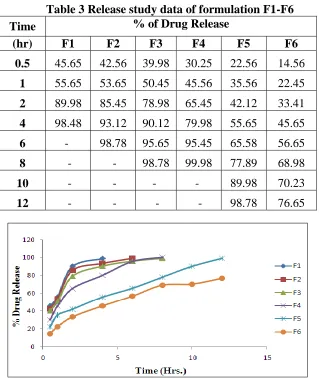
mucoadhesive microspheres equivalent to 100 mg drug were dispersed in 900 ml of 0.1 N HCI (pH=1.2) maintained at 37 ± 0.5°C and stirred at 55rpm. From the graphs (Fig 4) and data (Table 3) of in vitro drug release study, it was observed that formulation F 1 has low drug release and maximum amount of drug release found in formulation F5. The
kinetics of drug release from the
microspheres was studied by mathematical modeling the drug release to zero order, first order kinetics Table 4 and Fig.5 & 6.
Table 3 Release study data of formulation F1-F6
Time % of Drug Release
(hr) F1 F2 F3 F4 F5 F6
0.5 45.65 42.56 39.98 30.25 22.56 14.56
1 55.65 53.65 50.45 45.56 35.56 22.45
2 89.98 85.45 78.98 65.45 42.12 33.41
4 98.48 93.12 90.12 79.98 55.65 45.65
6 - 98.78 95.65 95.45 65.58 56.65
8 - - 98.78 99.98 77.89 68.98
10 - - - - 89.98 70.23
12 - - - - 98.78 76.65
Table 4 Release kinetics of optimized formulation of microsphere F5
Time (h) Square Root of Time(h)1/2
Log Time
Cumulative% Drug Release
Log Cumulative
% Drug Released
Cumulative % Drug Remaining
Log Cumulative
% Drug Remaining
0.5 0.707 -0.301 22.56 1.353 77.44 1.889
1 1 0 35.56 1.551 64.44 1.809
2 1.414 0.301 42.12 1.624 57.88 1.763
4 2 0.602 55.65 1.745 44.35 1.647
6 2.449 0.778 65.58 1.817 34.42 1.537
8 2.828 0.903 77.89 1.891 22.11 1.345
10 3.162 1 89.98 1.954 10.02 1.001
12 3.464 1.079 98.78 1.995 1.22 0.086
Fig 5 Zero order release kinetics graph of optimized formulations
Fig. 6 First order release kinetics graph of optimized formulations The In vitro drug release data of the
optimized formulation was subjected to goodness of fit test by linear regression analysis according to zero order, first order kinetic equation, in order to determine the mechanism of drug release. When the
Table 5 Comparative study of regression coefficient for selection of optimized Formulation F5
Release Kinetics Zero order First order
R2 Mucoadhesive
Microsphere 0.981 0.828
According to ICH guidelines, 3 months accelerated stability study at 40±2°c and 75±5% RH optimized formulations (F5) was carried out. It showed negligible change over time for parameters like appearance, drug content, dissolution and assay etc., No significant difference observed in the drug content between initial and formulations stored at 40±2°c & 75±5% RH for 3 months. Conclusion
There are a myriad techniques available for entrapment and controlled release of important drugs, but only a few pass all the requirements laid down for safe, effective and targeted drug delivery to humans. Chitosan is a very attractive alternative for use in human system because it has long been used as food additive. Controlled drug delivery systems aim to ensure sustained release of drugs in their therapeutic range and chitosan based microspheres are being increasingly used. In case of chitosan/TPP based controlled drug release preparations, chitosan and TPP concentration, pH of TPP and drug concentration are very important parameters for formation of non-fragile, spherical microspheres with good drug encapsulation and tunable in vitro drug release behavior. The stavudine loaded chitosan microspheres were prepared by ionic gelation using Na-TPP as the crosslinking agent. The in vitro dissolution study results indicated formulation F5 found to be the best formulation with controlled release of the drug over a period of 12 hours. The drug release from the microspheres followed zero order kinetics. The results of all studies have been shown to be satisfactory
Thus, Chitosan -TPP based release
formulations are an attractive alternative to traditional drug delivery systems.
References
1. Anil G, Satyanarayana T, Suresh Kumar
P and Pavani S: Formulation and evaluation of gastroretentive floating tablets of Venlafaxine hydrochloride. International Journal of Pharm & Ind. Res 2011; 1: 76-84.
2. Bindu MB, Zulkar NK, Ramalingam R,
Ravindernath A, Kusum B, Naga MM and David B: Formulation and evaluation
of mucoadhesive microspheres of
venlafaxine hydrochloride. Journal of Pharmaceutical Res. 2010; 3(11): 2597-2600.
3. Parmar H, Bakliwal S, Gujarathi N and
Pawar S: Different methods of
formulation and evaluation of
mucoadhesive microsphere. International Journal of App. Bio. and Pharma. Tech. 2010; 3(3); 1157-1167.
4. Velmurugana S and Ali M. Formulation
and evaluation of maraviroc
mucoadhesive microsheres by ionotropic gelation method. International Journal of Pharmaceutical Sciences 2013; 5(4): 294-302.
5. K. D. Tripathi, Essentials of Medical
Pharmacology, Jaypee Brothers’ Medical Publishers (P) Ltd, New Delhi, India, 2007.
6. A. R. Martin, Antiviral Agents; Text
Book of Organic Chemistry and
Pharmaceutical Chemistry, Lippincot-Reven, New York, NY, USA, 1998.
7. Kalyankar T. M., Nalanda T. Formulation
and Evaluation of Mucoadhesive
Pioglitazone Hcl Microspheres.
International Journal of Pharma World Research. 2010, 1(3):1- 14.
8. Venkatesh Gavini, Ganesh N.S,
Formulation and Evaluation of
mucoadhesive microspheres of
macromolecular polymers using
flurbiprofen as model drug. Der
pharmacia Lettre. 2012; 4(5): 1560-1566.
9. Nagahara N, Akiyama Y, Nakao M, Tada
M, Kitano M, Ogawa Y. Mucoadhesive microspheres containing amoxicillin for
clearance of Helicobacter pylori.
Antimicrob Agents Chemother. 1998; 42: 2492–2494.
10. Brahmankar DM and Jaiswal SB.
Biopharmaceutis and Pharmacokinetics: A Tretise, Vallabh Prakashan, New Delhi, 1st