Sex differences in tolerance to morphine antinociception in intra-nucleus
accumbens administration in rat
Maryam Beshkani1, Nasim Assar1, Parvaneh Najafizadeh2, Zahra Mousavi3*
1 Herbal Medicines Research Center, Pharmaceutical Sciences Branch, Islamic Azad University, Tehran, Iran
2 Department of Pharmacology, Iran University of Medical Sciences, Tehran, Iran, & Young Researchers & Elite Club, Pharmaceutical
Sciences Branch, Islamic Azad University, Tehran, Iran
3 Department of Pharmacology and Toxicology, Faculty of Pharmacy, Pharmaceutical Sciences Branch, Islamic Azad University, Tehran,
Iran, & Herbal Pharmacology Research Center, Tehran Medical Sciences Branch, Islamic Azad University, Tehran, Iran
Please cite this article as:
Beshkani M, Assar N, Najafizadeh P, Mousavi Z. Sex differences in tolerance to morphine antinociception in intra-nucleus accumbens administration in rat. Iranian J Pharmacol Ther. 2017 (August);15: 1-7.
ABSTRACT
Sex differences in analgesic responses and tolerance to morphine under the systemic and injection in some nuclei of brain have been reported, although the trend of these differences varies across studies. The purpose of the present study was to determine whether development tolerance to the analgesic effect of morphine differs between male and female in intra-nucleus accumbens administration of morphine in rat. In order to induce tolerance, adult male and female rats were injected with morphine (2.5, 5, 10 and 50 µg/0.5 µL; intra-accumbal infusions) for 4 consecutive days during which, non-tolerant group received a single dose of morphine (saline for 3 consecutive days and morphine on 4th day) and control vehicle group received saline (4 consecutive days). On day 4, tail flick and hot-plate test was done for pain evaluation in separated groups. The results of the study revealed 4 effects. 1) No significant morphine antinociceptive effect by tail flick test in both sexes, while significant antinociceptive effect of morphine were observed in the hotplate test. 2) No significant sex differences were observed in hotplate test after acute morphine injection to animals. 3) Rats showed significantly tolerance to morphine analgesic effects under protocol by hotplate test. 4) Female rats showed significantly more tolerance to morphine analgesic effects than males by hotplate test. These data demonstrate that there were sex differences in tolerance to morphine antinociception in intra-nucleus accumbens administration that is dependent, in part, on the nociceptive test.
Conflicts of Interest: Declared None
Funding: None
Keywords
Sex differences, Nucleus accumbens, Antinociception, Tolerance, Morphine, Rats
Corresponding to:
Zahra Mousavi,
Department of Toxicology & Pharmacology, Pharmaceutical Sciences Branch, Islamic Azad University (IAUPS), No 99, Yakhchal, Gholhak, Shariati St., Tehran, Iran
Email:
Received: 15 Jan 2017 Revised: 25 Feb 2017,
Accepted: 23 May 2017
INTRODUCTION
Animal studies have revealed that male and female show different sensitivities to the effect of opioids. For example, male rats are typically more sensitive than female to the antinociceptive properties of morphine in several different pain model [1-5]. There is also several evidence
demonstrating sex differences in the development of tolerance and dependence to opioid such as morphine [2, 4, 6, 7]. Male rats have been reported to show greater
antinociception than females following systemic
administration of morphine [3, 5, 8, 9]. Although, several
IRANIAN JOURNAL OF PHARMACOLOGY &THERAPEUTICS Copyright © 2017 by Iran University of Medical Sciences
Iranian J Pharmacol Ther. 2017 (August);15:1-7.
2 Beshkani et al.
factors such as different pharmacokinetics for drug and pain sensitivity may contribute to this difference, greater antinociception in male than female following intra cerebra ventricular (ICV) administration of morphine [10] point out a difference in the brains of male and female rats. It was shown that micro injection of morphine into the PAG or RVM nucleus of brain in male and female rat, antinociceptive effect produces greater in male rat rather than female[10-12].
Although these sex differences in morphine
antinociception and tolerance was not reported in others nucleus such as accumbens. We have hypothesized that the nucleus accumbens is a strong candidate to mediate sex difference in morphine induced antinociception since this
site play an important role on process of
reward/nociception/addiction mechanism. Therefore, the purpose of the present study was to determine whether development tolerance to the analgesic effect of morphine differs in intra–nucleus accumbens administration between male and female rats.
MATERIALS AND METHODS Animals
In the present study, male and female Wistar rats 200±20 g (Shahid Beheshti laboratories, Tehran, Iran) were housed in groups of six to eight and were allowed free access to food and water, except for a short duration of time when the animals were removed from their cages for testing. All experiments were conducted between 10 am and 4 pm with normal room light (12 hours of regular light/dark cycle) and temperature of 22±1°C. The procedures were carried out in accordance with the guidelines of Pharmaceutical Sciences Branch of Islamic Azad University for animal care and use. Additionally, efforts were made to minimize the animals’ suffering and to use only the number of animals necessary to produce reliable scientific data.
Drugs
Powdered morphine (Temad Co.) was dissolved in saline. The solutions were prepared immediately before use and intra accumbal injected in a volume of 0.5µL/Rat.
Experiments
In this study٫ tail-flick and hot plate tests were carried on separate groups. The objective of this experiment was to
determine whether tolerance developed to the
antinociceptive effect of microinjecting morphine into NAcc and whether there are sex differences in development morphine tolerance.
Nociceptive tests
Tail-flick test: The nociceptive threshold was measured by the tail-flick apparatus (Borj Sanat, Iran). The tail-flick latency (TFL) was also measured by exposing the dorsal surface of mice tail to radiant heat and recording the time in which animals removed the tail away from the noxious thermal stimulus. Their action time between the onset of heat
stimulus and movement of the tail was determined by an automatic sensor as TFL. The thermal intensity of radiant heat source was set to produce a baseline TFL between 3.0 and 4.0 (S). To avoid tissue damage٫ trial was automatically terminated if a response did not occur in 9.0 and 10.0 (S) (cut-off point). TFLs(s) were expressed as raw data. To evaluate the sensitivity of animals to nociceptive stimulus٫ the individual TFL was considered as a pain threshold before and after (15, 30 and 60 min) drug treatment.
Hot-Plate test
Animals were individually placed on a thermostatically controlled hot-plate (Borj Sanat, Iran), maintained at 55 ± 0.5 °C. Briefly, the animals were placed on the hot-plate apparatus and the time between their placement on platform and shaking or licking of the paws or jumping was recorded as the reaction time or latency of pain response. In order to avoid damage to paws of the animals, the standing time on the plate was limited to 20 seconds (cut-off time). Before treatment, the reaction time was taken once. The hot plate test was performed on all animals individually, 15, 30 and 60 min after treatment.
Experimental design
Male and female rats (200-220 gr) were anesthetized with (ketamine 100 mg/kg and Xylazine 10 mg/kg) and implanted with a guide cannula (23gange) aimed at NAcc. The rats were take recovery days for 7 days and then received morphine according to the protocol (in order to induce tolerance adult male and female rat were injected with morphine (2.5, 5, 10 50µg/0.5ml, 4 consecutive days) which non-tolerant group received a single dose of morphine (Saline for 3 consecutive and morphine on 4th day) and control vehicle groups received saline alone (4 consecutive days) (Fig. 1).
Surgery
Rats were anesthetized with ketamine/xylazine and placed in stereotaxic apparatus. Microinjections were performed by 30 gauge cannulae into the nucleus accumbens (1mm below the tip of the guide cannulae, AP, 1.8 mm; ML, 1.2 mm; DV, −7.2 mm) (Paxinos and Watson, 1986). Polyethylene tubing (PE-20) was used to attach injector cannula to the 1-µl Hamilton syringe. Drug solutions or vehicles were slowly administered in a total volume of 0.5µl/rat over a period of 60s into the nucleus. Injection needles were left in place for an additional 60s to facilitate diffusion of the drugs, and then the stylets were reinserted into the guide cannulae.
Statistical analysis
Comparisons between the groups were carried out using the analysis of variance (ANOVA) and post hoc Tukey’s test P<0.05 was considered to represent a significant difference.
RESULTS Tail flick test
Antinociceptive effects of morphine was initially examined by intra-accumbal administration using the
tail-flick test. Figure 2 shows that morphine did not significantly
increased tail-flick latency in a dose-dependent manner (2.5, 5, 10 and 50 µg/rat) 15 min after microinjection in male mice. When the time course of antinociception was examined, maximal mean TFL were observed 15 min after administration of morphine doses 10 and 50 µg/rat, and these doses were used to determine time course effect in male and
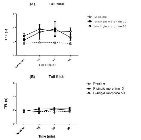
female rats. Figures 3A and 3B show that TFL values were
similar between morphine and saline groups, indicating that opioid agonist does not possess antinociceptive efficacy in intra-accumbal administration in both sexes. Although morphine (10mg/kg) significantly increased TFL in male rats 30min later to microinjection in compared to saline group. Figure 3 shows that there were significant sex differences in morphine effects on tail flick. Morphine
produced slightly increase in TFL 15 to 60 min post-injection in male rats but did not change tail flick response in female ones.
Hotplate test
Morphine microinjection in NAcc effect on inducing
morphine antinociception and tolerance were also
investigated in rats and sex differences were observed in this effect through hot plate test. Male and female rats received separately single doses of morphine (50µg/rat) and multiple doses (50 µg/rat, 3 consecutive days). Figure 5 shows the
Figure 1. Timeline of morphine tolerance induction experiments. After 7 days recovery from surgery and cannulation, intra-NAcc saline,
morphine according to the protocol (in order to induce tolerance rat were injected with morphine (2.5, 5, 10 50µg/0.5ml, 4 consecutive days) which non-tolerant group received a single dose of morphine (Saline for 3 consecutive and morphine on 4th day) and control vehicle groups received saline alone (4 consecutive days). Then nociceptive tests were performed 15, 30 and 60 min after the last injection.
Figure 2. Dose response was used to assess morphine
antinociceptive effect (2.5, 5, 10 and 50 μg/0.5 μl) in intra-accumbal microinjection in male rats. Nociception was assessed 15min after each injection.
ba se
lin
e 15 30 60
0 2 4 6
( A ) T a il flic k
T im e ( m in )
T
F
L
(
s
)
M - s a lin e M - s in g le m o r p h in e 1 0 M - s in g le m o r p h in e 5 0
Figure 3. A time course response was used to assess the
antinociceptive effect of morphine (10 and 50μg/0.5μl) in intra-accumbal microinjection in male (A) and female (B) rats. Nociception was assessed before, 15, 30 and 60 min after each injection. M=male, F=female.
4 Beshkani et al.
latency of male and female rats to respond on the hot-plate assays after single and multiple morphine microinjections in NAcc. After single morphine administration, females and male rats increased significantly latencies similarity. There
were no significant sex differences in morphine
antinociceptive effects in intra accumbal injection. In addition, following increasing period of morphine takings for 3 days, all rats showed tolerance to morphine antinociception, for the reason that latency come back to baseline value (Fig. 5).
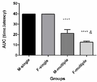
To illustration the sex differences in morphine tolerance, area under the curve (AUC), time-latency, of morphine antinociceptive effect per time was calculated. AUC was shown that morphine produced greater antinociception tolerance in females than males at some doses and time points on hot plate assay, and this sex difference was statistically significant in this assay (Fig. 6).
DISCUSSION
The present study was conducted to investigate, for the first time, the sexual difference after intra-NAcc microinjection of morphine on antinociception and tolerance to morphine using tail flick and hot plate tests in rats. Morphine doses, as well as, the antinociceptive model were selected according to administrated doses in previous studies which, despite the excepted primary doses (2.5 µg/0.5 mL), did not show any antinociceptive effect. However, increasing the dose level to 50µg/0.5mL revealed some antinociceptive effects. According to the present results on tail flick test, it was observed that the administration of different doses of morphine in the nucleus accumbens of both male and female rats induces significant antinociceptive effects in male rats only. Sensitivity to nociceptive stimulus in tail flick test was statistically significantly notable in male than female rats. Considering the rate of mortality in higher doses, the study was not continued in this test.
Moreover, the effect of morphine administration in the nucleus accumbens of both sexes' rats was studied using the hot plate test. However, the responses were not identical in the two models. The results indicated that 50µg/0.5 mL of morphine induced antinociceptive effects both in male and female rats similar to each other. Sensitivity to nociceptive stimulus in hot plate pain model had no statistically significant difference between male and female rats. In addition, daily administration of the same dose of morphine for 4 days could induce tolerance against the antinociceptive effect in both sexes. The tolerance was significantly higher in female rats.
The greater antinociception, although low, following microinjection of morphine into NAcc of male compared to female rats on tail flick test but not in hotplate test reported here is consistent with the sex differences reported following systemic or i.c.v. and RVM administration of morphine [3,
4, 10, 11, 13, 14]. Previous studies conducted by Kepler et
al., using morphine administration in PAG nucleus and tail
flick and jump tests in rats, reported the sexual difference in
this case, the male rats showed stronger antinociceptive effects than intact females [10]. Boyer et al. reported that sex
Figure 4. Sex differences in morphine antinociceptive effect
(50μg/0.5μl) in intra-accumbal microinjection in male and female rats by tail flick test. Nociception was assessed before, 15, 30 and 60 min after each injection. #P<0.05; ##P<0.001;
###P<0.0001 in compared to female-morphine group; M=male,
F=female.
Figure 5. Sex differences in tolerance to morphine
antinociceptive effect (50 μg/0.5 μl) in intra-accumbal microinjection in male and female rats by hot plate test. Nociception was assessed before, 15, 30 and 60 min after each injection. * Significantly different from respective sex group
(P<0.0001). # Significantly different from F-multiple group
(P<0.001); M=male, F=female.
Figure 6. Sex differences in AUC morphine antinociceptive
effect (50 μg/0.5 μl) in intra-accumbal morphine microinjection in male and female rats by hot plate test. Nociception was assessed before, 15, 30 and 60 min after each injection. * Significantly different from respective sex group
(P<0.0001); & Significantly different from M-multiple group
(P<0.05); M=male, F=female.
differences in morphine antinociception are mediated at least in part by the RVM. Microinjection of morphine into the RVM produced greater antinociception in male compared to female rats in tail withdrawal test. Male, but not female rats also showed a dose dependent decrease in locomotor activity [11]. In contrast, some previous studies showed no significant sex difference in antinociception after intra-PAG morphine microinjection [12, 15]. While in a study Tershner et al, DAMGO microinjection into vPAG was observed to be
more potent in femalecompared to male rats [16]. Although
sex differences following systemic administration of morphine could be caused by differences in morphine absorption or distribution, but direct administration of morphine into NAcc avoids this problem. However, there are
several probable reasons for this difference in
antinociception induced by morphine microinjection into NAcc of male compared to female rats such as dissimilar
sensitivity to noxious stimuli or differences in
responsiveness to morphine and or a functional and anatomical difference in NAcc of male and female rats.
It should be mentioned that an inconsistency was observed in the results of tail flick and hot plate tests in assessing the morphine antinociception effect and tolerance. As it has been already suggested, a different mechanism for pain inhibition is used for regulating phasic stimulation [17]. Different responses in these two models reflect the impact assessment model. On the other hand, tail flick test is a reflexive test which contains spinal mechanisms and thus, cannot efficiently show super spinal mechanisms as hot plate test does. Considering that tolerance is caused by disturbance in the relation of spinal and supra-spinal mechanisms, it is likely that a great part of mechanisms related to sexual difference in antinociception and tolerance are supra-spinal. This finding is consistent with the study of Ji et al., in which the sexual difference in systemic morphine injection in visceral pain was studied. They also stated that a great part of this sexual difference is due to supra-spinal and environmental mechanisms and spinal mechanism does not play an important role [18]
Therefore, the present findings are in agreement with the previous ones indicating that induction of painful stimuli and drug administration method can also be considered as important factors in pain behavior and morphine antinociceptive effect and morphine tolerance in exploring the role of sex hormones. In most previous studies in which other pain assessment approaches including formalin test, tail withdrawal, abdominal constriction, paw pressure and
shock jump were used, morphine induced more
antinociceptive effects in both male mice and rats, compared to female ones in systemic administration [3, 5, 6, 19]. However, there are studies in which no sexual difference in antinociceptive effects was observed or the antinociceptive effects in females were reported to be more than males [13, 20, 21]. It have been reported that morphine levels in CNS to be higher in male than in female mice and rats after systemic morphine injection [14, 22], but other studies report no sex differences in CNS or plasma morphine levels in rats [8, 23].
Since this difference was observed in CNS administration of morphine and other μ-agonists [10, 24, 25] and also morphine injection to RVM [11], PAG [16, 26] and in this study to NAcc, it seems that this phenomenon is centrally mediated. Thus, sex differences in opioid antinociception are probably caused by pharmacodynamics factors, such as sexually dimorphic brain nuclei at which opioids act. In previous study reported that selective lesions of µ receptor-expressing PAG neurons blocked systemic morphine analgesia in males only. Thus, as the PAG, constitute an essential circuit for opioid analgesia in pain-inhibitory pathways [27, 28], could integrate in the mediation of sex differences in morphine analgesia. The results of these studies demonstrate that both the anatomical organization, and functional activation, of the PAG-RVM circuit is sexually dimorphic, and may provide the anatomical substrate for sex-based differences in morphine analgesia [29]. The future studies could indicate if NAcc pathway is sexually dimorphic in its anatomical and functional organization and in its activation during pain sensitization pathway. NAcc is a critical component of the mesolimbic system and brain reward pathway with a key role in the mediation of rewarding effects of drug of abuse and natural rewards such as food and sexual behavioral [30-32]. Also based on previous study, NAcc is involved in opioid sensitization [33, 34]. Our previous study indicated that significant differences of glutamate level in nucleus accumbens of female rats versus males, so the involvement of excitatory amino acid neurotransmission in sex-related differences in morphine antinociception [4]. The results of the present study demonstrate for the first time that the actions of morphine on NAcc neurons were also found to be sexually dimorphic. Together, these results suggest that NAcc may provide the anatomical basis for observed sex differences in morphine analgesia and tolerance.
Also sexual difference in tolerance to morphine antinociception has been previously reported in systemic and central morphine administration in tail flick and formalin pain models. In our previous study was revealed that the sexual difference in morphine antinociceptive tolerance by chronic s.c. administration of morphine in the tail flick test in this case, female rats would express the tolerance faster than male rats [4]. There are other studies in which different accounts of sexual difference in the development of morphine tolerance have been reported. In a study conducted
by Holtman et al., it has been reported that during the
induction period of morphine tolerance more percentages of
female rats developed tolerance against morphine
antinociception effect, compared to male rats after i.p. administration using the tail flick test [35]. The same changes were reported in using the accumulative doses and
hotplate test (ED50 developed up to 6.9 and 3.7 in the
toleration of male and female rats, respectively [6].
Several factors including genes, sexually dimorphic in anatomical and functional organization involved in pain modulation, sexual hormones and environmental factors are involved in the expression of sexual difference in morphine
6 Beshkani et al.
antinociception and morphine tolerance[36]. It seems that sexual dimorphic in brain nuclei play a significant role in that regard. Thus, further investigation of the role of these brain nuclei such as NAcc is suggested.
Given the data obtained from most studies in the field,
researchers reached similar results, including that
antinociception in male rats is more than in female ones. Moreover, they found that female rats developed tolerance against morphine slower than male rats. Justifying this phenomenon, Krzanowska and Bodnar indicated that PAG is an important nucleus in which duplication of PPE mRNA is controlled by estradiol. Estradiol increases the duplication of PPE mRNA in female rats’ hypothalamus in estrous cycle, compared to male rats. In addition, endogenous opioid is increased following the increase of mRNA[26]. Ji et al. obtained similar results in the same nucleus. By injecting Mu-opioid receptor (MOR) antagonist, they observed more increase in morphine antinociception in female rats than in male ones. They suggested that MOR receptors in male rats are more than in female rats which makes MOR agonist stronger in the former. Finally, they concluded that estrous cycle and concentration of MOR receptor play a significant role in sexual difference in antinociception and tolerance. They showed that there was no sexual difference between the diestrous female and male rats and that the former was less sensitive to morphine than diestrous in the first phase of estrous. The least sensitivity level to antinociception effects of MOR agonist was observed 24 h after estrogen’s plasmatic peak [18]. Recent studies performed by Verzillo et al., in molecular level showed that MOR structure, as the major factor in antinociception, contains sexual dimorphic. They studied the effect of estrous’cycle and tissue and chronic morphine injection on mRNA variants and its consequent effect on sexual difference in antinociception and tolerance. They found that chronic morphine injection in spinal cord caused mRNA to tend to mRNA-1C1 and mRNA-1B2 and amplified MOEGs coupling only in male rats. Acute morphine injection in spinal cord increased heterodimer (MOR-KOR) in female rats, compared to male rats, which was observed more in estrous phase rather than in diestorus phase [37].
Another explanation is that outputs of accumbens nucleus (selected in the present study) are different from the outputs of RVM and PAG, the role of which in pain decline and antinociceptive supra-spinal mechanism have been already proved. Morphine administration in PAG and RVM cause on cell inhibition, and antinociception is indirectly applied by off cell. NAcc contains two core and shell sections. The former is more involved in motion system and the latter is related to limbic and motion system. These areas take many dopaminergic inputs from VTA at which limbic dopaminergic pathway begins. Many glutamatergic inputs also enter this nucleus. Neurons present in this nucleus are of cholinergic, dopaminergic and GABAergic types. Therefore, addictive drugs function in this nucleus by changing dopamine level and the reward system. Opioids allow more firing of dopaminergic neurons by inhibiting GABAergic
neurons, and they increase dopamine level in NAcc. Thus, NAcc plays a significant role in opioids effects. Morphine antinociception and tolerance in NAc nucleus in tail flick test have not been observed probably due to the difference in outputs of NAcc, RVM and PAG. Further researches are needed to study sexual difference in antinociception and tolerance of opioids in NAc nucleus by intra-cerebral
injection of
antagonists. Also it is recommended to studyon sexual difference in tolerance to opioids antinociception in NAcc nucleus by intra-cerebral injection of dopaminergic or GABAergic antagonists and studying the interaction dopaminergic and GABAergic systems with opioids.
CONCLUSION
In Conclusion, this study showed the sex differences in tolerance to morphine antinociception in intra-nucleus accumbens administration that is dependent, in part, on the nociceptive test. It seems that sexual dimorphic in brain nuclei play a significant role in that regard. Thus, further investigation of the role of these brain nuclei such as NAcc is suggested.
ACKNOWLEDGEMENT
This work was based on the Pharm-D thesis of Maryam Beshkani, Faculty of Pharmacy, Pharmaceutical Sciences Branch, Islamic Azad University, Tehran, Iran (IAUPS).
We thank Professor Rebecca M. Craft for review the article and her valuable comments.
CONFLICT OF INTEREST
The authors declare that this research does not have any conflict of interest with anyone or any institute.
REFERENCES
1. Craft RM. Sex differences in opioid analgesia: “from mouse to man”. Clin J Pain 2003;19:175-186.
2. Craft RM, Mogil JS, Aloisi AM. Sex differences in pain and analgesia: the role of gonadal hormones. Eur J Pain 2004;8:397-411.
3. Cicero TJ, Nock B, Meyer ER. Gender-related differences in the antinociceptive properties of morphine. J Pharmacol Experim Therap 1996;279:767-773.
4. Mousavi Z, Shafaghi B, Kobarfard F, Jorjani M. Sex differences and role of gonadal hormones on glutamate level in the nucleus accumbens in morphine tolerant rats: a microdialysis study. Eur J Pharmacol 2007; 554:145-149.
5. Aloisi AM, Albonetti ME, Carli G. Sex differences in the behavioural response to persistent pain in rats. Neurosci Let 1994;179:79-82. 6. Craft R, Stratmann J, Bartok R, Walpole T, King S. Sex differences in
development of morphine tolerance and dependence in the rat. Psychopharmacology 1999;143:1-7.
7. Badillo-Martinez D, Kirchgessner AL, Butler PD, Bodnar R: Monosodium glutamate and analgesia induced by morphine: test-specific effects. Neuropharmacology 1984, 23:1141-1149.
8. Cicero TJ, Nock B, Meyer ER. Sex-related differences in morphine’s antinociceptive activity: relationship to serum and brain morphine concentrations. J Pharmacol Experim Therap 1997;282:939-944. 9. Baamonde AI, Hidalgo A, Andres-Trelles F. Sex-related differences in
the effects of morphine and stress on visceral pain. Neuropharmacology 1989;28:967-970.
10. Kepler KL, Kest B, Kiefel JM, Cooper ML, Bodnar RJ. Roles of gender, gonadectomy and estrous phase in the analgesic effects of
intracerebroventricular morphine in rats. Pharmacol Biochem Behav 1989; 34:119-127.
11. Boyer JS, Morgan MM, Craft RM. Microinjection of morphine into the rostral ventromedial medulla produces greater antinociception in male compared to female rats. Brain Res 1998;796:315-318.
12. Bernal SA, Morgan MM, Craft RM. PAG mu opioid receptor activation underlies sex differences in morphine antinociception. Behav Brain Res 2007;177:126-133.
13. Bartok RE, Craft RM. Sex differences in opioid antinociception. Journal of Pharmacology and Experimental Therapeutics 1997;282:769-778.
14. Craft R, Kalivas P, Stratmann J. Sex differences in discriminative stimulus effects of morphine in the rat. Behav Pharmacol 1996. 15. Kanarek RB, Mandillo S, Wiatr C. Chronic sucrose intake augments
antinociception induced by injections of mu but not kappa opioid receptor agonists into the periaqueductal gray matter in male and female rats. Brain Res 2001;920:97-105.
16. Tershner SA, Mitchell JM, Fields HL. Brainstem pain modulating circuitry is sexually dimorphic with respect to mu and kappa opioid receptor function. Pain 2000;85:153-159.
17. Greenspan JD, Craft RM, LeResche L, Arendt-Nielsen L, Berkley KJ, Fillingim RB, et al. Studying sex and gender differences in pain and analgesia: a consensus report. Pain 2007;132:S26-S45.
18. Ji Y, Murphy AZ, Traub RJ. Sex differences in morphine-induced analgesia of visceral pain are supraspinally and peripherally mediated. Am J Physiol-Regul Integrat Compar Physiol 2006;291:R307-R314. 19. Craft RM. Sex differences in analgesic, reinforcing, discriminative, and
motoric effects of opioids. Experim Clin Psychopharmacol 2008; 16:376.
20. Ali B, Sharif S, Elkadi A. Sex Differences and the effect of gonadectomy on morphine‐induced antinociception and dependence in rats and mice. Clin Experim Pharmacol Physiol 1995;22:342-344. 21. Shekunova EV, Bespalov AY. Estrous cycle stage-dependent
expression of acute tolerance to morphine analgesia in rats. Eur J Pharmacol 2004;486:259-264.
22. Candido J, Lutfy K, Billings B, Sierra V, Duttaroy A, Inturrisi CE, et al. Effect of adrenal and sex hormones on opioid analgesia and opioid receptor regulation. Pharmacol Biochem Behav 1992;42:685-692. 23. Cicero TJ, Ennis T, Ogden J, Meyer ER. Gender differences in the
reinforcing properties of morphine. Pharmacol Biochem Behav 2000;65:91-96.
24. Argus MV, Matthews EK: Stratigraphic excavation techniques for paleontologists. J Paleontol 1991;17:119-127.
25. Kepler KL, Standifer KM, Paul D, Kest B, Pasternak GW, Bodnar RJ. Gender effects and central opioid analgesia. Pain 1991;45:87-94. 26. Krzanowska EK, Bodnar RJ. Morphine antinociception elicited from
the ventrolateral periaqueductal gray is sensitive to sex and gonadectomy differences in rats. Brain Res 1999;821:224-230. 27. Basbaum AI, Fields HL. Endogenous pain control mechanisms: review
and hypothesis. Ann Neurol 1978;4:451-462.
28. Basbaum AI, Fields HL. Endogenous pain control systems: brainstem spinal pathways and endorphin circuitry. Ann Rev Neurosci 1984; 7:309-338.
29. Loyd DR, Murphy AZ. Sex differences in the anatomical and functional organization of the periaqueductal gray‐rostral ventromedial medullary pathway in the rat: A potential circuit mediating the sexually dimorphic actions of morphine. J Compar Neurol 2006;496:723-738. 30. Russo SJ, Nestler EJ. The brain reward circuitry in mood disorders.
Nature Rev Neurosci 2013;14:609-625.
31. Morton G, Cummings D, Baskin D, Barsh G, Schwartz M. Central nervous system control of food intake and body weight. Nature 2006; 443:289-295.
32. Fiorino DF, Phillips AG. Facilitation of Sexual Behavior and Enhanced Dopamine Efflux in the Nucleus Accumbens of Male Rats afterd-Amphetamine-Induced Behavioral Sensitization. J Neurosci 1999; 19:456-463.
33. Azizi P, Haghparast A, Hassanpour-Ezatti M. Effects of CB1 receptor antagonist within the nucleus accumbens on the acquisition and expression of morphine-induced conditioned place preference in morphine-sensitized rats. Behav Brain Res 2009;197:119-124. 34. Vanderschuren LJ, Kalivas PW. Alterations in dopaminergic and
glutamatergic transmission in the induction and expression of behavioral sensitization: a critical review of preclinical studies. Psychopharmacology 2000;151:99-120.
35. Holtman JR, Sloan JW, Wala EP. Morphine tolerance in male and female rats. Pharmacol Biochem Behav 2004;77:517-523.
36. Lee CW-S, Ho K. Sex differences in opioid analgesia and addiction: interactions among opioid receptors and estrogen receptors. Molecul Pain 2013;9:45.
37. Verzillo V, Madia PA, Liu NJ, Chakrabarti S, Gintzler AR. Mu‐opioid receptor splice variants: sex‐dependent regulation by chronic morphine. J Neurochem 2014;130:790-796.