Visual light leak in high-frequency electromagnetic
radiation hole: A photon-electron theory for living tissue
for explaining myopia
Alex Lee(李大岷), Xianning West road 28#, Xi’an city, Shanxi province,
China.710,049.
E-mail: [email protected]
Explaining the epidemiology of myopia : 1.The photon of high-frequency
electromagnetic radiation can strike the eye to form a hole of a diameter in photon
diameter scale. 2.The visual light passes through the hole cannot be refracted on the
macula, this visual light ‘s image loses in the macula, which is the blurred vision.3.when
in the near distance, the undestroyed cornea and lens can collect more visual light and
then the cornea and lens refract the visual light to the macula. This is nearsightedness.
4.The particle property of photon of high-frequency electromagnetic radiation explains
elongation of myopic eye.5.Blue light and X-ray are two kinds of high-frequency
Abstract: The exact etiology of myopia remains elusive. The author proposes the
particle property of photon in living tissue and the photon-electron theory in living
tissue, if the frequency of the photon exceeds the threshold, the photon has enough
energy to strike the electron away and forms a hole of a diameter in photon
diameter scale in living tissue include the eye. Longer time of high-frequency
electromagnetic radiation will cause more holes and a larger radius of holes. The
visual light which passes through the radiated hole in the eye cannot be refracted on
the macula, this is myopia symptom of blurred vision, shortening the distance of the
eye and the object will include more visual light into cornea and lens, this is myopia
another symptom of nearsightedness. The particle property of photon causes
elongation of the eye and the macular holes. Blue light and X-ray are two kinds of
high-frequency electromagnetic radiation which can shot holes in eye and cause
TEXT
Myopia is a worldwide visual impairment1. Myopia has increased from 25% to 44%
between 1972 and 2004 in the United States1. In the cities of Asia, the prevalence is over
80%1. There will be 2.62 billion people in myopia by the year 2020, and about 4.76
billion people (49.8% of the world population) will become myopia by the end of 20502.
From 350 BC to now, literature has showed hypotheses about etiology of myopia such as
lack of enough outdoor activities and too long time of near work 1, literature suggested
that blue light from screen may correlate with myopia3,4, but the identical effect of blue
light remains unclear3-5. The specific causes of myopia remain undefined1,6,7.
A hypothesis of particle property of the high-frequency electromagnetic
radiation in living tissue
Albert Einstein proposed that light is not a wave passing through space, but a collection
of independent wave packets (photons), which has an energy hν 8. In 1923 the "Compton
scattering" showed the particle concept of electromagnetic radiation9. In 2017, an
experiment tested the records of light-by-light scattering at the Large Hadron Collider 10.
The electromagnetic radiation effect on cornea has been investigated on the wave
property but not the particle property11,12. The effects of ionizing radiation in living tissue
are controversial 13,14.
The first hypothesis: The photon has the particle-like property in living tissue.
According to photon-electron theory, when the photon’s frequency reaches or exceeds a
threshold, electrons are kicked out only by the hit of photons, this differs from the
classical electromagnetic theory8
The second hypothesis:
1. The photon of visual light’s frequency is lower than the threshold, which cannot hit the
electron away from its original position, the direction of visual light is changed by the
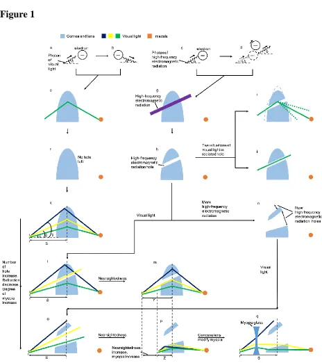
scattering of electron(Fig. 1. a-b ).
2. The photon of high-frequency electromagnetic radiation has enough frequency to hit
the electrons away and shot in a straight line in living tissue(Fig. 1. c-d ).
A hypothesis of massive photons of high-frequency electromagnetic
radiation producing holes in the eye
It has reported the holes produced by heavy-ion radiation. In 1983, literature showed a
membrane lesion in the heavy ion irradiated corneas 15, later in 2004, the Brookhaven
National Laboratory in the US made an experiment on cells irradiation by heavy ions,
which showed that holes may form as the result of heavy ion irradiation in liposomes and
that these holes are large enough to allow leakage of cell internal contents 16.
There is literature reported the hole results of electromagnetic radiation. In 1946,
literature reported an experiment result on the animal that the Cathode Ray have a
penetration without underlying reaction17. In 1983, an experiment showed the hole is the
physical damage to the lens after microwave radiation on the murine ocular lens18.In
2016, literature reported a corneal hole during radiation therapy for cancers of the head
and neck19.
1. The visual light can be focused on the macula by the scattering of electron without
hole left(Fig. 1. e-f ), because the frequency of the visual light is lower than the threshold,
when the photon of the visual light strikes the electron in eye, the direction of the light is
changed without kicking out the electron from its original position.
2. The high-frequency electromagnetic radiation can produce holes in living tissue
include the eye. Because the photon of the high-frequency electromagnetic radiation has
enough frequency to scatter the electron away from the original position in living tissue,
massive high-frequency photons can scatter massive electrons away from their original
positions in the living tissue, which leads to holes in living tissue(Fig. 1. g-h ). The
diameter of the hole irradiated by electromagnetic radiation is in the photon’s diameter
scale.
3. If the visual light gets into the radiation hole, there are two situations, the first situation
is the visual light touches the cornea and lens, this photon is refracted on the macula by
the cornea and lens, because the crystallins can scatter the photon of visual light
independently20, most light is refracted in the cornea, the exact focusing is made by the
soft lens with changing into different shapes11,20(Fig. 1. i ); Another situation is the visual
light passes through the hole without touching the cornea and lens, the focusing does not
happen, the visual light goes through the radiated hole in a straight line ,the macula
cannot get the visual light, the image of this visual light is lost in the macula(Fig. 1. j ).
Myopia
1. To illustrate the principle of this design, the writer ignores the pupil, vitreous body, etc.
The visual light refracted to retina forms a reversed image, this is omitted. The writer
2. To show the assumption, the scale of photon and photon radiated hole in the figures is
not the real scale.
3. To compare the visual acuity in a healthy eye and in an eye with radiated hole, it is
proposed that in a time, three visual light photons are in the incidence angles of α, β, γ, in
the following discussions, these three visual light photons are in the fixed incidence
angles of α,β, γ as the example visual light photons (Fig. 1. k).
4. Taking the distance of S for example, the S is the furthest vision distance for the three
chosen visual light photons for the health eye. We can see from Figure 1. k, the visual
light photon in the incidence angle of γ will not include into the eye when the distance is
further than S.
5. The three visual light photons fly into the eye in the distance of S, the healthy eye can
focus all the three visual light photons to the macula lutea21 (Fig. 1. k).
6. The three visual light photons fly into the eye in the distance of S, the eye with radiated
hole loses the photon of the incidence angle of β, the macula gets two photons but not
three photons, this means the visual acuity of the example three visual light photons
decreased (Fig. 1. l) .The vision in the eye with radiated hole is not distinct as in the
healthy eye in distance of S, because the macula gets three photons in Figure 1. k, the
macula gets two photons in Figure 1. l. This is one of myopia symptoms: blurred vision
in distance.
7. When the distance of the eye and the object is in Y which is closer than the S, the
photon of the incidence angle of β will get into the cornea and lens, the refraction
three photons(Fig. 1. m), the vision is distinct as in Figure 1. k, this is another myopia
symptoms: nearsightedness or distinct vision in close distance.
Development of myopia
1. From Figure 1. h to Figure 1. n, the eye is radiated by more high-frequency
electromagnetic radiation, more radiated holes appear in the eye, they are called the new
radiated holes in the following discussions(Fig. 1. n),which is different with the eye with
radiated hole in Figure 1. h.
2. In the distance of further than S, from Figure 1. o we can see, the eye with the new
radiated holes still cannot focus all the three visual light photons on the macula, because
the incidence angles are different which results in leakage of visual light photon in cornea
and lens. That means in a further distance than S, the eye with the new radiated holes
cannot have a clear vision as a healthy eye shown in Figure 1. k. This is myopia
symptom: if the distance was further than S, the eye with the new radiated holes still
cannot see clearly as a healthy eye.
3. In the distance of S, in the eye with the new radiated holes, the three visual light
photons pass through the new radiated holes without touching the cornea and lens, the
refraction does not happen, the visual lights go in the straight lines. The macula cannot
get the visual light photon, the eye loses the image of these three visual light photons
(Fig. 1. o). This is one of myopia symptoms: blurred vision in distance.
4. In the distance of between Z (Z˂Y˂S)and S, the different incidence angles will cause
the visual light photons excluded in cornea and lens in the eye with the new radiated
radiated holes without refraction(Fig. 1. p). This is one of myopia symptoms: blurred
vision in distance.
5.In the distance of Z, the distance of Z is the furthest distance for the eye with the new
radiated holes to include the three visual light photons to cornea and lens in the example,
three visual light photons are focused on the macula in the eye with the new radiated
holes by cornea and lens in the distance of Z (Fig. 1. p), this is another myopia symptom:
nearsightedness or distinct vision in close distance
Concluded from 1-5 above, in the distance of further than Z, the eye with the new
radiated holes cannot focus all the three visual light photons on the macula, in the
distance of Z and in the distance of the shorter than Z, the eye with the new radiation
holes can focus all the three visual light photons on the macula, these are myopia
symptom: blurred in distance, only having distinct vision in close distance.
Comparing Figure 1. l and Figure 1. o, we can find out that the eye with the new
radiated holes cannot focus all the three visual light photons on the macula in the distance
of S(Fig. 1. o), but the eye with radiated hole can focus two visual light photons on the
macula in the distance of S(Fig. 1. l), this is one symptom of the development of myopia:
fuzziness is more serious from Figure 1. l to Figure 1. o in the distance of S.
Comparing Figure 1. m and Figure 1. p, we can find out that the furthest distance for
including all the three visual light photons on the cornea and lens for the eye with the
new radiated holes is Z(Fig. 1. p), which is shorter than the furthest distance (Y) of
including all the three visual light photons on the cornea and lens for the eye with the
radiated hole (Fig. 1. m),this is another symptom of the development of myopia:
Conclusion from Figure 1. a to Figure 1. p: more radiation of high-frequency radiation,
more holes in eye, higher myopia.
The correction of myopia with wearing the concave lens
Myopia glass is a concave lens which can scatter the photon, using a concave lens can
help cornea and lens collecting more photons comparing with a non-concave lens in the
far distance of S for an eye with holes irradiated by high-frequency electromagnetic
radiation, the principle is shown in Figure 1. q. This is why myopic people can see
clearly in far distance by wearing myopia glass.
Eye elongation
Myopic eyes are longer compares with healthy eyes22, which caused by reduced collagen
synthesis and increased collagen degradation4. But according to this manuscript, massive
high-frequency photons radiate into the eye and hit electrons of the eye, if the radiation
incidence angle have a horizontal component which produces a propulsive force in the
axial length direction, the eye elongated by the shooting of high-frequency photons.
The radiated hole and the macular hole
1. The most common macular change in highly myopic eyes is macular holes 23-28. In this
article, larger radius of the radiated hole and more radiated holes correlate with the higher
myopia, because the high-frequency electromagnetic radiation can pass through the eye
ball, the high-frequency electromagnetic radiation penetrates not only the cornea and lens
but also the macula, according to the photon-electron theory in living tissue in this article,
there will appear some radiated holes in the macula, which is in accordance with the
2.The macular hole is a result of high myopia4, but according to this article, the hole in
the eye causes myopia, longer time of striking of photon of high-frequency
electromagnetic radiation causes larger radius of holes and causes higher degree of
myopia.
3.The macular hole is in micron scale 23-28, the author proposes the radiated hole is in
scale of photon scale.
Blue light
The blue light from computer, TV, phone or tablet is think to be no harmful to eye 29,but
according to this model, the author proposes the blue light from computer, TV, phone or
tablet is one of high-frequency electromagnetic radiation, which can strike the lens and
cornea to form a hole. The blue light is one cause of myopia.
X-ray
Although the Flat-Panel Display have replaced the Cathode Ray Tube Television Sets,
Video Displays and Computer Monitors for many years, but literature reports that there
were about 232 million Cathode Ray Tube Monitors and Television sets in the United
States by 2013,which may be reused 30. There will be about 0.27 to 5.86 million Cathode
Ray Tube Television in Peru by 2025 30. In Color Cathode Ray Tube Computer Monitors
and Color Cathode Ray Tube Television Set, three guns are used to shoot the electrons to
phosphor spots for producing red, green and blue31, during the shooting the electrons
colliding with the shadow mask, the phosphor layer, and the face panel glass produce
X-rays13,14,32, The mean energy of photons reaching the operator working in front of the
monitor was above 17 keV13. An experiment showed that the annual equivalent dose of
X-ray radiation of Cathode Ray Tube Computer Monitors and Cathode Ray Tube
Television Set is about 0.1 mSv/y higher than the background radiation14,the author
supposes the X-ray is one of the high-frequency electromagnetic radiation which causes
myopia.
Verification of this model
1.From experiment, we can find out that the blue light and X-ray can strike the lens and
cornea to form a hole in the radius of photon diameter scale.
2. In this kind of myopic eyeball, we will find out many holes which have a radius in the
photon’s radius scale.
3. Larger radius of the hole and more holes correlate with the higher myopia, according
to this article, the hole in the eye causes myopia, longer time of striking of photon of
high-frequency electromagnetic radiation causes larger radius of holes and causes higher
degree of myopia.
4. Testing myopic person’s living or working environment, we will find out
high-frequency electromagnetic radiation instrument.
Prevention of myopia:
Decreasing near work time and increasing the outdoor activity treated as the way of
prevention of myopia in children 4, but according to this article, prevention of myopia is
1. Using special glass which can avoid the eye from the high-frequency electromagnetic
radiation but can let visual light pass through, for example, the lead glass can decrease
X-ray beam radiation but let visual light pass 13,33,34.
2. Using special material which can block the machine from emitting high-frequency
electromagnetic radiation.
Curing myopia
When a person was shot by a bullet, we always need to cure the shot hole, the same as the
high-frequency electromagnetic radiation, we need to cure the hole in the eye to cure
Acknowledgements
References
1 Cooper, J. & Tkatchenko, A. V. A Review of Current Concepts of the Etiology and Treatment of Myopia. Eye & Contact Lens-Science and Clinical Practice44, 231-247,
doi:10.1097/icl.0000000000000499 (2018).
2 Liu, S. X., Ye, S., Wang, Q. F., Cao, Y. J. & Zhang, X. Breastfeeding and myopia: A cross-sectional study of children aged 6-12 years in Tianjin, China. Scientific Reports8, 10, doi:10.1038/s41598-018-27878-0 (2018).
3 Zhao, Z. C., Zhou, Y., Tan, G. & Li, J. Research progress about the effect and prevention of blue light on eyes. International Journal of Ophthalmology11, 1999-2003,
doi:10.18240/ijo.2018.12.20 (2018).
4 Wu, P. C., Huang, H. M., Yu, H. J., Fang, P. C. & Chen, C. T. Epidemiology of Myopia. Asia-Pacific Journal of Ophthalmology5, 386-393, doi:10.1097/apo.0000000000000236 (2016). 5 Zhang, T. et al. The Prevalence and Associations of Peripheral Retinopathy: Baseline Study of
Guangzhou Office Computer Workers. J. Ophthalmol., 6, doi:10.1155/2018/2358690 (2018). 6 de Jong, P. T. V. M. Myopia: its historical contexts. British Journal of Ophthalmology102,
1021-1027, doi:10.1136/bjophthalmol-2017-311625 (2018).
7 Warner, N. Update on myopia. Current Opinion in Ophthalmology27, 402-406, doi:10.1097/icu.0000000000000292 (2016).
8 Einstein, A. Quantum theory of radiation. Physikalische Zeitschrift18, 121-128 (1917). 9 Compton, A. H. J. P. R. "A quantum theory of the scattering of x-ray by light elements". 21,
0483-0502 (1923).
10 Collaboration, A. Evidence for light-by-light scattering in heavy-ion collisions with the ATLAS detector at the LHC. Nat Phys13, 852-858, doi:10.1038/nphys4208
http://www.nature.com/nphys/journal/v13/n9/abs/nphys4208.html#supplementary-information
(2017).
11 Meek, K. M. & Knupp, C. Corneal structure and transparency. Progress in Retinal and Eye Research49, 1-16, doi:10.1016/j.preteyeres.2015.07.001 (2015).
12 Meek, K. M. & Quantock, A. J. The Use of X-ray Scattering Techniques to Determine Corneal Ultrastructure. Progress in Retinal and Eye Research20, 95-137,
doi:10.1016/S1350-9462(00)00016-1 (2001).
13 Khaledi, N., Arbabi, A. & Dabaghi, M. X-RAY DOSE ESTIMATION FROM CATHODE RAY TUBE MONITORS BY MONTE CARLO CALCULATION. Health Phys.108, 401-406, doi:10.1097/hp.0000000000000221 (2015).
14 Perez-Vega, C., Zamanillo, J. M. & Ipina, J. S. Assessment of ionizing radiation from PC monitors and TV receivers. IEEE Trans. Consum. Electron.46, 1048-1051,
doi:10.1109/30.920460 (2000).
15 Nelson, A. C. & Tobias, C. A. Rapid development of corneal lesions in rats produced by heavy ions. Adv. Space Res.3, 195-209, doi:https://doi.org/10.1016/0273-1177(83)90190-4
(1983).
16 Koniarek, J. P., Thomas, J. L. & Vazquez, M. in Space Life Sciences: Radiation Risk Assessment and Radiation Measurement in Low Earth Orbit Vol. 34 Advances in Space Research-Series
(eds F. Cucinotta & G. Reitz) 1373-1377 (Pergamon-Elsevier Science Ltd, 2004).
17 Robbins, L. L. et al. SUPERFICIAL BURNS OF SKIN AND EYES FROM SCATTERED CATHODE RAYS. Radiology46, 1-23 (1946).
field and heating effects. Experimental Eye Research36, 75-90, doi:https://doi.org/10.1016/0014-4835(83)90091-X (1983).
19 Elghazi, T. et al. Unusual complication of radiation therapy, corneal perforation: about a case. The Pan African medical journal25, 64, doi:10.11604/pamj.2016.25.64.9980 (2016). 20 Bloemendal, H. et al. Ageing and vision: structure, stability and function of lens crystallins.
Progress in Biophysics and Molecular Biology86, 407-485, doi:https://doi.org/10.1016/j.pbiomolbio.2003.11.012 (2004).
21 Strauss, O. The retinal pigment epithelium in visual function. Physiol. Rev.85, 845-881, doi:10.1152/physrev.00021.2004 (2005).
22 Atchison, D. A. et al. Eye Shape in Emmetropia and Myopia. Investigative Ophthalmology & Visual Science45, 3380-3386, doi:10.1167/iovs.04-0292 (2004).
23 Kadonosono, K. et al. Treatment of retinal detachment resulting from myopic macular hole with internal limiting membrane removal. American Journal of Ophthalmology131, 203-207, doi:10.1016/s0002-9394(00)00728-5 (2001).
24 Lai, T. T. & Yang, C. M. LAMELLAR HOLE-ASSOCIATED EPIRETINAL PROLIFERATION IN LAMELLAR MACULAR HOLE AND FULL-THICKNESS MACULAR HOLE IN HIGH MYOPIA.
Retina-the Journal of Retinal and Vitreous Diseases38, 1316-1323, doi:10.1097/iae.0000000000001708 (2018).
25 Zheng, F. H., Wu, Z. H. & Leung, C. K. S. Detection of Bruch's Membrane Opening in Healthy Individuals and Glaucoma Patients with and without High Myopia. Ophthalmology125, 1537-1546, doi:10.1016/j.ophtha.2018.04.031 (2018).
26 You, Q. S. et al. Myopic Maculopathy Imaged by Optical Coherence Tomography The Beijing Eye Study. Ophthalmology121, 220-224, doi:10.1016/j.ophtha.2013.06.013 (2014). 27 Gu, Y. H. et al. Risk factors of rhegmatogenous retinal detachment associated with choroidal
detachment in Chinese patients. International Journal of Ophthalmology9, 989-993, doi:10.18240/ijo.2016.07.09 (2016).
28 Conart, J. B. et al. Outcomes of Macular Hole Surgery with Short-Duration Positioning in Highly Myopic Eyes A Case-Control Study. Ophthalmology121, 1263-1268,
doi:10.1016/j.ophtha.2013.12.005 (2014).
29 Vimont, C. Should You Be Worried About Blue Light? , < https://www.aao.org/eye-health/tips-prevention/should-you-be-worried-about-blue-light> (Aug. 24, 2017 ). 30 Gusukuma, M. & Kahhat, R. Electronic waste after a digital TV transition: Material flows and
stocks. Resour. Conserv. Recycl.138, 142-150, doi:10.1016/j.resconrec.2018.07.014 (2018). 31 For, P. & Holms, A. A Technical Research Report: The Cathode Ray Tube.
32 Cognetta, A. B., Green, W. H., Marks, M. M., Manausa, R. M. & Horenstein, M. G. Basal cell carcinoma and World War II–era cathode ray oscilloscope exposure. Journal of the American Academy of Dermatology52, S1-S7, doi:https://doi.org/10.1016/j.jaad.2004.07.001 (2005). 33 Hu, P. P. et al. Shielding Effect of Lead Glasses on Radiologists' Eye Lens Exposure in
Interventional Procedures. Radiat. Prot. Dosim.174, 136-140, doi:10.1093/rpd/ncw098 (2017).
Figure Legends
Figure 1. Principle of myopia and the modification of myopia
a. Incident photon of visual light with the incidence angle of δ.
b. The photon of visual light is scattered by the electron ,the refraction angle is ε.
c. Incident photon of high-frequency electromagnetic radiation with the incidence
angle of δ.
d. The photon of high-frequency electromagnetic radiation strikes out the electron
from the original position, the refraction does not happen, the photon goes in
straight line.
e. The incident visual light is refracted by cornea and lens on the macula.
f. No hole left in eye after refraction of visual light.
g. The high-frequency electromagnetic radiation strikes in a straight line in eye
without refraction.
h. The high-frequency electromagnetic radiation strikes out a hole in eye.
i. The visual light is refracted on the macula in the radiated hole while it touches the
j. The visual light is not refracted on the macula in the radiated hole while it does not
touch the cornea and lens.
k. Three visual light photons in incidence angles of: α, β, γ are all refracted on the
macula in healthy eye in the distance of S.
l. Two photons of visual light (incidence angles of α, γ)are refracted on the macula,
one photon of visual light (incidence angle of β)goes straightly through the
radiated hole in the distance of S.
m. All the three photons of visual light are refracted on the macula when shortening
the distance(Y<S) in the eye with radiated hole.
n. New high-frequency electromagnetic radiation holes appear in the eye with
radiated hole after radiated by more high-frequency electromagnetic radiation.
o. All the three photons of visual light are not refracted on the macula in the eye
with the new high-frequency electromagnetic radiation holes when the distance is
S.
distance(Z<Y<S).
q. In a distance of S, two photons of visual light are scattered by the concave lens on