Physiological studies investigating the effect of homeostatic plasticity of
the motor cortex on the expression of muscle strength.
By
Ashlyn K. Frazer.
College of Sport and Exercise Science Institute of Sport, Exercise and Active Living
Victoria University
Thesis submitted in fulfilment of the requirements for the degree of Doctor of Philosophy
1
Abstract
The effect of anodal transcranial direct current stimulation (tDCS) on corticospinal excitability and inhibition was studied throughout this thesis. The primary aim was to investigate the induction of homeostatic plasticity and its effect on muscle strength and to determine the influence of the brain-derived neurotrophic factor (BDNF)
polymorphism on the induction of homeostatic plasticity and muscle strength.
Study 1 investigated the effect of a single session of anodal tDCS on corticospinal excitability and inhibition of both the stimulated and non-stimulated primary motor cortex (M1) and whether the induction of homeostatic plasticity was influenced by the BDNF
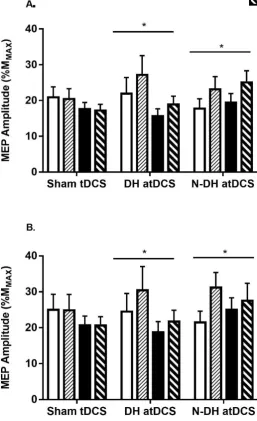
polymorphism. Corticospinal excitability increased for both the stimulated and non-stimulated hemisphere, irrespective of whether the dominant or non-dominant M1 was stimulated, showing functional connectivity between the motor cortices. Interestingly, there was a shift in lateralisation of corticospinal inhibition towards the right (non-dominant) M1 irrespective of which M1 was stimulated. The corticospinal responses following anodal tDCS were influenced by the BDNF polymorphism, with greater responses from Val/Val participants.
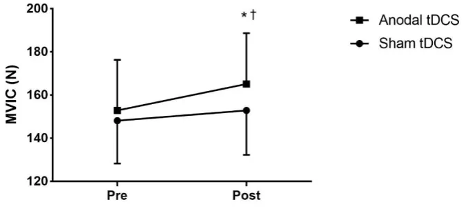
Given that the induction of corticospinal plasticity is thought to be important for motor function, Study 2 examined the effect of four consecutive sessions of anodal tDCS applied over the left M1 on corticospinal excitability/inhibition and transcranial magnetic stimulation (TMS) voluntary activation and whether these responses were influenced by the BDNF polymorphism. Following four consecutive days of anodal tDCS, corticospinal excitability and TMS voluntary activation (VATMS) of the wrist flexors increased, whilst
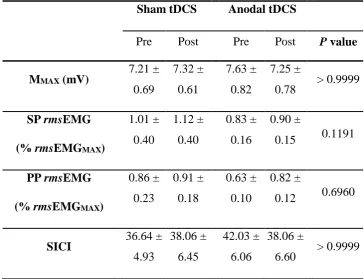
2 production. There were no changes in short-interval intracortical inhibition (SICI), however, the magnitude of the corticospinal responses was influenced by the BDNF
polymorphism, with the Val/Val showing greater responses.
3 It was concluded that the induction of homeostatic plasticity plays an important role in the development of muscle strength. Importantly, anodal tDCS does not appear to act focally upon the site of stimulation, but rather has a global effect on distant non-stimulated regions, demonstrating functional connectivity. Critically, the presence of the
4
Declaration
“I, Ashlyn K. Frazer declare that the PhD thesis entitled “Physiological studies investigating the effect of homeostatic plasticity of the motor cortex on the expression
of muscle strength” is no more than 100,000 words in length including quotes and
exclusive of tables, figures, appendices, bibliography, references and footnotes. This thesis contains no material that has been submitted previously, in whole or in part, for the award of any other academic degree or diploma. Except where otherwise indicated, this thesis is my own work”.
5
Acknowledgements
First and foremost, I would like to thank my Victoria University supervisors, Dr Jackie Williams and Associate Professor Michael Spittle, for allowing me to work independently and enabling me to complete my PhD in a timely manner. I would also like to thank Dr Dawson Kidgell for his continuous support and encouragement throughout the duration of my PhD studies. The guidance and dedication from all of you have ensured that this thesis is something I will be proud of forever.
I especially wish to acknowledge all my participants for their commitment over the study period. I know that, for many of you, giving your time was a personal favour and I am grateful as, without you, my PhD would not exist.
My sincere thanks to Mum, Dad and my partner for all the support you have shown me over the years. Without your love and understanding, the completion of my PhD would not have been possible.
6
Publications and Abstracts
Publications arising from thesis
Chapter 4
Frazer, A, Williams, J, Spittle, M, Rantalainen, T & Kidgell, D 2016, ‘Anodal Transcranial Direct Current Stimulation of the Motor Cortex Increases Cortical
Voluntary Activation and Neural Plasticity’, Muscle and Nerve, vol. 54, no. 5, pp. 903-913.
Chapter 6
Frazer, A, Williams, J, Spittle, M, & Kidgell, D 2016, ‘Cross-education of muscular strength is facilitated by homeostatic plasticity’, European Journal of Applied Physiology. (Accepted January 5th 2017).
Abstract arising from thesis
Chapter 4
7
Table of Contents
Abstract ... 1
Declaration ... 4
Acknowledgements ... 5
Publications and Abstracts ... 6
List of Figures ... 12
List of Tables ... 15
List of Abbreviations ... 16
Chapter 1 : Introduction ... 19
1.1 Primary aim of the research ... 24
1.2 Secondary aim of the research ... 24
1.3 Specific aims of the research ... 25
1.4 Primary hypothesis of the research ... 25
Chapter 2 : Review of Literature ... 26
2.1 Organisation of primary motor cortex ... 28
2.2 Corticospinal tract ... 30
2.2.1 Techniques used to measure motor cortical function ... 31
2.2.2 Transcranial magnetic stimulation ... 32
2.2.2.1 Single-pulse TMS ... 32
2.2.2.2 Paired-pulse TMS ... 35
2.2.2.3 TMS voluntary activation (VATMS) ... 36
2.3 Corticospinal plasticity ... 36
2.3.1 Mechanisms of corticospinal plasticity ... 37
2.3.1.1 Long-term potentiation ... 38
2.3.1.2 NMDA receptor... 38
2.3.2 Brain-derived neurotrophic factor and corticospinal plasticity ... 39
2.4 Experimentally-induced corticospinal plasticity ... 41
2.4.1 Transcranial direct current stimulation ... 42
2.4.2 NIBS and functional connectivity ... 44
2.4.3 The induction of homeostatic plasticity and its effect on motor performance .. 45
2.4.4 Improvements in motor performance following tDCS in the absence of training ... 47
8
2.5 Use-dependent corticospinal plasticity ... 52
2.5.1 Corticospinal plasticity following a single session of strength training ... 54
2.5.5.1 Corticospinal adaptations to strength training and TMS ... 54
2.5.5.2 BDNF and the induction of use-dependent corticospinal plasticity ... 56
2.5.2 Cross-education ... 57
2.5.2.1 Evidence of cross-education... 58
2.5.2.2 Mechanisms of cross-education and the potential use of tDCS ... 59
2.6 Conclusion ... 62
Chapter 3 : Anodal tDCS increases bilateral cortical excitability irrespective of hemispheric dominance ... 64
3.1 Introduction ... 65
3.2 Methods ... 68
3.2.1 Participants ... 68
3.2.2 Experimental approach ... 68
3.2.3 Root mean square electromyography and maximal voluntary isometric contraction ... 71
3.2.4 Surface electromyography ... 71
3.2.5 Transcranial magnetic stimulation ... 72
3.2.6 Maximum compound muscle action potential ... 73
3.2.7 Transcranial direct current stimulation ... 73
3.2.8 BDNF genotyping ... 74
3.2.9 Data analysis ... 76
3.2.10 Statistical analysis ... 77
3.3 Results ... 79
3.3.1 Pre-stimulus rmsEMG, active motor threshold stimulus intensity, maximal compound wave, and visual analogue scale ... 79
3.3.2 Corticospinal excitability ... 81
3.3.3 Corticospinal inhibition ... 86
3.3.4 Laterality index ... 88
3.4 Discussion ... 90
Chapter 4 : Anodal transcranial direct current stimulation of the motor cortex increases TMS voluntary activation and corticospinal plasticity ... 95
4.1 Introduction ... 96
4.2 Methods ... 99
9
4.2.2 Experimental approach ... 99
4.2.3 Voluntary strength testing ... 100
4.2.4 Surface electromyography ... 101
4.2.5 Transcranial magnetic stimulation and TMS voluntary activation (VATMS) ... 101
4.2.6 Maximum compound muscle action potential ... 102
4.2.7 Transcranial direct current stimulation ... 103
4.2.8 BDNF analysis ... 103
4.2.9 Data analysis ... 105
4.2.10 Statistical analysis ... 106
4.3 Results ... 107
4.3.1 Pre-stimulus rmsEMG, maximal compound wave, and visual analogue scale 107 4.3.2 Maximal voluntary isometric contraction ... 109
4.3.3 Corticospinal excitability ... 110
4.3.4 Corticospinal inhibition ... 114
4.3.5 SICI ... 116
4.3.6 TMS voluntary activation (VATMS) ... 116
4.4 Discussion ... 119
4.4.1 Repeated sessions of anodal tDCS increased isometric strength and TMS voluntary activation (VATMS) ... 119
4.4.2 Corticospinal excitability and inhibition following accumulated bouts of anodal tDCS in different BDNF genotypes ... 120
Chapter 5 : Augmenting the corticospinal responses to strength training through homeostatic plasticity ... 125
5.1 Introduction ... 126
5.2 Methods ... 131
5.2.1 Participants ... 131
5.2.2 Experimental approach ... 131
5.2.3 Voluntary strength testing ... 133
5.2.4 Strength training protocol ... 133
5.2.5 Surface electromyography ... 134
5.2.6 Transcranial magnetic stimulation ... 134
5.2.7 Maximum compound muscle action potential ... 134
5.2.8 Transcranial direct current stimulation ... 135
10
5.2.10 Data analysis ... 135
5.2.11 Statistical analysis ... 135
5.3 Results ... 137
5.3.1 Pre-stimulus rmsEMG, maximal compound wave, and visual analogue scale 137 5.3.2 Maximal voluntary isometric contraction force ... 139
5.3.3 Corticospinal excitability ... 140
5.3.4 Corticospinal inhibition ... 145
5.3.5 Corticospinal excitability and inhibition in different BDNF genotypes ... 148
5.4 Discussion ... 150
5.4.1 Anodal tDCS induces homeostatic plasticity (Experiment 2) ... 150
5.4.2 Anodal tDCS prior to strength training increases corticospinal excitability and decreases intracortical inhibition ... 151
5.4.3 Corticospinal excitability and intracortical inhibition is not differentially modulated following strength training in different BDNF genotypes ... 154
Chapter 6 : Cross-education of muscle strength is facilitated by homeostatic plasticity ... 156
6.1 Introduction ... 157
6.2 Methods ... 162
6.2.1 Participants ... 162
6.2.2 Experimental approach ... 162
6.2.3 Voluntary strength testing ... 165
6.2.4 Strength training protocol ... 165
6.2.5 Surface electromyography ... 165
6.2.6 Transcranial magnetic stimulation ... 165
6.2.7 Maximum compound muscle action potential ... 166
6.2.8 Transcranial direct current stimulation ... 166
6.2.9 BDNF genotyping ... 166
6.2.10 Data analysis ... 166
6.2.11 Statistical analysis ... 166
6.3 Results ... 168
6.3.1 Pre-stimulus rmsEMG, maximal compound wave, and visual analogue scale 168 6.3.2 Dynamic strength of the untrained limb ... 170
6.3.3 Corticospinal excitability ... 171
11
6.3.5 Cross-activation ... 177
6.3.6 Dynamic strength of the untrained limb, cross-activation, corticospinal excitability and inhibition in different BDNF genotypes ... 178
6.4 Discussion ... 181
6.4.1 Anodal tDCS applied to the ipsilateral M1 induces homeostatic plasticity .... 181
6.4.2 Anodal tDCS applied to the ipsilateral M1 prior to strength training augments the cross-transfer of strength ... 182
6.4.3 Corticospinal responses of the ipsilateral M1 following ipsilateral tDCS and cross-education ... 183
6.4.4 Corticospinal excitability and intracortical inhibition is differentially modulated following ipsilateral anodal tDCS and strength training in different BDNF genotypes ... 186
Chapter 7 : General Discussion and Conclusion ... 188
7.1 The induction of homeostatic plasticity ... 189
7.1.1 The influence of the BDNF polymorphism on the induction of homeostatic plasticity ... 196
7.2 The functional consequences of homeostatic plasticity ... 198
7.2.1 The influence of the BDNF polymorphism on experimentally-induced and use-dependent plasticity ... 202
7.3 Conclusion ... 204
References... 207
Appendices ... 240
Appendix A: Edinburgh Handedness Inventory ... 240
12
List of Figures
Figure 2.1: Cytoarchitecture of the primary motor cortex. ... 30
Figure 2.2: The components of an MEP. ... 33
Figure 2.3: Example of interhemispheric communication between the right and left M1. ... 60
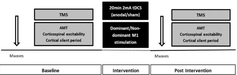
Figure 3.1: Schematic representation of Study 1 experimental design ... 69
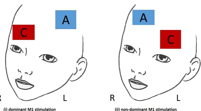
Figure 3.2: Schematic representation of the two tDCS electrode arrangements used .... 70
Figure 3.3A-B: Changes in MEP amplitude for the stimulated and non-stimulated hemispheres. ... 83
Figure 3.4A-B: (A) Changes in MEP amplitude of the stimulated hemisphere with different BDNF genotypes. (B) Raw EMG responses of the stimulated hemisphere with different BDNF genotypes. ... 85
Figure 3.5A-B: Changes in corticospinal silent period duration for the stimulated and non-stimulated hemispheres. ... 87
Figure 4.1: Schematic representation of Study 2 experimental design. ... 100
Figure 4.2: Example of PCR analysis for five unidentified subjects. ... 104
Figure 4.3: Changes in MVIC strength of the right wrist flexors following four
consecutive sessions of sham and anodal tDCS.. ... 109
Figure 4.4A-C: Changes in MEP amplitude following four consecutive sessions of (A) sham tDCS, (B) anodal tDCS (C) with different BDNF genotypes. ... 113
Figure 4.5A-C: Changes in corticospinal silent period duration following four consecutive sessions of (A) sham tDCS, (B) anodal tDCS (C) different BDNF
13
Figure 4.6A-C: (A) Raw force traces for three levels of wrist flexor voluntary contraction force (B) Raw traces of the superimposed twitches produced by cortical stimulation (C) Raw EMG responses produced by cortical stimulation. ... 117
Figure 4.7: Changes in VATMS following four consecutive sessions of sham and anodal
tDCS. ... 117
Figure 5.1A-B: Schematic representation of the design of Study 3 (Experiment 1 and 2). ... 132
Figure 5.2: Changes in MVIC strength of the right biceps brachii muscle following anodal tDCS + ST and sham tDCS + ST.. ... 139
Figure 5.3A-B: Changes in MEP amplitude following (A) sham tDCS + ST (B) anodal tDCS + ST.. ... 141
Figure 5.4A-B: AUC for corticospinal excitability and corticospinal inhibition ... 143
Figure 5.5: Changes in MEP amplitude before and after 20 min of anodal and sham tDCS (Experiment 2). ... 144
Figure 5.6A-B: Changes in corticospinal silent period duration following (A) sham tDCS + ST (B) anodal tDCS + ST ... 146
Figure 5.7: Changes in corticospinal silent period duration before and after 20 min of anodal and sham tDCS (Experiment 2). ... 147
Figure 5.8: Changes in MEP amplitude of the stimulated hemisphere before and after 20 min of anodal and sham tDCS (Experiment 2)... 149
Figure 6.1A-B: Schematic representation of Study 4 (Experiment 1 and 2)... 164
Figure 6.2: Changes in 1RM strength of the left biceps brachii muscle following
14
Figure 6.3A-B: Changes in MEP amplitude following (A) ipsilateral sham tDCS + ST (B) ipsilateral anodal tDCS + ST ... 172
Figure 6.4: Changes in MEP amplitude before and after 20 min of ipsilateral anodal and sham tDCS (Experiment 2). ... 173
Figure 6.5A-B: Changes in corticospinal silent period duration following (A) ipsilateral sham tDCS + ST (B) ipsilateral anodal tDCS + ST. ... 175
Figure 6.6: Changes in corticospinal silent period duration before and after 20 min of ipsilateral anodal and sham tDCS (Experiment 2). ... 176
Figure 6.7: Changes in MEP amplitude at 130% AMT during a contralateral MVIC following ipsilateral anodal tDCS + ST and ipsilateral sham tDCS + ST... 177
15
List of Tables
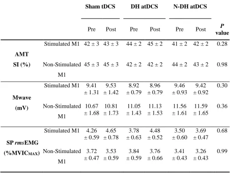
Table 3-1: Mean (± SE) for AMT stimulus intensity, MMAX and single-pulse TMS
pre-stimulus rmsEMG for the stimulated and non-stimulated M1 prior to and following sham and anodal tDCS (dominant and non-dominant M1 stimulation). ... 80
Table 3-2: Mean (± SE) for laterality index prior to and following sham and anodal tDCS (dominant and non-dominant hemisphere stimulation arrangements). ... 89
Table 4-1: Mean (± SE) for MMAX, single-pulse and paried-pulse TMS pre-stimulus rmsEMG at 130% AMT and SICI prior to and following four consecutive sessions of sham and anodal tDCS. ... 108
Table 4-2: Mean (± SE) for MEP amplitudes expressed at percentage of MMAX at
110-210% AMT prior to and following four consecutive sessions of sham and anodal tDCS. ... 111
Table 4-3: r2 values for the estimated resting twitch values for each participant ... 118
Table 5-1: Mean (± SE) for AMT stimulus intensity, MMAX and single-pulse TMS
pre-stimulus rmsEMG prior to and following sham tDCS + ST and anodal tDCS + ST. ... 138
Table 6-1: Mean (± SE) for AMT stimulus intensity, MMAX and single-pulse TMS
16
List of Abbreviations
1RM: One-repetition maximum
AMT: Active motor threshold
AMT SI: Active motor threshold stimulus intensity
AUC: Area under the curve
BDNF: Brain-derived neurotrophic factor
Ca2+: Calcium
CMEPS: Cervicomedullary motor–evoked potentials
CNS: Central nervous system
CST: Corticospinal tract
DH atDCS: Anodal tDCS applied over the dominant M1
ECR: Extensor carpi radialis
EEG: Electroencephalography
EMG: Electromyography
FCR: Flexor carpi radialis
FDI: First dorsal interosseus
fMRI: Functional magnetic resonance imaging
GABA: Gamma-Aminobutyric acid
H-reflex: Hoffman reflex
ICF: Intracortical facilitation
IHI: Interhemispheric inhibition
iM1: Ipsilateral primary motor cortex
ISI: Interstimulus interval
17
JTT: Jebsen-Taylor Hand Function Test
LI: Laterality index
LICI: Long-interval intracortical inhibition
LQ: Laterality quotient
LTD: Long-term depression
LTP: Long-term potentiation
MEG: Magnetoencephalography
Mg2+: Magnesium
M1: Primary motor cortex
MEP: Motor evoked potential
MMAX: Maximum compound wave
MT: Motor threshold
MVIC: Maximal voluntary isometric contraction
N-DH atDCS: Anodal tDCS applied over the non-dominant M1
NIBS: Non-invasive brain stimulation
NIRS: Near-infrared spectroscopy
NMDA receptor: N-methyl-D-aspartate receptor
PAS: Paired associative stimulation
PET: Positron emission tomography
PP: Paired-pulse
PTP: Post-tetanic potentiation
rmsEMG: Root mean square of the surface EMG
RMT: Resting motor threshold
18
sEMG: Surface electromyography
SICF: short-interval intracortical facilitation
SICI: Short-interval intracortical inhibition
SP: Single-pulse
ST: Strength training
STP: Short-term potentiation
TBS: Theta burst stimulation
tDCS: Transcranial direct current stimulation
TMS: Transcranial magnetic stimulation
VAS: Visual analogue scale
19
20 The human primary motor cortex (M1) is highly modifiable and plays a critical role in the acquisition of motor behaviours (Pascual-Leone et al. 1995, Classen et al. 1998, Rioult-Pedotti et al. 1998, Butefisch et al. 2000, Muellbacher et al. 2002). The modifiable nature of the M1 is termed plasticity and involves changes in synaptic efficacy of pyramidal neuron connections, involving the processes of long-term potentiation (LTP) and long-term depression (LTD) (Hess & Donoghue 1996, Sanes & Donoghue 2000). Improvements in synaptic efficacy are mediated by the N-methyl-D-aspartate (NMDA) receptor which operates as a channel responsible for the induction of LTP and LTD (Castro-Alamancos et al. 1995). However, it should be noted that the M1 contains a large number of pyramidal tract neurons which descend and synapse monosynaptically onto the ventral horn of the spinal cord forming the corticospinal tract (CST) (Nathan et al. 1990). Therefore, in the context of this thesis, modifications of the intrinsic circuitry of the M1 (i.e., increases in corticospinal excitability and decreases in inhibition), which improve neural transmission along the CST, will be termed corticospinal plasticity.
The induction of corticospinal plasticity has been shown to occur following both experimentally-induced plasticity using non-invasive brain stimulation (NIBS) protocols (e.g. transcranial direct current stimulation [tDCS]) (Nitsche & Paulus 2000, Nitsche & Paulus 2001, Nitsche et al. 2005, Sale et al. 2007, Kidgell et al. 2013) and use-dependent activities that involve repetitive activity (i.e., strength training and skill training) targeted at improving motor performance (Hortobagyi et al. 2011, Selvanayagam et al. 2011, Leung et al. 2015, Nuzzo et al. 2016).
21 whilst cathodal tDCS decreases corticospinal excitability (Nitsche & Paulus 2000, Nitsche & Paulus 2001). This increased/decreased level of excitability following a low/high level of synaptic activity is termed homeostatic plasticity. Despite the substantial evidence establishing the capacity for tDCS to modulate the excitability of neurons within the M1, it has recently been shown to induce effects in distant brain areas caused by activity of interconnected brain zones (Sale et al. 2015). This concept is termed
functional connectivity and is based upon the working hypothesis that changes in localised brain activity can influence distant, but functionally related areas, which is an essential function of the healthy brain (Sale et al. 2015). Thus, it has been suggested that anodal tDCS may be a plausible method to induce homeostatic plasticity of distant interconnected structures [i.e., the ipsilateral hemisphere] (Pavlova et al. 2014). Therefore, this concept was examined in Chapter 3 (Study 1) by investigating the bilateral effects of uni-hemisphere anodal tDCS.
experimentally-22 induced plasticity and its effect on muscle strength to explore the role of plasticity in regulating the expression of muscle strength.
To date, there has been significant attention surrounding the application of tDCS before and/or during motor training (known as motor priming), which is based upon the assumption that enhanced neural activity within the M1 will facilitate the mechanisms associated with LTP or LTD (Ziemann & Siebner 2008). Two sub-divisions of motor priming include gating and homeostatic plasticity which differ according to the timing of when tDCS is applied (i.e., during or before motor training) (Siebner 2010). The theory of gating occurs instantaneously and describes the influx of calcium ions into the targeted corticospinal neurons resulting in the disinhibition of intracortical inhibitory circuits (Ziemann & Siebner 2008, Siebner 2010). Gating is attained concurrently with motor training and has been shown to facilitate motor performance (Nitsche et al. 2003d, Boggio et al. 2006, Galea & Celnik 2009, Hunter et al. 2009, Reis et al. 2009, Stagg et al. 2011, Hendy & Kidgell 2014). Of relevance to this thesis is the principle of homeostatic plasticity whereby the resting state of corticospinal neurons are altered (increased/decreased level of excitability following a low/high level of synaptic activity) due to changes in postsynaptic glutamate receptor activity (Ziemann & Siebner 2008, Siebner 2010) prior to motor training.
23 commonly seen following acute (single session) and short-term (i.e., < 3 weeks) strength training, has been attributed to changes in corticospinal plasticity in the absence of muscle hypertrophy (Beck et al. 2007, Griffin & Cafarelli 2007, Kidgell & Pearce 2010, Kidgell et al. 2010b, Hortobagyi et al. 2011, Leung et al. 2015, Nuzzo et al. 2016). A fundamental example of corticospinal plasticity underpinning improvements in strength is the cross-education phenomenon, whereby strength training of one limb results in an increase in strength of the opposite untrained limb (Munn et al. 2004, Carroll et al. 2006). Given that anodal tDCS has been shown to modulate NMDA receptor activity, and subsequently produce a shift in the resting membrane potential (Nitsche & Paulus 2000), it would be likely that anodal tDCS could be used to increase synaptic activity within the M1 prior to strength training. Whilst Chapter 5 (Study 3) investigated the effect of priming the M1 using anodal tDCS prior to a single bout of strength training on corticospinal responses, Chapter 6 (Study 4) determined whether priming the ipsilateral M1 was a plausible method to enhance the cross-transfer of strength.
24
BDNF secretion and LTP/LTD processes suggests the importance of this factor in regulating corticospinal plasticity (Cheeran et al. 2008, Li Voti et al. 2011). To date, no studies have examined whether the BDNF polymorphism differentially regulates the induction of corticospinal plasticity and the acquisition of strength following anodal tDCS and strength training. Therefore, a collective secondary aim of this thesis (Studies 1-4) was to investigate the role of the BDNF polymorphism as a potential regulator of the efficacy of experimentally-induced and use-dependent plasticity protocols used to modulate the expression of muscle strength in healthy adults.
Overall, the purpose of this thesis was to systematically examine the corticospinal responses to various homeostatic-inducing protocols (anodal tDCS alone and in combination with strength training) to investigate the corticospinal responses that may contribute to the expression of strength.
1.1 Primary aim of the research
1. To systematically investigate the induction of homeostatic plasticity and its effect on muscle strength.
1.2 Secondary aim of the research
25
1.3 Specific aims of the research
1. To examine the effect of a single session of anodal tDCS on indices of corticospinal excitability and inhibition of both the stimulated and non-stimulated hemisphere (Study 1).
2. To examine the effect of repeated sessions of anodal tDCS on muscle strength, VATMS,
and indices of corticospinal plasticity (Study 2).
3. To examine the effect of inducing homeostatic plasticity of the M1 using anodal tDCS prior to a single bout of strength training on corticospinal excitability and inhibition (Study 3).
4. To examine the effect of inducing homeostatic plasticity of the ipsilateral M1 using anodal tDCS prior to a single bout of strength training on the cross-transfer of strength and corticospinal excitability/inhibition of the ipsilateral M1 (Study 4).
5. To investigate the role of the BDNF polymorphism as a potential regulator of the efficacy of experimentally-induced and use-dependent plasticity protocols used to the expression of muscle strength (Studies 1-4).
1.4 Primary hypothesis of the research
1. It was hypothesised that the induction of homeostatic plasticity (i.e., increased corticospinal excitability, decreased corticospinal inhibition, increased VATMS) would
contribute to the expression of muscle strength.
2. It was hypothesised that the presence of the BDNF polymorphism would influence the corticospinal responses to anodal tDCS, but would not affect VATMS and the expression
26
Chapter 2
:
Review of
28 M1 and CST have been identified as integral functional structures of experimentally-induced and use-dependent corticospinal plasticity and, therefore, will be reviewed in further detail.
2.1 Organisation of primary motor cortex
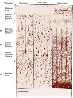
The pyramidal and stellate cellular arrangement of the M1 forms a laminar appearance when viewed vertically (Rothwell 1994). Pyramidal tract cells are found within several layers within the M1, with the largest known as Betz cells located in the internal pyramidal layer (layer V). The origins of the pyramidal tract cells are grouped and distributed intermittently forming the horizontal component of layer V (Mountcastle 1997). These cells are among the largest in the central nervous system (CNS) and have long axons which descend and synapse monosynaptically onto the ventral horn of the spinal cord (Mountcastle 1997).
29 they generally have shorter axons which do not leave the cortex and are responsible for making inhibitory synapses (Rothwell 1994).
The M1 is structurally organised into six layers which consist of horizontally and vertically orientated bands. As shown in Figure 2.1, the layers of the M1 are: (I) Molecular layer- this is the most superficial layer containing axons and
dendrites.
(II) External granular layer- consists mainly of stellate cells and small pyramidal cells giving this layer a granular stripped appearance. (III) External pyramidal layer- composed mostly of medium and large
pyramidal cells which are the main source of corticocortical fibres. (IV) Internal granular layer- consists predominately of stellate cells with
some pyramidal cells present.
(V) Internal pyramidal layer- thickest area containing the giant pyramidal cells called Betz cells which are the source of efferent (output) fibres. (VI) Multiform layer- deepest layer consisting of fusiform and pyramidal cells
30
Figure 2.1: Cytoarchitecture of the primary motor cortex (Kandel et al. 2000, pp. 265).
2.2 Corticospinal tract
31 remaining 10%, that do not cross, form the anterior CST which controls movements of the trunk. Most of these fibres eventually cross just before terminating in the ventral horn of the spinal cord. The majority of axons enter the ventral horn and terminate in the intermediate and ventral areas on interneurons and motor neurons (Rothwell 1994). Changes within the M1 and along the CST can be quantified using several non-invasive techniques targeting direct and indirect activation of pyramidal tract cells.
2.2.1 Techniques used to measure motor cortical function
Several non-invasive human brain mapping techniques including TMS, fMRI, PET, NIRS, MEG and EEG have been used to investigate functional and structural reorganization within the M1 (Siebner & Rothwell 2003, Schaechter 2004, Imfeld et al. 2009). PET and fMRI techniques examine the brain’s hemodynamic response to a task by assessing changes in regional cerebral blood flow indicating changes in neuron activation (Greenberg et al. 1979, Schaechter 2004). Both these neuroimaging methods enable non-invasive time course feedback of spatial and temporal dynamics of cortical activation, providing evidence of representational plasticity in the human brain (Hallett 2000, Siebner & Rothwell 2003). NIRS is a similar technique but examines changes in the absorption of near-infrared light by haemoglobin-containing elements in the cerebral blood to measure brain activation; however it has limited use due to poor spatial resolution (Schaechter 2004).
32
2.2.2 Transcranial magnetic stimulation
Over the past several decades, TMS has emerged as a robust, non-invasive tool used to examine physiological and pathophysiological functions of the CNS. Exploration into excitatory and inhibitory circuits within the M1 has provided valuable objective insight into the possible mechanisms of corticospinal plasticity (Rossini & Rossi 2007). TMS stimulates underlying cortical tissue via a brief electrical current that is passed through a coil which is placed over the individual’s scalp (Hallett 2000). The magnetic field created by the circulating electrical current depolarises neurons or their axons (Hallett 2000) and causes a muscle response of the target muscle (Merton 1980). Single- and paired-pulse TMS can be used to measure various parameters of corticospinal plasticity involving changes confined to the M1 and changes along the CST and is currently used within clinical and research settings (Hallett 2000).
2.2.2.1 Single-pulse TMS
33
Figure 2.2: The components of an MEP recorded by surface electromyography (sEMG) from single-pulse TMS (Pearce & Kidgell 2011).
34 curve (AUC) can be calculated using the method of trapezoidal integration during the construction of corticospinal excitability recruitment curves (Talelli et al. 2008, Carson et al. 2013). AUC has high validity and provides additional insight into the input-output properties of the CST (Carroll et al. 2001a, Carson et al. 2013).
Motor threshold (MT) is the minimum amount of stimulation needed to produce an MEP (Hallett 2000). Threshold can be examined at rest (resting motor threshold [RMT]) or during a tonic voluntary contraction (active motor threshold [AMT]) (Kidgell et al. 2010b) and has previously been shown to change following motor training and NIBS (Pascual-Leone et al. 1995, Khedr et al. 2010). Contraction of the muscle reduces the threshold required to elicit an MEP due to the increased level of excitability of neurons at both a cortical and spinal level (Di Lazzaro et al. 2003, Di Lazzaro et al. 2004). Latency is measured from time of stimulation to the onset of the MEP, reflecting the conduction time from the M1 to the motor neuron pool in the spinal cord (Kidgell & Pearce 2011). During tonic voluntary contraction, latency is reduced by 2-3 ms due to facilitation of the spinal motor neurons (Rossini et al. 1994). Silent period is defined from the onset of the MEP to the return of EMG, characterised by a flat lining or non-activity of sEMG (Cantello et al. 1992, Wilson et al. 1993, Chen et al. 1999, Kidgell & Pearce 2011). Initially, the silent period is due to spinal cord refractoriness, however, the latter part is a result of cortical inhibition (Wilson et al. 1993, Chen et al. 1999, Hallett 2000). This measure is regulated by neurons that use Gamma-Aminobutyric acid (GABA), in particular GABAB (Chen et al. 1999, Werhahn et al. 2007) and represents the overall
35 which does not measure cortico-cortical inputs. To quantify changes specifically within the M1, paired-pulse TMS can be used.
2.2.2.2 Paired-pulse TMS
Paired-pulse TMS enables the examination of inhibitory and excitatory mechanisms specific to the M1 (Kujirai et al. 1993, Fisher et al. 2002, Rothwell et al. 2009, Di Lazzaro et al. 2012b). Short-interval intracortical inhibition (SICI), as described by Kujirai et al. (1993), involves a sub-threshold conditioning stimulus followed by a supra-threshold test stimulus, separated by an interstimulus interval (ISI) of 1-5 ms and is used to study intracortical inhibition (Chen 2004). This results in the suppression of I-waves (indirect activation of pyramidal cells) evoked by the test stimulus, providing a measure of the intrinsic cortical connections mediated by GABAA receptors (Kujirai et
36
2.2.2.3 TMS voluntary activation (VATMS)
In addition to single and paired-pulse TMS techniques that examine the excitatory and inhibitory pathways within the M1 (Chen et al. 1999, Siebner & Rothwell 2003), motor cortical drive can also be assessed (Todd et al. 2003, Lee et al. 2008, Lee et al. 2009). This measure provides information regarding the net motor output from the M1 (i.e., VATMS), identifying potential sites of neural drive impairment (Todd et al. 2003,
Todd et al. 2004). To quantify sub-maximal M1 output, the level of neural drive to the muscle is determined by the presence of a superimposed twitch force that is produced by single-pulse TMS during a maximal voluntary isometric contraction (MVIC) (Lee et al. 2008). The superimposed twitch represents the single-pulse TMS eliciting extra force from the muscle during a MVIC due to sub-maximal motor output from the M1, while the absence of a superimposed twitch suggests maximal output from the M1 (i.e., maximal neural drive) (Todd et al. 2003, Todd et al. 2004, Lee et al. 2008, Goodall et al. 2009). TMS has been shown to be a reliable and valid measurement of voluntary activation in the human wrist extensor, knee extensor and elbow flexor muscle (Todd et al. 2003, Todd et al. 2004, Lee et al. 2008, Goodall et al. 2009, Sidhu et al. 2009). This technique provides an additional measure of corticospinal efficiency, demonstrating changes in motor cortical output via the recruitment of motor units used in force generation.
2.3 Corticospinal plasticity
37
plasticity and involves reorganisation of neural assemblies that control movement at both a cortical and spinal level. Although the mechanisms involved in the induction of plasticity have been well described (Classen et al. 1998, Ziemann et al. 1998a, Carroll et al. 2001c, Kleim et al. 2002), less is understood about the potential role of the brain-derived neurotrophic factor (BDNF) polymorphism modulating corticospinal plasticity and its link to muscle function. This is of interest as individual responses to plasticity-inducing protocols are highly variable, making it difficult to draw conclusions regarding the efficacy of various protocols. In the following sections, the mechanisms of corticospinal plasticity will be reviewed before considering the potential modulatory role of the BDNF polymorphism.
2.3.1 Mechanisms of corticospinal plasticity
38
2.3.1.1 Long-term potentiation
Enhancement of synaptic connections can involve short-term potentiation (STP), which lasts 5-20 minutes, and LTP, which can last from hours to days (Bliss & Collingridge 1993). LTP is an activity-dependent process resulting in the long-lasting enhancement of synaptic transmission that provides the basis for information storage within the brain (Bear & Malenka 1994, Hess et al. 1996).
LTP is characterised by three distinct properties including cooperativity, associativity and input-sensitivity (Bliss & Collingridge 1993). Cooperativity refers to the range of threshold intensities required for the induction of activity-dependent potentiation. The threshold necessary for the induction of LTP is dependent on the interaction between intensity and pattern of tetanic stimulation. Unless stimulation is ‘strong’, LTP will not be triggered, resulting in STP and post-tetanic potentiation (PTP) being induced (McNaughton et al. 1978, Malenka 1991). Importantly, LTP is associative, meaning a weak input can only be potentiated if it is active at the same time as a strong input (McNaughton et al. 1978, Collingridge & Bliss 1987). These three properties are mediated by the NMDA receptor which is located on the post-synaptic dendrites of excitatory synapses (Collingridge & Bliss 1987).
2.3.1.2 NMDA receptor
39 (Mg2+), allowing the influx of calcium (Ca2+) into the post-synaptic dendritic spine (Bear & Malenka 1994). The level of depolarization will consequently determine whether the cooperativity threshold will be enough to induce LTP. Failure to induce LTP is a result of inadequate reduction of the Mg2+ block, rather than the insufficient release of glutamate to activate the NMDA receptors (Bliss & Collingridge 1993).
Bliss and Collingridge (1993) demonstrated the necessity of NMDA receptor activation for the induction of LTP. This finding prompted investigation into the potential relationship between NMDA receptor activation and the induction of experimental and use-dependent corticospinal plasticity (Liebetanz et al. 2002, Nitsche et al. 2003a). Using pharmacological agents, Butefisch (2000) demonstrated that use-dependent plasticity of the hand area of the M1 following motor training was significantly reduced when the NMDA receptor was blocked. Similarly, the necessity of NMDA receptor activation for the induction of experimentally-induced plasticity was confirmed when the administration of the NMDA receptor antagonist, dextromethorphan was found to inhibit the long-lasting effects of tDCS (Liebetanz et al. 2002, Nitsche et al. 2003a). Collectively, these findings show that the NMDA receptor is an important operating mechanism in the formation of use-dependent and experimentally-induced corticospinal plasticity via activating LTP processes. In addition, other cellular mechanisms involving neurotrophic factors (i.e., neurotrophins, glial cell-line derived neurotrophic factor family ligands, and neuropoietic cytokines) that interact with the NMDA receptor and the induction of LTP have also been recognized in shaping corticospinal plasticity.
2.3.2 Brain-derived neurotrophic factor and corticospinal plasticity
40
BDNF is a neurotrophin which is involved in a variety of CNS functions including but not limited to cell survival, proliferation and synaptic growth (Antal et al. 2010). In humans, a naturally occurring single nucleotide polymorphism results in the substitution of valine to methionine at codon 66 (val66met), which has been associated with reduced episodic memory and increased risk of neuropsychiatric disorders (Egan et al. 2003, Pezawas et al. 2004, Bath & Lee 2006). The distribution of the polymorphism varies widely between regions and ethnicities, with approximately 30-50% of people worldwide identified as either heterozygous (Val/Met) or homozygous (Met/Met) for the Met substitution. Expression of the Met allele is more commonly found among Asian (51% in Japan) compared to Caucasian populations (30% in America) (Shimizu et al. 2004) with evidence suggesting those of Caucasian descent have larger associated cognitive and behavioural consequences (Bath & Lee 2006). Abnormal cortical morphology is a shared characteristic among carriers of the Met variant form of BDNF (Bath & Lee 2006). Smaller hippocampal volumes and poorer performance on memory tasks have been revealed, determining the anatomical and functional consequences of the BDNF
polymorphism (Egan et al. 2003, Pezawas et al. 2004).
41 theoretical model. In addition, it is unclear what modulatory effect the BDNF
polymorphism may have on experimentally-induced and use-dependent corticospinal plasticity. Therefore, a secondary aim of this thesis was to examine the potential impact of the BDNF polymorphism on the efficacy of tDCS and acute bouts of strength training to induce corticospinal plasticity.
2.4 Experimentally-induced corticospinal plasticity
Several NIBS methods have been used to assess the potential underlying mechanisms and regulators of corticospinal plasticity, including tDCS, theta burst stimulation (TBS), paired associative stimulation (PAS), I-wave periodicity TMS (iTMS) and repetitive transcranial magnetic stimulation (rTMS) (Nitsche et al. 2003b, Siebner & Rothwell 2003, Sale et al. 2007, Thickbroom 2007, Kidgell et al. 2016). Such NIBS techniques have been shown to modify levels of corticospinal excitability and inhibition which have been attributed to LTP and LTD (Thickbroom et al. 2006). More recently, tDCS has emerged as a common NIBS technique used to modulate corticospinal excitability/inhibition with the aim of modifying motor behaviour in both healthy and clinical populations (Ridding & Ziemann 2010, Nitsche & Paulus 2011). More specifically, tDCS has been used in combination with motor training, which has evolved into a popular paradigm known as ‘motor priming’. Motor priming is thought to facilitate motor learning and involves the application of tDCS either prior or during motor learning (Stoykov & Madhavan 2015). Two established priming theories have been proposed which include gating and homeostatic plasticity (Siebner 2010). Gating occurs concurrently with motor training (i.e., tDCS while training), while homeostatic plasticity
42 Despite extensive research examining the indices of corticospinal plasticity of the stimulated M1 following anodal tDCS (Nitsche & Paulus 2000, Nitsche & Paulus 2001, Bastani & Jaberzadeh 2012, Kidgell et al. 2013, Pellicciari et al. 2013), little is understood about the bilateral effects (i.e., non-stimulated M1) of uni-hemisphere stimulation. Given that other NIBS techniques have been shown to modulate not only the intended stimulated tissue but also distal areas of the brain including the contralateral hemisphere (Gilio et al. 2003), it would appear evident that the bilateral effects of anodal tDCS must be explored to ensure the feasibility of tDCS as a priming method for inducing homeostatic plasticity prior to motor training. Furthermore, individual corticospinal responses to anodal tDCS are highly variable and the expression of the BDNF polymorphism has been identified as a potential contributing factor (Cheeran et al. 2008, Antal et al. 2010). Differential modulation of corticospinal plasticity between different BDNF genotype carriers is of interest when examining the induction of LTP, which is an essential physiological process involved in corticospinal plasticity and motor learning (Cheeran et al. 2008, Cirillo et al. 2012). Therefore, the following discussion will examine the induction of homeostatic plasticity following tDCS, the use of tDCS in the absence of motor training and prior to motor training (motor priming) to enhance motor performance, and the potential regulatory role of the BDNF polymorphism.
2.4.1 Transcranial direct current stimulation
43 of parameters have been shown to influence the efficacy of tDCS including current strength, electrode size, stimulation duration and orientation of the electrode field (Nitsche et al. 2008). Orientation includes the position and polarity of electrodes, which determines the direction of modulation (increase/decrease corticospinal excitability). Anodal stimulation (positively charged electrode) results in neuronal depolarisation and an increase in corticospinal excitability. Cathodal stimulation has the opposite effect whereby hyperpolarization of neurons occurs leading to decreased corticospinal excitability (Nitsche & Paulus 2000).
44 recently been shown that NIBS techniques, including tDCS, not only exerts a neuro-modulatory effect over the stimulated region, but also distal areas connected to the region of stimulation (Gilio et al. 2003, Lang et al. 2004).
2.4.2 NIBS and functional connectivity
45 et al. 2003, Schambra et al. 2003, Shin & Sohn 2011) and decrease interhemispheric inhibition (IHI) between the left and right M1 (Gilio et al. 2003). Likewise, Lang et al. (2004) found that 10 min of anodal and cathodal tDCS at 1 mA modulated transcallosal inhibition. Interestingly, this finding was not accompanied by a bilateral increase in M1 excitability, with only an increase in MEP amplitude seen in the stimulated M1. Importantly, it should be highlighted that a key methodological component of the studies investigating NIBS techniques and the concept of functional connectivity is that many used a dominant M1 arrangement whereby the stimulated hemisphere was the dominant M1 (left) and non-stimulated hemisphere was the non-dominant M1 (right). Notably, it has previously been shown that a hemispheric imbalance exists (dominant vs non-dominant) as demonstrated by the non-dominant hemisphere having a lower motor threshold, higher MEPs (De Gennaro et al. 2004) and shorter corticospinal silent period durations (Priori et al. 1999). A potential difference in hemispheric baseline characteristics poses an interesting question as to whether the magnitude of bilateral corticospinal plasticity is affected by the direction of stimulation (dominant vs non-dominant M1 stimulated), and if there is a greater scope for the induction of corticospinal plasticity of the non-dominant hemisphere.
2.4.3 The induction of homeostatic plasticity and its effect on motor performance
46 before or during motor training, with the working hypothesis that enhanced neural activity within the M1 will facilitate the mechanisms associated with LTP or LTD (Ziemann & Siebner 2008). Two theories have been proposed to underlie the response of corticospinal output neurons following priming protocols including gating and homeostatic plasticity (Siebner 2010). The theory of gating occurs instantaneously and describes the influx of calcium ions to the targeted corticospinal neurons resulting in the disinhibition of intracortical inhibitory circuits (Ziemann & Siebner 2008, Siebner 2010). Gating is attained concurrently with motor training and has been shown to facilitate motor performance tasks such as hand function using the Jebsen-Taylor Hand Function Test (JTT), maximal strength, movement speed, reaction time and speed-accuracy trade-off (Nitsche et al. 2003d, Boggio et al. 2006, Galea & Celnik 2009, Hunter et al. 2009, Reis et al. 2009, Stagg et al. 2011, Hendy & Kidgell 2014). For example, Christova et al. (2015) showed a significant reduction in SICI following the application of anodal tDCS during grooved pegboard training. However, it appears that the efficacy of priming during training may be limited to fine motor skill training tasks (i.e., pegboard). In support of this notion, Hendy et al. (2013) investigated the use of anodal tDCS applied to the active M1 during training to enhance maximal voluntary strength. Interestingly, there was no difference in strength gain between conditions, suggesting that strength training appeared to have a powerful effect on modulating mechanisms associated with LTP, therefore, potentially limiting further corticospinal responses induced by anodal tDCS.
47 Importantly, the lack of interactions observed between conditions by Hendy et al. (2013, 2014), suggests that a critical consideration to maximise the effectiveness of anodal tDCS as a M1 priming technique is the timing of application (i.e., during or prior the training). Given that anodal tDCS has been shown to modulate NMDA receptors, and subsequently produce a shift in the resting membrane potential (Nitsche & Paulus 2000), it would be conceivable that anodal tDCS is a promising priming tool to increase synaptic activity prior to a single bout of strength training to further augment the acute corticospinal responses to strength training (Leung et al. 2015). In addition, the application of anodal tDCS to the ipsilateral M1 prior to unilateral strength training may result in a shift of the resting membrane potential and increase synaptic activity of the ipsilateral M1, which may in turn further promote bilateral activation of both motor cortices and enhance the cross-transfer of strength; however, no studies have examined this. For further discussion of the corticospinal responses to strength training and the cross-transfer of strength, please see Section 2.5.
2.4.4 Improvements in motor performance following tDCS in the absence of training
48 of motor sequencing tasks have all been reported (Boggio et al. 2006, Vines et al. 2006, Cogiamanian et al. 2007, Tanaka et al. 2009, Tanaka et al. 2011, Kidgell et al. 2013).
In healthy adults, accumulated bouts of anodal tDCS have been shown to improve motor performance with retention lasting up to three months following stimulation (Boggio et al. 2007, Reis et al. 2009). Although the underlying physiological changes were not examined, the induction of LTP has been suggested to underlie the improvement in motor performance (Reis et al. 2009). Previously, changes in corticospinal excitability have been examined over a five day period whereby participants were exposed to daily anodal tDCS stimulation (Alonzo et al. 2012). Corticospinal excitability was shown to significantly increase but, unfortunately, no motor performance outcome was used to assess any functional effects of the tDCS intervention. A recent study investigating the effect of repeated sessions of anodal tDCS demonstrated an increase in corticospinal excitability accompanied by an increase in muscle strength (Frazer et al. 2016). Interestingly, there was no change in SICI, however a reduction in corticospinal silent period was reported suggesting that accumulated bouts of anodal tDCS appear to modulate GABAB rather than GABAA neurons (Frazer et al. 2016). Because the
corticospinal silent period that follows the excitatory MEP is caused by activation of long-lasting GABAB mediated inhibition and reflects the temporary suppression in motor
cortical output (Werhahn et al. 2007), it appears that cumulative bouts of anodal tDCS specifically target neural circuits that use GABAB as their neurotransmitter (Frazer et al.
49 To date, the TMS literature has primarily focused on the acute effects of tDCS modulating corticospinal excitability and the subsequent change in motor performance. Although these studies have provided valuable insight into possible acute physiological mechanisms, motor output from the M1 can also be quantified via VATMS which provides
further insight into the mechanisms regulating corticospinal plasticity and the expression of strength. The level of neural drive to a muscle during exercise is commonly termed ‘voluntary activation’ (Gandevia et al. 1995) and can be estimated by interpolation of a single supramaximal electrical stimulus to the motor nerve during an isometric voluntary contraction (Merton 1954). Although twitch interpolation assesses neural drive to a muscle during exercise, it cannot provide insight into the precise location of any neural drive impairment (cortical or sub-cortical) (Lee et al. 2009, Carroll et al. 2011). In light of this, TMS has been employed to measure net motor output from the M1 (i.e., VATMS)
identifying potential sites of neural drive impairment (Todd et al. 2003, Todd et al. 2004). This technique can provide additional information regarding corticospinal efficiency following anodal tDCS by demonstrating changes in motor cortical output via the recruitment of motor units used in force generation.
At present, studies have concentrated on the reliability and validity of TMS to measure VATMS in various muscle groups (Todd et al. 2003, Todd et al. 2004, Lee et al.
2008, Sidhu et al. 2009). However, translation of this technique into applied research settings, such as assessing changes in corticospinal plasticity following accumulated bouts of anodal tDCS, has been only examined once (Frazer et al. 2016). Interestingly, an increase in VATMS and strength was observed, suggesting that accumulated bouts of
50 muscle strength (Frazer et al. 2016). Although, the use of tDCS to induce homeostatic plasticity (i.e., modify corticospinal excitability) and acutely improve motor performance is well established (Nitsche et al. 2008, Vines et al. 2008, Kidgell et al. 2013, Frazer et al. 2016), the efficacy of tDCS may also be influenced by individual genetic variations such as the BDNF polymorphism (Antal et al. 2010, Puri et al. 2015, Frazer et al. 2016). Therefore, it appears vital to identify individual variants that may impact on the effectiveness of tDCS protocols.
2.4.5 BDNF and the induction of experimentally-induced corticospinal plasticity
Corticospinal responses to various NIBS techniques have been shown to differ significantly between individuals (Cheeran et al. 2008, Chang et al. 2014, Hwang et al. 2015). Genetic factors, including the role of BDNF, have been reported as potential contributors to the variability of results observed within the literature. Evidence using TMS to evaluate the efficacy of NIBS techniques generally suggest that the presence of the BDNF polymorphism significantly impacts corticospinal plasticity and motor performance. This is highlighted by the response of Met allele carriers being different to
51 (Chang et al. 2014, Hwang et al. 2015). However, the influence of BDNF on NIBS protocols is not always consistent with some studies showing no difference in corticospinal plasticity between Val66Val and Val66Met carriers following rTMS and iTBS (Li Voti et al. 2011, Nakamura et al. 2011). Importantly, it should be noted that the protocol duration used in these studies may not have been sufficient to activate cellular processes of activity-dependent BDNF secretion (Li Voti et al. 2011, Nakamura et al. 2011).
Interestingly, only a limited number of studies have investigated the impact of the
52 forms of experimentally-induced plasticity and specifically what mechanisms are involved. Furthermore, given the BDNF polymorphism has been shown to shape an individual’s responsiveness to both experimentally-induced (i.e., tDCS) and use-dependent (i.e., motor skill training) plasticity protocols (Kleim et al. 2006, Cheeran et al. 2008, Antal et al. 2010), it would be critical to identify whether this genetic factor may also influence the effectiveness of using tDCS as a priming protocol prior to motor training to augment the corticospinal responses to a single bout of strength training.
2.5 Use-dependent corticospinal plasticity
It is well established that the modifiable nature of the M1 plays an important role in motor learning and performance (Pascual-Leone et al. 1995, Classen et al. 1998, Rioult-Pedotti et al. 1998, Butefisch et al. 2000, Muellbacher et al. 2002). Corticospinal plasticity has been shown to occur because of repetitive activity and is involved in the improvement in motor behaviour. TMS and fMRI imaging studies have established the M1’s involvement during the early phase of skill acquisition following several different motor skill training paradigms including repetitive ballistic training, visuo-motor tracking and peg board training (Garry et al. 2004, Adkins et al. 2006, Carroll et al. 2008, Selvanayagam et al. 2011). Emerging evidence suggests that the induction of use-dependent corticospinal plasticity may differ according to a variation in the BDNF gene
(val66met). Certainly, the influence of the BDNF polymorphism on the induction of corticospinal plasticity has been observed following use-dependent paradigms such as motor skill training (Kleim et al. 2006, Cirillo et al. 2012).
54 discussion will examine the corticospinal responses after a single strength training session, the phenomena of cross-education and the potential influence of the BDNF
polymorphism on the induction of use-dependent corticospinal plasticity.
2.5.1 Corticospinal plasticity following a single session of strength training
Strength gains in the absence of muscle hypertrophy following a period of training has often been attributed to changes within the CNS (Carroll et al. 2001b, Carroll et al. 2011). Although underlying neural mechanisms inevitably play a role in the development of strength, it is unclear which specific pathways are involved and how they mediate the expression of muscle strength following a single session of strength training. Currently, studies are inconclusive regarding the role corticospinal plasticity plays in the development of strength and expression of muscle strength (following a single session), and whether the BDNF polymorphism contributes to this variability. Therefore, the following will discuss the corticospinal responses to a single session of strength training, as measured by TMS, and the potential regulatory role of the BDNF polymorphism.
2.5.5.1 Corticospinal adaptations to strength training and TMS
55 first dorsal interosseus (FDI) at 80% MVC. To address these conflicting results, Nuzzo et al. (2016) used TMS and cervicomedullary motor–evoked potentials (CMEPs) to examine changes in synatpic efficacy and motor neuron output following a single bout of ballistic strength training involving high force and high rate of force development contractions of the elbow flexor muscles. The results showed an increase in motor-evoked potentials (MEPs) and CMEPs 15 min post training and increased CMEP and MEP twitch forces (Nuzzo et al. 2016). Given the combination of the robust techniques used in this study, it would appear that changes in synaptic efficacy within neural pathways that control specific muscles are likely to influence muscle activation and consequently improve force production (Carroll et al. 2001b). However, the influence of the BDNF
polymorphism on the induction of corticospinal plasticity following a single session of strength training has yet to be examined and may in fact be a potential variable contributing to these mixed findings.
To date, many studies have primarily used single-pulse TMS to assess changes in corticospinal excitability following a single session of strength training. Although this measure provides valuable insight into the overall excitability of the corticospinal tract, changes in cortical inhibition have been proposed to attenuate M1 output via GABA receptor mediated interneuron transmission (McCormick 1989). Importantly, reductions in SICI, as mediated by GABAA receptors have also been shown to decrease following
56 similar (Leung et al. 2015). Interestingly, corticospinal silent period duration has been shown to decrease following short-term strength training of the upper and lower limb (Kidgell & Pearce 2010, Latella et al. 2012, Hendy & Kidgell 2013) however, this is yet to be examined following a single session of strength training. Furthermore, the established role of BDNF mediating learning and memory related processes (Egan et al. 2003, Hariri et al. 2003) suggests that the presence of the polymorphism may also influence the induction of use-dependent plasticity following skill or strength training.
2.5.5.2 BDNF and the induction of use-dependent corticospinal plasticity
57 to the idea that the BDNF polymorphism may also influence the corticospinal responses to strength training.
2.5.2 Cross-education
A fascinating phenomenon that provides profound evidence for the involvement of the CNS in the rapid development of strength following short-term strength training is cross-education. Cross-education describes the process whereby strength training of one limb results in an increase in strength of the opposite untrained limb (Munn et al. 2004, Carroll et al. 2006). Although this phenomenon has been well documented following short-term (i.e., three weeks) and moderate-term (i.e., eight weeks) unilateral strength training (Farthing et al. 2005, Lee et al. 2009, Goodwill et al. 2012, Latella et al. 2012), there are few studies that have examined the acute effects of cross-education (single session) (Hendy & Kidgell 2014, Leung et al. 2015). The exact physiological mechanisms that underlie the cross-education of strength is unknown, however the ipsilateral M1 to the trained limb has been suggested to play a significant role (Kobayashi & Pascual-Leone 2003, Hortobágyi et al. 2003b, Zijdewind et al. 2006, Perez & Cohen 2008, van Duinen et al. 2008, Howatson et al. 2011). For example, when rTMS is applied to the ipsilateral M1, the cross-transfer effect is abolished (Lee et al. 2010) but, when anodal tDCS has been applied to the ipsilateral M1 during strength training, the cross-transfer effect is augmented (Hendy & Kidgell 2014). Also, there have been no studies that have examined the induction of homeostatic plasticity of the ipsilateral M1 and the effect it may have on the cross-education of muscle strength. Again, given the importance of the BDNF
58 the regulatory mechanisms (corticospinal response and BDNF polymorphism) underpinning the cross-education effect is necessary to maximise clinical applications of the cross-education phenomenon.
2.5.2.1 Evidence of cross-education
59
2.5.2.2 Mechanisms of cross-education and the potential use of tDCS
60
61 Activation of the untrained ipsilateral M1, in addition to the trained M1, appears to contribute to the strength gain of the untrained contralateral limb. This would suggest an opportunity to promote further activation of the ipsilateral cortex via external means and potentially enhance cross-education of muscle strength. Given that tDCS is known to induce homeostatic plasticity (Liebetanz et al. 2002, Nitsche et al. 2004a, Nitsche et al. 2004b, Ridding & Ziemann 2010), this NIBS tool may be useful to exploit the ‘spill-over’ effect. Indeed, the application of anodal tDCS to the ipsilateral M1 prior to unilateral strength training may result in a shift of the resting membrane potential and increase synaptic activity of the ipsilateral M1, which may in turn further promote bilateral activation of both motor cortices and enhance the cross-education of muscle strength. However, this hypothesis remains to be tested and forms a central point of this thesis.
Given the emerging evidence that suggests the induction of homeostatic plasticity may differ according to a variation in the BDNF gene (val66met) (Kleim et al. 2006, Cirillo et al. 2012, Frazer et al. 2016), and the notion that tasks involving force generation (i.e., strength training) may share similar underlying neural substrates to motor skill training (Carroll et al. 2001b, Leung et al. 2015), underscores the importance of future targeted research. It is possible that the BDNF polymorphism may influence the magnitude of bilateral activation (increase in corticospinal excitability) of both motor cortices, as described by the cross-activation hypothesis, which may in turn affect the level of strength transfer to the untrained limb. Certainly, if individuals with the BDNF
cross-62 education of strength and the ipsilateral corticospinal responses to a single session of strength training. Understanding the potential underlying regulatory factors is necessary to maximise the clinical applications of cross-education.
2.6 Conclusion
A considerable amount of research has been dedicated to exploring how the CNS mediates motor performance and whether this can be enhanced in an experimental and/or use-dependent manner. Experimentally-induced and use-dependent plasticity-inducing paradigms have both shown promising results regarding the enhancement of motor performance in healthy and clinical populations (Ridding & Ziemann 2010, Carroll et al. 2011). Consequently, the focus of investigation has shifted towards the combination of both plasticity-inducing protocols, known as motor priming, to further facilitate motor performance. Although preliminary data demonstrate potential functional benefits of motor priming, little is understood regarding the bilateral effects of NIBS (i.e., tDCS) and how this paradigm may be translated into cross-education models. Furthermore, individual responses have been shown to be highly variable, creating uncertainty regarding the therapeutic value of using plasticity-inducing protocols to improve motor performance. The BDNF polymorphism has been identified as a potential regulator involved in the induction of corticospinal plasticity (Cheeran et al. 2008, Antal et al. 2010), but little is known as to what extent this factor may impact on the efficacy of experimentally-induced and use-dependent plasticity protocols used to improve motor performance.