China
Received May 10, 2017; Accepted July 21, 2017; Epub August 15, 2017; Published August 30, 2017
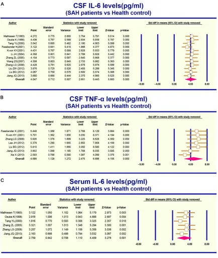
Abstract:Objective: We conducted this study to explore the correlation of interleukin-6 (IL-6) and tumor necrosis factor-alpha (TNF-α) levels in cerebrospinal fluid (CSF) and serum in the patients with subarachnoid hemorrhage (SAH). Methods: Computerized and manual search strategies were performed to retrieve relevant studies. Studies met our inclusion and exclusion criteria were enrolled. Statistical analyses were performed by Comprehensive Meta-analysis 2.0 (Biostat Inc., Englewood, New Jersey, USA). Results: A total of seventeen studies were finally included in this meta-analysis, including 449 SAH patients. The results revealed that IL-6 and TNF-α levels in CSF and serum in SAH patients were higher than those in the health control group (CSF: IL-6: SMD = 4.047, 95% CI = 2.651-5.443, P < 0.001; TNF-α: SMD = 4.686, 95% CI = 2.475-6.896, P < 0.001; Serum: IL-6: SMD = 2.759, 95% CI = 1.110-4.409, P = 0.001; TNF-α: SMD = 1.737, 95% CI = 1.358-2.116, P < 0.001). Further analysis about cerebral vasospasm (CVS) risk indicated that the IL-6 and TNF-α levels in CSF and serum in the CVS group were significantly higher than those in the non-CVS group (CSF: IL-6: SMD = 2.025, 95% CI = 1.048-3.003, P < 0.001; TNF-α: SMD = 0.969, 95% CI = 0.583-1.355, P < 0.001; Serum: IL-6: SMD = 4.310, 95% CI = 1.271-7.350, P = 0.005; TNF-α: SMD = 1.021, 95% CI = 0.549-1.492, P < 0.001). Conclusion: Our evidences suggested that IL-6 and TNF-α levels in CSF and serum may be correlated with SAH, which provides a certain of significant guidance for early diagnosis and monitoring of SAH.
Keywords: Correlation, cerebrospinal fluid, serum, subarachnoid hemorrhage, IL-6, TNF-α
Introduction
Subarachnoid hemorrhage (SAH) is a devastat-ing neurological disease that can result from a ruptured aneurysm and has high morbidity and mortality rates that exceeds 50% [1]. Generally, SAH accounts for 5% of all stroke cases, affect-ing more than 30,000 individuals annually in North America, and half of the patients with SAH are younger than 55 years [2]. It was reported that SAH patients were at risk of developing cerebral vasospasm (CVS), cerebral
ischemia, or ischemic neurological deficits [3].
Among which, CVS is the second leading factor causing massive disability and death in SAH patients [4]. Approximately 20% of patients with CVS have ischemic complications in spite of receiving therapy, and more than half of SAH patients die from CVS [5]. SAH is related to medical conditions that include intracranial
artery dissections, arteriovenous malforma-tion, mycotic aneurysms, reversible cerebral vasoconstriction syndrome, bleeding disorders, and vasculitis [6, 7]. Despite great advances in the early detection, diagnosis, and proper treat-ment of SAH, the overall outcome of SAH patients remains poor [8]. Currently, neuroim-aging and biochemical markers in
cerebrospi-nal fluid (CSF) and serum/plasma are two areas
of extensive biomarker research [9]. One study
has shown that the inflammatory response and
cytokine release of interleukin-6 (IL-6) and
tumor necrosis factor-alpha (TNF-α) are related
to the severity of illness, occurrence of CVS and clinical outcomes in SAH patients [10].
IL-6, a pleiotropic inflammatory cytokine, has a
molecular weight of 26 kD and a biological half-life of less than one hour [11]. Generally, IL-6
-sis, and acute phase responses, which are
reactions to many types of inflammatory stimuli [12]. TNF-α, which is produced by activated astrocytes and macrophages, is a pro-inflam -matory cytokine that plays a role in the
self-propagation of neuronal inflammation [13]. TNF-α exerts it function in the inflammatory
response, and it can also initiate a pro-apoptot-ic pathway, with vascular endothelial cells act-ing as the major target [14]. A clinical study demonstrated an association between clinical
status and inflammation by measuring the inflammatory markers in CSF or serum [15]. Elevated IL-6 and TNF-α levels have been con -sistently detected in the CSF and serum of SAH patients, and they have been used as evidence of vasospasm via the following observations: transcranial Doppler ultrasound, delayed
isch-emic deficit and poor SAH outcome [16].
Previous studies have shown that the
associa-tion between SAH and inflammaassocia-tion may medi
-ate poor outcomes, the elev-ated levels of TNF-α
and IL-6 in CSF and serum provided evidence of vasospasm and poor outcomes in SAH [17, 18]. However, several studies that attempted to
cor-relate TNF-α and IL-6 levels in serum and CSF yielded conflicting results [19, 20]. Therefore,
the aim of this study was to explore the
correla-tion of IL-6 and TNF-α levels in CSF or serum
with SAH.
Materials and methods
Search strategy
Relevant studies published before April, 2017 that addressed the correlation of IL-6 and
TNF-α levels in CSF or serum with SAH were
obtained by searching multiple independent computerized databases (PubMed, China Bio- Medicine (CBM), Web of Science, China Na- tional Knowledge Infrastructureand Cochrane Library). Keywords and free words were com-bined to do the search strategy, such as SAH, primary subarachnoid hemorrhage, acute hem-orrhagic cerebrovascular disease, CSF, cyto-kines, interleukin and tumor necrosis factor, etc.
Study selection
The inclusion criteria were as follows: (1) res- earch topics: the correlation of IL-6 level or
TNF-α level in CSF or serum with SAH; (2) study sub -jects: patients were clinically diagnosed with
SAH; (3) outcomes: IL-6 or TNF-α level in CSF or
serum were investigated between groups. The exclusion criteria were as follows: (1) articles with only an abstract and summary; (2) animal studies; (3) repetition of published documents;
(4) articles with insufficient data; (5) only the
latest complete study was considered when selected studies were published by the same author.
Data extraction and quality assessment
Relevant information from selected articles was extracted by two independent
investiga-tors and recorded on a predefined form. Specifically, the following data were obtained: first author, time of publication, country, ethnic -ity, language, age, gender, number of cases, control group and detection method. The two investigators evaluated the quality of the included studies in light of the critical appraisal skill program (CASP) criteria (http://www.casp-uk.net/). The CASP criteria are standardized as the following 11 aspects: whether the study addresses a clearly focused issue (CASP01); whether an appropriate method was employed to answer their question (CASP02); whether the cases were recruited in an acceptable way (CASP03); whether the controls were selected in an acceptable way (CASP04); whether the exposures were accurately measured to mini-mize bias (CASP05); what confounding factors have the authors accounted for or have the authors considered as potential confounding factors in the design or analysis (CASP06); whether the results of this study are complete (CASP07); the precision of the results (CASP08); whether the results are reliable (CASP09); whether the results be can applied to the local population (CASP10); and whether the results
of the study fit with other available evidence
(CASP11). Discrepancies in the CASP scores for each of the included articles were further addressed by a third reviewer through group discussion and consultation.
Statistical analysis
Our meta-analysis was performed using Co- mprehensive Meta-analysis 2.0 (Biostat Inc., Englewood, New Jersey, USA). The standard
mean difference (SMD) with its 95% confidence
interval (95% CI) was utilized for evaluating the
associations of IL-6 or TNF-α levels in CSF or
per-formed to determine the significance of the
overall effect [21]. A forest plot was drawn to
reflect the values of SMD and 95% CI between
the study groups. To assess the heterogeneity across studies, we calculated Cochran’s Q-sta-
tistic (significance level of P ≤ 0.05) and I2 test
[22, 23]. The degrees of heterogeneity were low, moderate and high, which correspond to I2
values of 25%, 50% and 75%, respectively. For the presence of heterogeneity, a
random-effects model was utilized; otherwise, a
fixed-effects model was applied [24]. Multiple meta-regression analysis was used to evaluate the potential heterogeneous sources, and the Monte Carlo simulation method was used to perform corrections for multiple tests [22, 25]. A sensitivity analysis was conducted to assess whether the results were affected after exclu-sion of any single selected study. The funnel plots, classic fail-safe N and the Egger’s linear regression test were constructed to determine whether a publication bias was present [26-28]. All tests were two-sided, and P < 0.05 was
considered statistically significant.
Results
Included studies
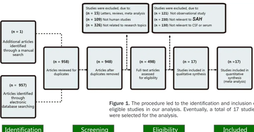
Figure 1 presents the procedure used to iden-tify the inclusion of eligible studies in our analy-sis. We initially retrieved 958 studies through our manual and electronic database search. There were 10 duplicates, 15 letters or reviews,
109 non-human studies and 326 non-related articles that were considered ineligible and thus excluded. The remaining 498 studies were further reviewed, among which 481 were excluded because 121 were non-observational studies, 230 were irrelevant to SAH and 130 were irrelevant to CSF or serum. Finally, 17 studies were enrolled in this study, which included a total of 449 SAH patients [18, 29-45]. These selected studies were published between 1993 and 2013 and included sub-jects in both Asian (n = 13) and Caucasian (n = 4) populations. Among these studies, 1 was performed in Sweden, 1 in Poland, 2 in Germany, 10 in China, 2 in Korea and 1 in Japan. Table 1 illustrated the baseline charac-teristics of these 17 studies in detail.
Correlation between IL-6 and TNF-α levels in
CSF and SAH
Among the 17 selected studies, 12 reported a correlation between IL-6 levels in CSF and SAH.
Significant heterogeneity was found among
studies (I2 = 96.843%, P
h< 0.001); therefore, a
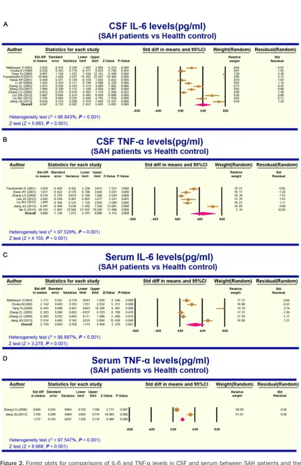
random-effects model was utilized. The results of the meta-analysis showed that higher levels of IL-6 were found in the CSF patients with SAH when compared with the health control group (SMD = 4.047, 95% CI = 2.651-5.443, P < 0.001; Figure 2A). An analysis of the incidence
of CVS revealed IL-6 levels in CSF were signifi -cantly higher in the CVS group than that in the non-CVS group (SMD = 2.025, 95% CI =
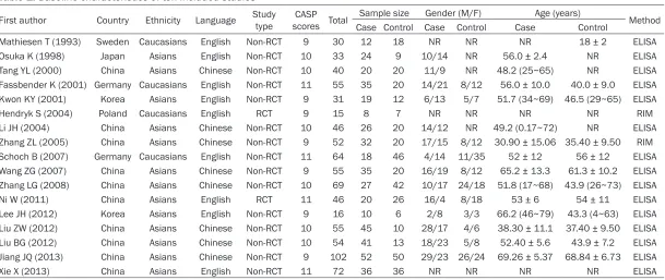
[image:3.612.94.516.71.290.2]Table 1. Baseline characteristics of ten included studies
First author Country Ethnicity Language Study type scores TotalCASP Sample size Gender (M/F) Age (years) Method
Case Control Case Control Case Control
Mathiesen T (1993) Sweden Caucasians English Non-RCT 9 30 12 18 NR NR NR 18 ± 2 ELISA
Osuka K (1998) Japan Asians English Non-RCT 10 33 24 9 10/14 NR 56.0 ± 2.4 NR ELISA
Tang YL (2000) China Asians Chinese Non-RCT 10 40 20 20 11/9 NR 48.2 (25~65) NR ELISA
Fassbender K (2001) Germany Caucasians English Non-RCT 11 55 35 20 14/21 8/12 56.0 ± 10.0 40.0 ± 9.0 ELISA
Kwon KY (2001) Korea Asians English Non-RCT 9 31 19 12 6/13 5/7 51.7 (34~69) 46.5 (29~65) ELISA
Hendryk S (2004) Poland Caucasians English RCT 9 15 8 7 NR NR NR NR RIM
Li JH (2004) China Asians Chinese Non-RCT 10 46 26 20 14/12 NR 49.2 (0.17~72) NR ELISA
Zhang ZL (2005) China Asians Chinese Non-RCT 9 52 32 20 17/15 8/12 30.90 ± 15.06 35.40 ± 9.50 RIM
Schoch B (2007) Germany Caucasians English Non-RCT 11 64 18 46 4/14 11/35 52 ± 12 56 ± 12 ELISA
Wang ZG (2007) China Asians Chinese Non-RCT 9 55 35 20 16/19 8/12 65.2 ± 13.3 61.3 ± 10.2 ELISA
Zhang LG (2008) China Asians Chinese Non-RCT 10 69 27 42 10/17 24/18 51.8 (17~68) 43.9 (26~73) ELISA
Ni W (2011) China Asians English RCT 11 46 20 26 16/4 8/18 53 ± 6 54 ± 11 ELISA
Lee JH (2012) Korea Asians English Non-RCT 9 16 10 6 2/8 3/3 66.2 (46~79) 43.3 (4~63) ELISA
Liu ZW (2012) China Asians Chinese Non-RCT 10 55 45 10 28/17 4/6 38.30 ± 11.1 37.40 ± 9.50 ELISA
Liu BG (2012) China Asians Chinese Non-RCT 10 54 41 13 18/23 5/8 52.40 ± 5.6 43.9 ± 7.2 ELISA
Jiang JQ (2013) China Asians Chinese Non-RCT 9 102 52 50 29/23 26/24 69.26 ± 5.37 68.84 ± 6.73 ELISA
Xie X (2013) China Asians English Non-RCT 11 72 36 36 NR NR NR NR ELISA
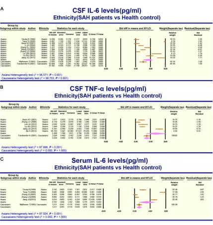
3.003, P < 0.001; Figure 3A). According to the subgroup analysis of ethnicity, SAH patients had higher CSF levels of IL-6 than the health control group in the Asian populations (SMD = 3.244, 95% CI = 1.873-4.615, P < 0.001), but no statistical correlation was found in the Caucasian populations (SMD = 10.392, 95% CI = -6.188-26.973, P = 0.219; Figure 4A). Seven studies examined the correlation between
TNF-α levels in CSF and SAH. A random-effects
model was applied to examine the existence of
significant heterogeneity (I2 = 97.529%, P h <
0.001). The results indicated that TNF-α levels
in CSF were markedly higher in patients with SAH than in the health control group (SMD = 4.686, 95% CI = 2.475-6.896, P < 0.001; Figure 2B). According to the incidence of CVS in
SAH patients, TNF-α levels in CSF in the CVS
group were remarkably higher than those in the non-CVS group (SMD = 0.969, 95% CI = 0.583-1.355, P < 0.001; Figure 3B). A subgroup
analy-sis based on ethnicity suggested that TNF-α
[image:7.612.92.521.61.513.2]levels in CSF in SAH patients were higher than those in the health control group in both the Asian and Caucasian populations (Asian popu-lations: SMD = 5.448, 95% CI = 2.768-8.129, P
< 0.001; Caucasian populations: SMD = 3.024, 95% CI = 2.236-3.813, P < 0.001; Figure 4B).
Correlation between IL-6 and TNF-α level in
serum and SAH
The correlation between IL-6 level in serum and
SAH was reported in 6 studies. The significant
heterogeneity found among studies led to the selection of a random-effects model (I2 =
[image:8.612.91.525.71.583.2]96.897%, Ph< 0.001). The results of the meta-analysis demonstrated that higher level of IL-6 in serum was found in SAH patients than in the health control group (SMD = 2.759, 95% CI = 1.110-4.409, P = 0.001; Figure 2C). Further analysis concerning CVS risk showed that the
serum level of IL-6 was higher in the CVS group than in the non-CVS group (SMD = 4.310, 95% CI = 1.271-7.350, P = 0.005; Figure 3C). An
ethnicity-stratified analysis indicated that
serum levels of IL-6 were higher in SAH patients than in the health control group in both the Asian and Caucasian populations (Asian popu-lations: SMD = 3.122, 95% CI = 1.064-5.179, P
= 0.003; Caucasian populations: SMD = 1.211, 95% CI = 0.543-1.878, P < 0.001; Figure 4C). Two studies examined the correlation between
TNF-α level in serum and SAH. A
random-effects model was applied to identify the
exis-tence of significant heterogeneity (I2 = 97.547%, Ph < 0.001). The results indicated that serum
levels of TNF-α were markedly higher in SAH
patients than in the health control group (SMD = 1.737, 95% CI = 1.358-2.116, P < 0.001; Figure 2D). According to the CVS risk found in
SAH patients, serum levels of TNF-α in the CVS
group were remarkably higher than those in the non-CVS group (SMD = 1.021, 95% CI = 0.549-1.492, P < 0.001; Figure 3D).
Source of heterogeneity
The sensitivity analysis showed that all of the studies included in the meta-analysis had no
significant effect on the SMD values for the cor
-relation of IL-6 or TNF-α levels in serum or CSF
with SAH (Figure 5A-C). The funnel plot was symmetrical, which suggested no obvious pub-lication bias. The classic fail-safe N together with the Egger’s linear regression test further
confirmed that a significant publication bias
was found among the included studies (both P
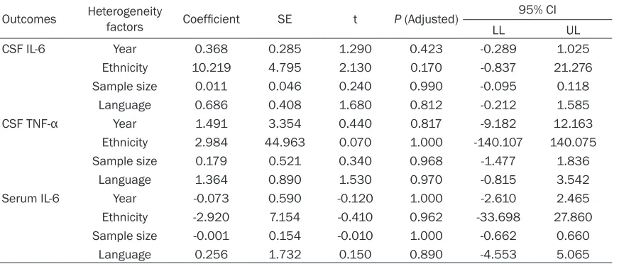
< 0.05) (Figure 6A-C). Multiple meta-regression analysis showed that age, ethnicity, sample size and language were neither the main sourc-es of heterogeneity, nor the key factors affect-ing the overall effect (P > 0.05, Table 2).
Discussion
The present meta-analysis was constructed to investigate the correlation of the levels of IL-6
and TNF-α in CSF or serum with SAH. Our
meta-analysis results suggest that the levels of IL-6 in the CSF and serum were higher in SAH patient than in the health control group, indicat-ing that these two cytokines in CSF and serum may be associated with SAH. This can be explained by the fact that when SAH occurred,
a local inflammatory reaction in the brain tissue
activated a massive release of cytokines,
including IL-6 and TNF-α. The identification of
related signal transduction molecules also acti-vated a variety of kinases in cells, which
increased the flow velocity in the basilar artery
in the brain and further increased the incidence of CVS in SAH patient [40].
One of the findings in this meta-analysis showed
[image:10.612.91.527.87.273.2]that the levels of IL-6 in the CSF and serum of SAH patient were higher than those in the health control group, suggesting the increased level of IL-6 in CSF and serum may be associ-ated with the onset of SAH, especially CVS. It is well-known that higher IL-6 levels are closely associated with various diseases, such as type Table 2. Meta-regression analysis of potential source of heterogeneity
Outcomes Heterogeneity factors Coefficient SE t P (Adjusted) 95% CI
LL UL
CSF IL-6 Year 0.368 0.285 1.290 0.423 -0.289 1.025
Ethnicity 10.219 4.795 2.130 0.170 -0.837 21.276
Sample size 0.011 0.046 0.240 0.990 -0.095 0.118
Language 0.686 0.408 1.680 0.812 -0.212 1.585
CSF TNF-α Year 1.491 3.354 0.440 0.817 -9.182 12.163
Ethnicity 2.984 44.963 0.070 1.000 -140.107 140.075
Sample size 0.179 0.521 0.340 0.968 -1.477 1.836
Language 1.364 0.890 1.530 0.970 -0.815 3.542
Serum IL-6 Year -0.073 0.590 -0.120 1.000 -2.610 2.465
Ethnicity -2.920 7.154 -0.410 0.962 -33.698 27.860
Sample size -0.001 0.154 -0.010 1.000 -0.662 0.660
Language 0.256 1.732 0.150 0.890 -4.553 5.065
Furthermore, a series of reactions initiated by
IL-6 play significant roles in stimulating intracra -nial vascular spasm, causing increases in vas-cular resistance and critical pressure, as well
as reductions in blood flow velocity and blood flow, thus aggravating cerebral ischemia, hem
-orrhage, inflammatory reactions and vascular
spasm and greatly exacerbating the clinical symptoms of SAH patients [34]. In accordance with our results, SAH patients generally showed elevated IL-6 levels in serum, CSF, cerebral
extracellular fluid, and peripheral veins [50].
Furthermore, Schoch et al. provided strong
evi-dence that IL-6 was significantly elevated in
SAH patients with unfavorable outcome [37].
Another finding in our meta-analysis indicated that the levels of TNF-α in the CSF and serum of
SAH patient were higher than those in the health control group, suggesting the increased
levels of TNF-α in CSF and serum may be asso
-ciated with the onset of SAH. TNF-α is a pro-inflammatory cytokine that is associated with
SAH-related endothelial cell apoptosis,
oxida-tive stress, and recruitment of inflammatory
mediators of vasospasm [51]. Additionally,
TNF-α plays an important role in the inflamma -tory cascade and host defense against
infec-tions, which was identified as a potential thera -peutic target for autoimmune disease [52]. One study provided evidence that apoptosis occurred when endothelial cells were exposed
to specific inducers, such as TNF-α; therefore, TNF-α together with the caspase-dependent
cascades were believed to play important roles in SAH-induced apoptosis [53]. In fact,
increased circulating TNF-α is related to more
severe secondary brain injury in other condi-tions, including intra-cerebral hemorrhage and
ischemic stroke [54]. At the cellular level, TNF-α
may be involved in SAH via the pleiotropic
culating inflammatory marker related to poor
SAH outcomes in humans.
Given the influence of ethnicity on the levels of IL-6 and TNF-α in SAH, subgroup analyses were
constructed based on ethnicity. The
ethnicity-stratified analysis showed that the levels of TNF-α in CSF and IL-6 in serum were higher in
SAH patients than in the health control group in both Asian and Caucasian populations. However, the CSF levels of IL-6 were higher in SAH patients than in the health control group in the Asian populations, but no statistical corre-lation was found in the Caucasian popucorre-lations. These differences between ethnicity subgroups could be attributed to geographical, environ-mental and genetic factors.
Some limitations should be addressed while interpreting the results of the present meta-analysis. First, the 17 studies included in the analysis involved only Caucasians and Asians, whereas other ethnicities were inapplicable in the meta-analysis, which could contribute to a selection bias. Second, the lack of original data from some of the included studies restricted a further assessment. For example, an analysis
of the ethnicity subgroups for TNF-α level in serum was not conducted because of insuffi -cient data. Moreover, with the restrictions of the search parameters and a lack of persons
who were proficient in other language, only
studies published in English and Chinese were included into our meta-analysis, studies pub-lished in other languages were not included.
In conclusion, our findings demonstrated that the levels of IL-6 and TNF-α in CSF and serum
Disclosure of conflict of interest
None.
Address correspondence to: Dr. Tao Ma, Department of Neurology, Wuxi No.2 People’s Hospital, Nanjing Medical University, 68 Zhongshan Road, Wuxi 214002, Jiangsu Province, P. R. China. Tel: 15251643918; E-mail: [email protected]
References
[1] Fujii M, Yan J, Rolland WB, Soejima Y, Caner B and Zhang JH. Early brain injury, an evolving frontier in subarachnoid hemorrhage research.
Transl Stroke Res 2013; 4: 432-446.
[2] Bederson JB, Connolly ES Jr, Batjer HH, Dacey RG, Dion JE, Diringer MN, Duldner JE Jr, Har-baugh RE, Patel AB, Rosenwasser RH; Ameri-can Heart Association. Guidelines for the man-agement of aneurysmal subarachnoid hemo- rrhage: a statement for healthcare profession-als from a special writing group of the Stroke Council, American Heart Association. Stroke 2009; 40: 994-1025.
[3] Alaraj A, Charbel FT and Amin-Hanjani S. Peri-operative measures for treatment and preven-tion of cerebral vasospasm following sub-arachnoid hemorrhage. Neurol Res 2009; 31: 651-659.
[4] Zwienenberg-Lee M, Hartman J, Rudisill N, Madden LK, Smith K, Eskridge J, Newell D, Ver-weij B, Bullock MR, Baker A, Coplin W, Mericle R, Dai J, Rocke D, Muizelaar JP; Balloon Pro-phylaxis for Aneurysmal Vasospasm (BPAV) Study Group. Effect of prophylactic translumi-nal balloon angioplasty on cerebral vasospasm and outcome in patients with Fisher grade III subarachnoid hemorrhage: results of a phase II multicenter, randomized, clinical trial. Stroke 2008; 39: 1759-1765.
[5] Tjahjadi M, Konig R, Wirtz CR, Woischneck D and Kapapa T. Cerebral vasospasm and health-related quality of life after subarach-noid hemorrhage. World Neurosurg 2013; 80: 113-120.
[6] Cabral KP, Fraser GL, Duprey J, Gibbons BA, Hayes T, Florman JE and Seder DB. Prothrom-bin complex concentrates to reverse warfarin-induced coagulopathy in patients with intracra-nial bleeding. Clin Neurol Neurosurg 2013; 115: 770-774.
[7] Cuvinciuc V, Viguier A, Calviere L, Raposo N, Larrue V, Cognard C and Bonneville F. Isolated acute nontraumatic cortical subarachnoid hemorrhage. AJNR Am J Neuroradiol 2010; 31: 1355-1362.
[8] Fountas KN, Tasiou A, Kapsalaki EZ, Paterakis KN, Grigorian AA, Lee GP and Robinson JS Jr.
Serum and cerebrospinal fluid c-reactive pro -tein levels as predictors of vasospasm in aneu-rysmal subarachnoid hemorrhage. Clinical ar-ticle. Neurosurg Focus 2009; 26: E22. [9] Lad SP, Hegen H, Gupta G, Deisenhammer F
and Steinberg GK. Proteomic biomarker dis-covery in cerebrospinal fluid for cerebral vaso -spasm following subarachnoid hemorrhage. J Stroke Cerebrovasc Dis 2012; 21: 30-41. [10] Chaichana KL, Pradilla G, Huang J and
Tamar-go RJ. Role of inflammation (leukocyte-endo -thelial cell interactions) in vasospasm after subarachnoid hemorrhage. World Neurosurg 2010; 73: 22-41.
[11] Brown JM, Grosso MA and Harken AH. Cyto-kines, sepsis and the surgeon. Surg Gynecol Obstet 1989; 169: 568-575.
[12] Hoge J, Yan I, Janner N, Schumacher V, Cha-laris A, Steinmetz OM, Engel DR, Scheller J, Rose-John S and Mittrucker HW. IL-6 controls the innate immune response against Listeria monocytogenes via classical IL-6 signaling. J Immunol 2013; 190: 703-711.
[13] Jiang Y, Liu DW, Han XY, Dong YN, Gao J, Du B, Meng L and Shi JG. Neuroprotective effects of anti-tumor necrosis factor-alpha antibody on apoptosis following subarachnoid hemorrhage in a rat model. J Clin Neurosci 2012; 19: 866-872.
[14] Beeftink MM, Ruigrok YM, Rinkel GJ and van den Bergh WM. Relation of serum TNF-alpha and TNF-alpha genotype with delayed cerebral ischemia and outcome in subarachnoid hem-orrhage. Neurocrit Care 2011; 15: 405-409. [15] Hopkins SJ, McMahon CJ, Singh N, Galea J,
Hoadley M, Scarth S, Patel H, Vail A, Hulme S, Rothwell NJ, King AT and Tyrrell PJ. Cerebrospi-nal fluid and plasma cytokines after subarach -noid haemorrhage: Csf interleukin-6 may be an early marker of infection. J Neuroinflamma -tion 2012; 9: 255.
[16] Zemke D, Farooq MU, Mohammed Yahia A and Majid A. Delayed ischemia after subarachnoid hemorrhage: result of vasospasm alone or a broader vasculopathy? Vasc Med 2007; 12: 243-249.
[17] Muroi C, Bellut D, Coluccia D, Mink S, Fujioka M and Keller E. Systemic interleukin-6 concen-trations in patients with perimesencephalic non-aneurysmal subarachnoid hemorrhage. J Clin Neurosci 2011; 18: 1626-1629.
[18] Chou SH, Feske SK, Atherton J, Konigsberg RG, De Jager PL, Du R, Ogilvy CS, Lo EH and Ning M. Early elevation of serum tumor necrosis factor-alpha is associated with poor outcome in subarachnoid hemorrhage. J Investig Med 2012; 60: 1054-1058.
the impact of between-study heterogeneity in multivariate meta-analyses. Stat Med 2012; 31: 3805-3820.
[23] Peters JL, Sutton AJ, Jones DR, Abrams KR and Rushton L. Comparison of two methods to de-tect publication bias in meta-analysis. JAMA 2006; 295: 676-680.
[24] Zintzaras E, Ioannidis JP. Heterogeneity testing in meta-analysis of genome searches. Genet Epidemiol 2005; 28: 123-137.
[25] Ferrenberg AM, Swendsen RH. New Monte Carlo technique for studying phase transitions.
Phys Rev Lett 1988; 61: 2635-2638.
[26] Sterne JA, Egger M. Funnel plots for detecting bias in meta-analysis: guidelines on choice of axis. J Clin Epidemiol 2001; 54: 1046-1055. [27] Wikstrom EA, Naik S, Lodha N and Cauraugh
JH. Balance capabilities after lateral ankle trauma and intervention: a meta-analysis. Med Sci Sports Exerc 2009; 41: 1287-1295. [28] Egger M, Davey Smith G, Schneider M and
Minder C. Bias in meta-analysis detected by a simple, graphical test. BMJ 1997; 315: 629-634.
[29] Mathiesen T, Andersson B, Loftenius A and von Holst H. Increased interleukin-6 levels in cere-brospinal fluid following subarachnoid hemor -rhage. J Neurosurg 1993; 78: 562-567. [30] Osuka K, Suzuki Y, Tanazawa T, Hattori K,
Ya-mamoto N, Takayasu M, Shibuya M and Yoshi-da J. Interleukin-6 and development of vaso-spasm after subarachnoid haemorrhage. Acta Neurochir (Wien) 1998; 140: 943-951. [31] Tang YL QX, Sheng WL and Wang J. Serum and
cerebrospinal fluid il-6 levels in patients with subarachnoid hemorrhage. Chinese Journal of Nervous and Mental Diseases 2000; 26: 120. [32] Fassbender K, Hodapp B, Rossol S, Bertsch T,
Schmeck J, Schutt S, Fritzinger M, Horn P, Va-jkoczy P, Kreisel S, Brunner J, Schmiedek P, Hennerici M. Inflammatory cytokines in sub -arachnoid haemorrhage: association with ab-normal blood flow velocities in basal cerebral arteries. J Neurol Neurosurg Psychiatry 2001; 70: 534-537.
spinal fluid interleukin-6 levels in patients with subavachnoid hemorrhage. Stroke and Ner-vous Diseases 2005; 12: 33-35.
[37] Schoch B, Regel JP, Wichert M, Gasser T, Vol-bracht L and Stolke D. Analysis of intrathecal interleukin-6 as a potential predictive factor for vasospasm in subarachnoid hemorrhage. Neu-rosurgery 2007; 60: 828-836.
[38] Wang ZG JY, Qu CC and Wang ZG. Dynamical changes in concentration of il-6 and sicm-1 in cerebrospinal fluid in the patients with aneu -rysml subarachnoid hemorrhage. Chinese Journal of Gerontology 2007; 27: 1691-1692. [39] Zhang LG ZMaWD. The relationship between
the changes of tnf-α, il-6 and et after sub -arachnoid hemorrhage and secondary cere-bral vasospasm. Shandong Medical Journal 2008; 48: 34-35.
[40] Ni W, Gu YX, Song DL, Leng B, Li PL and Mao Y. The relationship between il-6 in csf and occur-rence of vasospasm after subarachnoid hem-orrhage. Acta Neurochir Suppl 2011; 110: 203-208.
[41] Lee JH, Park DH, Back DB, Lee JY, Lee CI, Park KJ, Kang SH, Cho TH and Chung YG. Compari-son of cerebrospinal fluid biomarkers between idiopathic normal pressure hydrocephalus and subarachnoid hemorrhage-induced chronic hy-drocephalus: a pilot study. Med Sci Monit 2012; 18: PR19-25.
[42] Liu BG GX, He LM, Long W and Xiao G. Mea-surement of il-1β, il-6 and tnf-α levels in cere -brospinal fluid of patients with aneurysmal subarachnoid hemorrhage and its significance. Journal of Clinical Research 2012; 29: 404-406.
[43] Liu ZW KW, Shi XF, Yin XL, Jia J, Yi LZ, Zhou LQ, Ouyang XH, Jiu ZS and Chen GC. Study of cere-brospinal fluid interleukin-6 levels in patients with aneurysmatic subarachnoid hemorrhage.
Chin J Clinicians (Electronic Edition) 2012; 06: 1140-1143.
with subarachnoid hemorrhage. Chinese Jour-nal of Gerontology 2013; 33: 2288-2289. [45] Xie X, Wu X, Cui J, Li H and Yan X. Increase
icam-1 and lfa-1 expression by cerebrospinal fluid of subarachnoid hemorrhage patients: In -volvement of tnf-alpha. Brain Res 2013; 1512: 89-96.
[46] Kao HW, Lee KW, Kuo CL, Huang CS, Tseng WM, Liu CS and Lin CP. Interleukin-6 as a prog-nostic biomarker in ruptured intracranial aneu-rysms. PLoS One 2015; 10: e0132115. [47] Heikkila K, Ebrahim S, Lawlor DA. Systematic
review of the association between circulating interleukin-6 (il-6) and cancer. Eur J Cancer 2008; 44: 937-945.
[48] Ostrowski RP, Colohan AR and Zhang JH. Mo-lecular mechanisms of early brain injury after subarachnoid hemorrhage. Neurol Res 2006; 28: 399-414.
[49] Li J, Liang X, Wang Q, Breyer RM, McCullough L and Andreasson K. Misoprostol, an anti-ulcer agent and PGE2 receptor agonist, protects against cerebral ischemia. Neurosci Lett 2008; 438: 210-215.
[50] Sarrafzadeh A, Schlenk F, Gericke C and Va-jkoczy P. Relevance of cerebral interleukin-6 after aneurysmal subarachnoid hemorrhage.
Neurocrit Care 2010; 13: 339-346.
[51] Vecchione C, Frati A, Di Pardo A, Cifelli G, Car-nevale D, Gentile MT, Carangi R, Landolfi A, Carullo P, Bettarini U, Antenucci G, Mascio G, Busceti CL, Notte A, Maffei A, Cantore GP, Lem-bo G. Tumor necrosis factor-alpha mediates hemolysis-induced vasoconstriction and the cerebral vasospasm evoked by subarachnoid hemorrhage. Hypertension 2009; 54: 150-156.
[52] Mariette X, Matucci-Cerinic M, Pavelka K, Tay-lor P, van Vollenhoven R, Heatley R, Walsh C, Lawson R, Reynolds A and Emery P. Malignan-cies associated with tumour necrosis factor inhibitors in registries and prospective obser-vational studies: a systematic review and me-ta-analysis. Ann Rheum Dis 2011; 70: 1895-1904.
[53] Cahill J, Calvert JW and Zhang JH. Mechanisms of early brain injury after subarachnoid hemor-rhage. J Cereb Blood Flow Metab 2006; 26: 1341-1353.
[54] Hanafy KA, Grobelny B, Fernandez L, Kurtz P, Connolly ES, Mayer SA, Schindler C and Badja-tia N. Brain interstiBadja-tial fluid tnf-alpha after sub -arachnoid hemorrhage. J Neurol Sci 2010; 291: 69-73.
[55] Zhou ML, Shi JX, Hang CH, Cheng HL, Qi XP, Mao L, Chen KF and Yin HX. Potential contribu-tion of nuclear factor-kappaB to cerebral vaso-spasm after experimental subarachnoid hem-orrhage in rabbits. J Cereb Blood Flow Metab 2007; 27: 1583-1592.