Functional differences between Tcf1 isoforms
in early Xenopus development
GIULIETTA ROËL, OLAF VAN DEN BROEK and OLIVIER DESTRÉE*
Hubrecht Institute, Utrecht, the NetherlandsABSTRACT In Xenopus gastrula stage embryos, four isoforms of Tcf1 (B, C, D and E) are present with high amino acid sequence conservation compared to fish, mice and human. We studied pos-sible functional differences between these Tcf1 isoforms during early Xenopus development. After overexpression of single Tcf1 isoforms, two distinct phenotypes were observed. Overexpression of the B or D isoforms of Tcf1, which both lack a C-clamp, enhances early canonical Wnt signaling and induces ectopic dorsal mesoderm at the expense of ventrolateral mesoderm prior to gastrulation, causing severe antero-dorzalization of embryos. Overexpression of the E-isoform, which contains a complete C-clamp, does not induce ectopic dorsal mesoderm, but rather leads to severe caudal truncation. Overexpression of the C-isoform, which contains a partial C-clamp, induces a similar phenotype. Mutation of a single amino acid in the C-clamp, known to produce a hypomorphic mu-tant in D. melanogaster, led to a gain of function in inducing ectopic organizer tissue, as observed after overexpression of the B or D isoforms of Tcf1. Depletion of the C-clamp exon from the zygotic mRNA pool, by injection of a morpholino oligo that targets the splice acceptor site of the exon containing the C-clamp, caused a severe shortening of the AP-axis. Furthermore, embryos showed poor development of the CNS, paraxial mesoderm and primary blood vessels. In situ hybridization analysis showed that Lef1 expression was downregulated at the mid-hindbrain boundary, in the otic vesicles and the branchial arches. The results indicate that in post-gastrula stage Xenopus embryos, the E-tail of Tcf1 is required for expression of Lef1 and for blood vessel formation.
KEY WORDS:
Tcf1, isoforms, Wnt, Lef1, angiogenesis, embryo, Xenopus
Introduction
Lef/Tcf transcription factors play essential roles during embryonic development, organogenesis and in tissue homeostasis. Interac-tion of Lef/Tcf proteins with beta-catenin mediates Wnt-induced transcriptional activation of target genes (Logan and Nusse, 2004; Cadigan and Waterman, 2012; Schuijers et al., 2014). This signal transduction system has an ancient origin (Kraus et al., 2016). Lef/ Tcf proteins can also act as transcriptional repressors by binding to Groucho/Transducin-Like Enhancer of split (TLE) proteins that function as co-repressors by interacting with histone deacetylases whose activity leads to the generation of transcriptionally silent chromatin (Roose et al., 1998; Li et al., 2014).
All Lef/Tcfs contain a high mobility group (HMG) box that can bind to a core eight nucleotide binding site (Dooijes et al., 1993; Van de Wetering et al., 1992). It is not until the binding of the co-activator beta-catenin that the promoters of many Wnt target genes get activated (Logan and Nusse, 2004; Clevers, 2006; Cadigan
www.intjdevbiol.com
*Address correspondence to: Olivier Destrée. Hubrecht Institute, Uppsalalaan 8, 3584 CT Utrecht, the Netherlands. Tel: 00-312-9441-0621.
e-mail: [email protected]
Submitted: 31 August, 2016. Accepted: 16 September 2016.
ISSN: Online 1696-3547, Print 0214-6282
© 2017 UPV/EHU Press Printed in Spain
Abbreviations used in this paper: HMG, high mobility group; TLE, transducin-Like
enhancer of split.
and Waterman, 2012; Schuijers et al., 2014). In vertebrates, the Lef/Tcf family consists of four members: Tcf1 (Tcf7), Lef1, Tcf3 (Tcf7l1) and Tcf4 (Tcf7l2).
Lef/Tcfs encode multiple splice variants (van de Wetering et al., 1996; Howng et al., 2004; Weise et al., 2010) and conserved func-tional domains have been determined. In Xenopus, Tcf4 produces alternative peptides flanking exon VI that are important for activation or repression of the promoters of Wnt target genes (Pukrop et al., 2001) and that are determining in inducing a secondary axis upon overexpression in Xenopus embryos (Gradl et al., 2002). Alterna-tive expression of different isoforms of Tcf4 was demonstrated to be essential for specifying the CNS (Kunz et al., 2004).
and Waterman, 2012; Wallmen et al., 2012; Hoverter et al., 2014; Ravindranath and Cadigan, 2014). This domain was first discovered in TCF1 and TCF4 (Atcha et al., 2003; Atcha et al., 2007) and is also present in invertebrate TCFs (Cadigan and Waterman, 2012). The C-clamp from TCF/Pangolin is a zinc-binding domain with the four conserved cysteines coordinating a zinc ion (Ravindranath and Cadigan, 2014). It is sufficient for binding to helper sites, that are critical for activation of Wnt/b-catenin regulated cis-regulatory
modules (Chang et al., 2008; Hoverter et al., 2012). The C-clamp of TCF/Pangolin also binds to the HMG domain and inhibits this domain from binding to its cognate DNA site (Ravindranath and Cadigan, 2014).
The C-clamp of TCF1 is required for the strong activation of Lef1 promoter constructs in COS cells (Atcha et al., 2003). Aberrant activation of full length Lef1 has been shown to be Wnt regulated in colorectal cancers, which may thereby link the incidence of splice variants to cancer progression (Hovanes et al., 2001; Li et al., 2006). In mammals TCF1 is subject to extensive alternative splicing resulting in at least seven different C-terminal isoforms (van de Wetering et al., 1996). The Tcf1 isoforms described in Xenopus and zebrafish show high conservation in their amino acid sequence (Roël et al., 2003; Veien et al., 2005). No studies have been re-ported on possible functional differences between Tcf1 isoforms during early development of Xenopus. Here, we show that the effects of ectopic XTcf1 isoform over-expression depend on the presence or absence of the C-clamp. Overexpression of isoforms that do not contain the C-clamp led to antero-dorsalization of the injected embryos due to expanded dorsal mesoderm formation. Inactivation of the C-clamp, in a way that was shown to produce a hypomorphic Wingless mutant in flies (Van de Wetering et al., 1997; Ravindranath and Cadigan, 2014), led to loss of specificity, the obtained phenotype was similar to that of embryos with over-expressed isoforms missing the C-clamp. Furthermore, we show that the C-clamp of XTcf1 is required for the expression of Lef1 in the vascular system and the brain during early development.
Results
Differences in response to ectopic overexpression of XTcf1 isoforms depend on C-terminal sequences
Because no studies were reported on the role of different XTcf1 isoforms during early development, we initiated a functional analysis
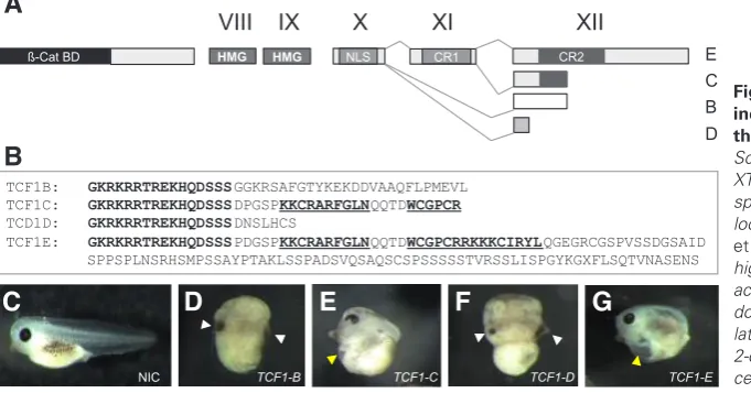
of the four C-terminal isoforms that were shown to be present in Xenopus embryos (Roël et al., 2003). Fig. 1a shows a schematic drawing of the genomic organisation of XTcf1. In gray are depicted the conserved domains which together comprise the C-clamp (Atcha et al., 2003; Cadigan and Waterman, 2012). XTcf1E contains the complete C-clamp, while XTcf1C only contains the first 20 residues of the 29 amino acid motif. Alternative splicing of exon X to exon XII results in isoforms XTcf1B and XTcf1D. This occurs at different splice acceptor sites and in different reading frames in exon XII, which generates unique amino acid sequences 3’ of the nuclear localization signal (Fig. 1B, in bold).
Expression constructs were made for all four XTcf1 isoforms and capped mRNAs were generated by in vitro transcription. Injec-tion at the 2-cell stage of 200 pg of XTcf1 mRNA in X. tropicalis embryos resulted in two distinct phenotypes (Fig. 1C - 1G). Over-expression of isoforms XTcf1B and Xtcf1D, which both lack the C-clamp, resulted in a strong antero-dorsalization of the injected embryos (Fig. 1D and F), with a DAI index of 8 to 9 (according to Kao and Elinson, 1998). This was frequently accompanied by (partial) axis duplication as indicated by the presence of a second cement gland (Fig. 1D and F, white arrowhead), for statistics see Fig. 3D. Overexpression of isoforms XTcf1C and XTcf1E, did not lead to antero-dorsalization but resulted in a caudal truncation of the embryos (Fig. 1E and G). Doubling the amounts of XTcf1C or XTcf1E mRNA produced similar results. A caveat for these and further data is that different (mutant) XTcf1 mRNAs and proteins may be unstable or misfolded.
We analyzed the expression patterns of the organizer genes Chordin and Goosecoid as well as the ventro-lateral genes Wnt8 and Cdx1 during gastrulation of XTcf1 injected embryos to investigate if the differences in phenotypes observed at tailbud stages are due to defects at earlier stages (Fig. 2). From the expression patterns of Chordin and Goosecoid in the XTcf1B and XTcf1D injected embryos it became clear that the antero-dorsalization seen at later stages was dependent on ectopic ‘early’ WNT signaling on the ventral side of the embryo (Smith et al., 1991). Ectopic dorsal mesoderm originated in the embryos at the expense of ventro-lateral tissues, as indicated by the absence of Wnt8 and Cdx1. Embryos injected with either XTcf1C or XTcf1E did not show changes in expression patterns of Chordin and Goosecoid, indicating that mesoderm induction and gastrulation were relatively normal.
To further investigate how the specificity of XTcf1 is dependent
Fig. 1. Overexpression of single XTcf1 isoforms induces two distinct phenotypes dependent on the presence or absence of a (partial) C-clamp. (A) Schematic drawing of the genomic organisation of XTcf1 showing exons X to XII generating different XTcf-1 splice variants. The two conserved motifs CR1 and CR2 located on exons XI and XII comprise the C-clamp (Atcha
et al., 2003). NLS: nuclear localization signal. HMG:
high-mobility group DNA binding domain. (B) amino acid sequences of the different splice variants of XTcf1 downstream of exon X. (C-G) X. tropicalis embryos after lateral injection of the four different Tcf1 isoforms at the 2-cell stage. NIC, non-injected control; white arrow head, cement gland; yellow arrow head, heart.
TCF1B: GKRKRRTREKHQDSSSGGKRSAFGTYKEKDDVAAQFLPMEVL TCF1C: GKRKRRTREKHQDSSSDPGSPKKCRARFGLNQQTDWCGPCR
TCD1D: GKRKRRTREKHQDSSSDNSLHCS
TCF1E: GKRKRRTREKHQDSSSPDGSPKKCRARFGLNQQTDWCGPCRRKKKCIRYLQGEGRCGSPVSSDGSAID SPPSPLNSRHSMPSSAYPTAKLSSPADSVQSAQSCSPSSSSSTVRSSLISPGYKGXFLSQTVNASENS
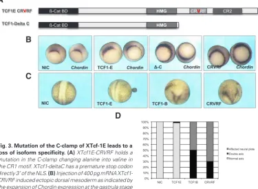
on its C-terminus we generated two constructs (Fig. 3A). Substitution of an alanine for a valine in the C-clamp leads to a hypomorhic mu-tation in D. melanogaster (Van de Wetering et al., 1997). Therefore we made one construct containing an alanine to valine transition in XTcf1E (CRVRF). The other construct car-ried a premature stop directly 3’ of the exon that contains the nuclear localization signal and lacks the C-terminus (deltaC). Injection of XTcf1-deltaC induced circular orga-nizers (Fig. 3), similar to XTcf1B and Xtcf1D (cf Fig. 2). Embryos injected with XTcf1-CRVRF also showed an expansion of the expression of Chordin (Fig. 3B).
To quantify the different effects of XTcf1 isoforms, double axis forma-tion was analysed at neurula stages (Fig. 3D). Injection of XTcf1E-deltaC mRNA in the most animal part of X. laevis embryos prior to the first cleavage induced an ectopic axis in 51% of the embryos (33/65) while injection of XTcf1E induced an ectopic axis in only 2% (1/62). Mutation of the C-clamp in XTcf1E-CRVRF led to an increase in double axis formation to 29% (18/62). In the remaining cases where no obvious ectopic axis had formed all neural plates were broadened. This phenomenon was never observed in XTcf1E injected embryos and may represent intrinsic differences between the isoforms which cause these defects at later stages.
Isoforms XTcf1 E and C are re-quired for Lef1 expression and angiogenesis
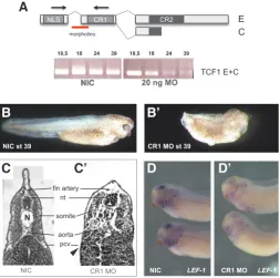
To study the function of the C-clamp of XTcf1 during early Xeno-pus development we generated a specific knockdown of C-clamp containing XTcf1 isoforms by in-jecting a morpholino targeting the splice acceptor site of exon X that contains the C-clamp. This way we generated a zygotic loss of the C-clamp of XTcf1 (Fig. 4A). RT-PCR analysis with the reverse primer on the exon containing the C-clamp re-vealed a clear downregulation of the C-clamp after gastrula stages (Fig. 4A). C-clamp-depleted embryos
Fig. 2. Effect of XTcf1 isoform overexpression on the expression of organizer genes and ventrolateral genes during gastrulation after injection of single isoforms of XTcf1. Whole mount in situ hybridisation of gastrula stage embryos after injection of different XTcf1 isoforms. Overexpression of isoforms B and D induces ectopic dorsal mesoderm at the expense of ventrolateral tissue, as indicated by the expansion of Chordin and Goosecoid and the absence of Wnt8 and Cdx1.
Fig. 3. Mutation of the C-clamp of XTcf-1E leads to a loss of isoform specificity. (A) XTcf1E-CRVRF holds a mutation in the C-clamp changing alanine into valine in the CR1 motif. XTcf1-deltaC has a premature stop codon directly 3’ of the NLS. (B) Injection of 400 pg mRNA XTcf1-CRVRF induced ectopic dorsal mesoderm as indicated by the expansion of Chordin expression at the gastrula stage
0%
(Figure 4B and B’) typically showed a shortening of the AP axis, aberrant pigment formation and abnormal development of the eyes. Sectioning of the embryos showed that somites, notochord and CNS formed, but were reduced and not well differentiated (Fig. 4C and C’). The most striking effects were on angiogenesis as indicated by the absence of the major blood vessels: aorta, posterior car-dinal veins and fin artery (Fig. 4D and D’). This phenotype is very similar to that observed after depletion of Lef1 (Roël et al., 2009). It has been shown that the E-tail of TCF1 is required to activate the LEF1 promoter in different cell lines (Atcha et al., 2003). To test in vivo if Lef1 expression is regulated by the C-clamp of XTcf1 we injected low amounts of C-clamp morpholino (10 ng) and analyzed Lef1 expression in stage 32 embryos by in situ hybridization. At that stage, Lef1 is a robust marker for the mid-hindbrain boundary, the otic vesicles and the branchial arches (Molenaar et al., 1998). In C-clamp-depleted embryos, Lef1 expression was downregulated in these tissues, indicating a role for the C-clamp of XTcf1 in the activation of the Lef1 promoter in vivo.
Discussion
Differences in response to ectopic overexpression of XTcf1 isoforms
In X. laevis and X. tropicalis gastrula stage embryos four isoforms of XTcf1 (B, C, D and E) are present that show high amino acid sequence conservation compared to fish, mice and human (Roël et al., 2003). Of these XTcf1 isoforms only the E-isoform contains a complete C-clamp (Fig 1). The Xtcf1C isoform contains only the first 20 residues of the 29 amino acid C-clamp and therefore lacks the second basic amino acid stretch and the fourth cysteine (Ravindranatha and Cadigan, 2014). TCF1C isoforms are also found in fish (Veien et al., 2005) and human (van de Wetering et al., 1996). The expression pattern of XTcf1 has been described (Roël et al., 2003) but it is not known where and when the different isoforms are expressed. We show that overexpression of XTcf1B
and XTcf1D causes severe antero-dorsalization, while overexpres-sion of XTcf1C or XTcf1E does not induce ectopic dorsal mesoderm but causes caudal truncation of the injected embryos.
The results presented here for XTcf1B and XTcf1D confirm and extend earlier results (Janssens et al., 2013). Activation of direct target genes after Tcf1 B or D overexpression, like Foxd3 (Janssens et al., 2013), is supposed to take place through Wnt-responsive elements, after recruitment of b-catenin (Logan and
Nusse, 2004; Cadigan and Waterman, 2012; Schuijers et al., 2014). Overexpression of XTcf1B leads to the upregulation of many genes expressed at the dorsal side of the embryo as found by microarray analysis of early gastrula stage embryos (van den Broek and Destrée, unpublished results) including known direct beta catenin/Tcf target genes like siamois, nr3, dkk1, foxd3 and the indirect targets chordin and goosecoid (Fig. 2). On the other hand, genes known to be expressed in the ventrolateral zone of gastrula stage embryos, like wnt8 and cdx1, were downregulated (Fig. 2) amongst others, like sizzled, mespo, delta2 and cdx4.
Our over-expression studies show that XTcf1E and XTcf1C induce a different phenotype and a different response of target genes compared to XTcf1B and XTcf1D. XTcf1E contains a complete C-clamp with all four essential cysteines present (Ravindranath et al., 2014). C-clamp containing isoforms of TCFs have been shown to be potent transcriptional regulators with an expanded transcriptome directed by C-clamp-Helper site interactions (Hoverter et al., 2014). The C-clamp enables targeting to a greater number of gene loci for stronger occupancy and transcription regulation (Hoverter et al., 2014). To our surprise, classical dorsal genes such as Chordin, and Goosecoid were not hyper-activated at gastrula stages by overexpression of XTcf1E (Fig. 2), but at tailbud stages severe caudal truncation was observed (Fig. 1). Wallmen et al., (2012) have demonstrated that in embryonic stem cells Tcf1/ Tcf4-dependent gene responses to Wnt are primarily mediated by C-clamp-containing Tcf1E and Tcf4E splice variants. Furthermore these authors showed that overexpressed Tcf1E cannot invade silent chromatin. These results could explain why overexpressed XTcf1E does not (hyper)activate endogenous targets at gastrula stages. The late effects of XTcf1E overexpression observed at tailbud stages may be explained by the findings of Ravindranath and Cadigan (2014) that the C-clamp binds to the HMG domain and inhibits it’s ability to bind DNA. This way ‘late’ Wnt respon-sive genes may be targeted resulting in their downregulation. To explain the effects of XTcf1C overexpression, which are similar to those of XTcf1E, is more difficult. The last cysteine of the C-clamp is required for activation of a W-CRM reporter in Drosophila cell culture (Ravindranath and Cadigan, 2014). A C-clamp containing this mutation was still able to a minor extent to bind in vitro to a
Fig. 4. Depletion of XTcf1 C and E isoforms from the zygotic mRNA pool by inhibiting splicing using a morpholino oligo (MO). (A) position of the splice morpholino targeting the exon that carries the CR1 motif. RT-PCR analysis of the mRNA of non-injected control (NIC) embryos and embryos injected with 20 ng splice MO using PCR primers amplifying exon X and XI. (B) Control stage 39 embryo. (B’) Phenotype of an embryo after injection of 20 ng splice MO. (C) Histological sections of stage 39 NIC embryo showing clear vessels. (C’) CR1 MO injected embryo showing lack of major blood vessels (cf Roël et al., 2009). Arrow indicates expected level of the posterior cardinal vein (pcv). (D) In situ hybridization of XLef1 in NIC stage 32 embryo and XTcf1- CR1 splice MO (10 ng) injected embryo (D’). XLef1 expression is downregulated in the branchial arches and the midbrain.
HMG-Helper site probe (Ravindranath and Cadigan, 2014). Pos-sibly, overexpressed XTcf1C, though with limited binding capac-ity, still has inhibitory activity. A Tcf1C isoform is also present in zebrafish embryos at 24 hours postfertilization (Veien et al., 2005), suggesting a conserved function.
Our overexpression data contrast with the findings of Standley et al., (2006) that depletion of maternal XTcf1 abrogates expres-sion of Wnt target genes ventrally and laterally, and activates their expression dorsally. Zygotic knockdown of XTcf1 has different effects (Liu et al., 2005). Together these results indicate that a delicate timing and localization of expression of different XTcf1 isoforms is necessary during early and later phases of Wnt signaling in the Xenopus embryo.
Isoforms XTcf1 E and C are required for Lef1 expression and angiogenesis
Possible target genes of XTcf1E may differ to a great extent from the targets of XTcf1B or XTcf1D (Hoverter et al., 2014). We opted for Lef1 as a possible target of XTcf1 after gastrulation because Lef1 was able to rescue XmyoD expression after zygotic XTcf1 knockdown (Liu et al., 2005) and LEF1 is a target of TCF1 in colon cancer (Atcha et al., 2003).
We show that Lef1 expression is downregulated, as determined by in situ hybridization, in Xenopus postgastrula stage embryos by loss of function of the C-clamp of Tcf1. The phenotype of the resulting embryos at tailbud stages (Fig. 4) is similar to that seen after depletion of Lef1: a strong reduction of caudal structures, mesoderm derivatives and underdeveloped CNS (Roël et al., 2009). Histological analysis of XTcf1 C-clamp depleted embryos showed that somites, notochord and CNS formed, but that these tissues are reduced in size and are poorly differentiated. The most striking effects were on angiogenesis as shown by the absence of the major blood vessels: the aorta, posterior cardinal veins and fin artery. These effects are very similar to those observed after deple-tion of Lef1 (Fig. 4 and Roël et al., 2009). Our results indicate that expression of the (partial) C-clamp containing isoforms of XTcf1, XTcf1C and XTcf1E, is required in post-gastrula stage Xenopus embryos for expression of Lef1 and for blood vessel formation.
E-tail specific activation of the LEF1 promoter was observed in different cell lines (Atcha et al., 2003). In COS cells two Wnt response elements are required for a strong activation of the Lef1 promoter and activation of the promoter occurs exclusively by TCF forms that contain the C-clamp (Atcha et al., 2003). In human EAhy926 endothelial cells, Lef1 promotes endothelial cell invasion and regulates matrix metalloproteinase-2 expression (Planutiene et al., 2011). Phng et al. (2009) showed that The Notch-regulated ankyrin repeat protein (Nrarp) acts as a molecular link between Notch- and Lef1-dependent Wnt signaling in endothelial cells to control stability of new vessel connections in mouse and zebrafish.
Further study is necessary to determine if crosstalk between Tcf1 and Lef1 is of general importance for angiogenesis in the embryo, during organogenesis and in tumor formation.
Materials and Methods
In situ hybridizationProbes were generated via RT-PCR and cloned in pGEM-T easy vectors (Promega) or generated by in vitro transcription directly from an RT-PCR with T7 overhang on the reverse primer. Digoxygenin-labelled antisense probes were synthesized by use of the Ambion Maxiscript kit
with digoxigenin-UTP (Roche). Whole-mount in situ hybridization proce-dures were as described (Molenaar et al., 1998) with the modification that hybridization was at 65C and no RNase treatment was performed when probes were used from X. tropicalis.
Morpholino oligo and mRNA injections
XTcf1 expression constructs were cloned in pT7TS after RT-PCR. Con-structs were sequenced to check for a full length product. Capped mRNA was synthesized with Ambion mMessage mMachine kit after linearization with XbaI. Embryos were injected with 200 pg (X. tropicalis) or 400 pg (X. laevis) of mRNA. Embryos were fixed in MEMPFA (100 mM MOPS, 2 mM EGTA, 1 mM MgSO4, 4% paraformaldehyde) and methanol. C-clamp-splice morpholino oligo (sequence: GCCAGGGTCTGAGTATAGCAACATG) was injected at 10 or 20 ng in X. tropicalis embryos after natural mating, later-ally, at the 2-cell stage.
Mutagenesis of the expression constructs
Mutagenesis of the C-clamp exon was performed in two separate PCRs. Two antisense primers with a mismatch in the middle to substitute the alanine for a valine were used. PCR 1 contained the Rev primer (GGTTGAGGCCAAAGCGAACTC-TGCATTTCTTAGG), PCR 2 contained the Fwd primer (CCTAAGAAATGCAG-AGTTCGCTTTGGCCTCAACC). After the initial PCRs the two products were put together without primers for twenty cycles. Hereafter, the initial fwd primer from PCR1 and rev primer from PCR 2 were added and another 30 cycles were performed. Afterwards, the construct was sequenced to ensure a full length product. The delta C construct was prepared via PCR with the reverse primer TTAGTCTGAACTAGAA-TCCTGGTGTTTTTC.
RT-PCR analysis of the C-clamp-exon
Total RNA was isolated via phenol-chloroform extraction. RT-PCR was performed with the Promega M-MLV RT-PCR kit. Primer used on the C-clamp-exon was GAGGCCAAAGCGAGCTCTGCATTTCTT.
Sectioning of embryos
Fixed embryos were embedded in Technovit 8100 and sectioned at 15 mm.
Acknowledgements
G.R. and O.D. were supported by the Earth and Life Sciences Foundation ALW, subsidized by the Netherlands Organization for Scientific Research NWO and the EU (QLRT-2000-01275). O.v.d.B. and O.D. were supported by the IOP Genomics program (IGE01010), which was subsidized by the Dutch Ministry of Economic Affairs.
References
ATCHA A, MUNGUIA JE, LI TW, HOVANES K, WATERMAN ML (2001). A new beta-catenin-dependent activation domain in T cell factor. J Biol Chem 278: 16169-16175. ATCHA FA, SYED A, WU B, HOVERTER NP, YOKOYAMA NN, TING JH, MUNGUIA JE, MANGALAM HJ, MARSH JL, WATERMAN ML (2007) A unique DNA binding domain converts T-cell factors into strong Wnt effectors. Mol Cell Biol 27: 8352-8363. CADIGAN KM, WATERMAN ML (2012). TCF/LEFs and Wnt Signaling in the Nucleus.
Cold Spring Harb Perspect Biol 4: pii: a007906
CHANG MV, CHANG JL, GANGOPADHYAY A, SHEARER A, CADIGAN KM (2008). Activation of wingless targets requires bipartite recognition of DNA by TCF. Curr Biol 18: 1877-1881.
CLEVERS H (2006). Wnt/beta-catenin signaling in development and disease. Cell 127: 469-480.
DOOIJES D, VAN DE WETERING, M, KNIPPELS L, CLEVERS H (1993). The Schizo-saccharomyces pombe mating-type gene mat-Mc encodes a sequence-specific DNA-binding high mobility group box protein. J Biol Chem 268: 24813-24917. GRADL D, KÖNIG A, WEDLICH D (2002). Functional diversity of Xenopus lymphoid
HOWNG SL, HUANG FH, HWANG SL, LIEU AS, SY WD, WANG C, HONG YR (2004). Differential expression and splicing isoform analysis of human Tcf-4 transcription factor in brain tumors. Int J Oncol 25: 1685-1692.
HOVANES K, LI TW, MUNGUIA JE, TRUONG T, MILOVANOVIC T, LAWRENCE MARSH J, HOLCOMBE RF, WATERMAN ML (2001). Beta-catenin-sensitive isoforms of lymphoid enhancer factor-1 are selectively expressed in colon cancer. Nat Genet 28: 53-57.
HOVERTER, N.P., TING, J.H., SUNDARESH, S., BALDI, P., WATERMAN, M.L. (2012). A WNT/p21 circuit directed by the C-clamp, a sequence-specific DNA binding domain in TCFs. Mol Cell Biol 32: 3648-3662.
HOVERTER NP, ZELLER MD, MCQUADE MM, GARIBALDI A, BUSCH A, SELWAN EM, HERTEL KJ, BALDI P, WATERMAN ML (2014). The TCF C-clamp DNA binding domain expands the Wnt transcriptome via alternative target recognition. Nucleic Acids Res 42: 13615-13632.
JANSSENS S, VAN DEN BROEK O, DAVENPORT IR, AKKERS RC, LIU F, VEEN-STRA GJ, HOPPLER S, VLEMINCKX K, DESTRÉE O (2013). The Wnt signaling mediator tcf1 is required for expression of foxd3 during Xenopus gastrulation. Int J Dev Biol 57: 49-54.
KRAUS Y, AMAN A, TECHNAU U, GENIKHOVICH G (2016). Pre-bilaterian origin of the blastoporal axial organizer. Nat Commun 7: 11694-11702.
KUNZ M, HERRMANN M, WEDLICH D, GRADL D (2004). Autoregulation of canonical Wnt signaling controls midbrain development. Dev Biol 273: 390-401. MOLENAAR M, ROOSE J, PETERSON J, VENANZI S, CLEVERS H, DESTRÉE O
(1998). Differential expression of the HMG box transcription factors XTcf-3 and XLef-1 during early Xenopus development. Mech Dev 75: 151-154.
LI TW, TING JH, YOKOYAMA NN, BERNSTEIN A, VAN DE WETERING M, WATER-MAN ML (2006). Wnt activation and alternative promoter repression of LEF1 in colon cancer. Mol Cell Biol 26: 5284-5299.
LIU F, VAN DEN BROEK O, DESTRÉE O, HOPPLER S (2005). Distinct roles for Xenopus Tcf/Lef genes in mediating specific responses to Wnt/beta-catenin signalling in mesoderm development. Development 132: 5375-5385.
LOGAN CY, NUSSE R (2004). The Wnt signaling pathway in development and disease. Annu Rev Cell Dev Biol 20: 781-810.
PHNG LK, POTENTE M, LESLIE JD, BABBAGE J, NYQVIST D, LOBOV I, ONDR JK, RAO S, LANG RA, THURSTON G, GERHARDT H (2009). Nrarp coordinates endothelial Notch and Wnt signaling to control vessel density in angiogenesis. Dev Cell 16: 70-82.
PLANUTIENE M, PLANUTIS K, HOLCOMBE RF (2011). Lymphoid enhancer-binding factor 1, a representative of vertebrate-specific Lef1/Tcf1 sub-family, is a Wnt-beta-catenin pathway target gene in human endothelial cells which regulates matrix metalloproteinase-2 expression and promotes endothelial cell invasion.
Vasc Cell 3: 28-38.
PUKROP T, GRADL D, HENNINGFELD KA, KNÖCHEL W, WEDLICH D, KÜHL M (2001). Identification of two regulatory elements within the high mobility group box transcription factor XTCF-4. J Biol Chem 276: 8968-8978.
RAVINDRANATH A, CADIGAN KM (2014). Structure-Function analysis of the C-clamp of TCF/Pangolin in Wnt/b-catenin signaling. Plos One 9: e86180.
ROËL G, VAN DEN BROEK O, SPIEKER N, PETERSON-MADURO J, DESTRÉE O (2003). Tcf-1 expression during Xenopus development. Gene Expr Patterns 3: 123-126.
ROËL G, GENT YY, PETERSON-MADURO J, VERBEEK F, DESTRÉE O (2009). Lef1 plays a role in patterning the mesoderm and ectoderm in Xenopus tropicalis. Int J Dev Biol 53: 81-89.
ROOSE J, MOLENAAR M, PETERSON J, HURENKAMP J, BRANTJES H, MOERER P, VAN DE WETERING M, DESTRÉE O, CLEVERS H (1999). The Xenopus Wnt effector XTcf-3 interacts with Groucho-related transcriptional repressors. Nature 395: 608-612.
SCHUIJERS J, MOKRY M, HATZIS P, CUPPEN E, CLEVERS H (2014). Wnt-induced transcriptional activation is exclusively mediated by TCF/LEF. EMBO J 33: 146-156. STANDLEY HJ, DESTRÉE O, KOFRON M, WYLIE C, HEASMAN J (2006). Mater-nal XTcf1 and XTcf4 have distinct roles in regulating Wnt target genes. Dev Biol 289: 318-328.
VAN DE WETERING M, CAVALLO R, DOOIJES D, VAN BEEST M, VAN ES J, LOU-REIRO J, YPMA A, HURSH D, JONES T, BEJSOVEC A, PEIFER M, MORTIN M, CLEVERS H (1997). Armadillo coactivates transcription driven by the product of the Drosophila segment polarity gene dTCF. Cell 88: 789-799.
VAN DE WETERING M and CLEVERS H (1992). Sequence-specific interaction of the HMG box proteins TCF-1 and SRY occurs within the minor groove of a Watson-Crick double helix. EMBO J 11: 3039-3044.
VAN DE WETERING M, CASTROP J, KORINEK V, CLEVERS H (1996). Extensive alternative splicing and dual promoter usage generate Tcf-1 protein isoforms with differential transcription control properties. Mol Cell Biol 16: 745-752.
VEIEN ES, GRIERSON MJ, SAUND RS, DORSKY RI (2005). Expression pattern of zebrafish tcf7 suggests unexplored domains of Wnt/beta-catenin activity. Dev Dyn 233: 233-239.
WALLMEN B, SCHREMPP M, HECHT A (2012). Intrinsic properties of Tcf1 and Tcf4 splice variants determine cell-type-specific Wnt/b-catenin target gene expression.
Nucleic Acids Res 40: 9455-9469.
The Wnt signaling mediator tcf1 is required for expression of foxd3 during Xenopus gastrulation
Sylvie Janssens, Olaf Van Den Broek, Ian R. Davenport, Robbert C. Akkers, Fei Liu, Gert Jan C. Veenstra, Stefan Hoppler, Kris Vleminckx and Olivier Destrée
Int. J. Dev. Biol. (2013) 57: 49-54 http://dx.doi.org/10.1387/ijdb.120191kv
5 yr ISI Impact Factor (2013) = 2.879
XRASGRP2 is essential for blood vessel formation during Xenopus development Kan Suzuki, Shuji Takahashi, Yoshikazu Haramoto, Yasuko Onuma, Kentaro Nagamine, Koji Okabayashi, Kohei Hashizume, Tadashi Iwanaka and Makoto Asashima
Int. J. Dev. Biol. (2010) 54: 609-615 http://dx.doi.org/10.1387/ijdb.092929ks
Lef1 plays a role in patterning the mesoderm and ectoderm in Xenopus tropicalis Giulietta Roël, Yoony Y.J. Gent, Josi Peterson-Maduro, Fons J. Verbeek and Olivier Destrée Int. J. Dev. Biol. (2009) 53: 81-89
http://dx.doi.org/10.1387/ijdb.072395gr
Genetic interaction between Lef1 and Alx4 is required for early embryonic development Kata Boras-Granic, Rudolf Grosschedl and Paul A. Hamel
Int. J. Dev. Biol. (2006) 50: 601-610 http://dx.doi.org/10.1387/ijdb.062153kb