Available Online at www.ijcsmc.com
International Journal of Computer Science and Mobile Computing
A Monthly Journal of Computer Science and Information Technology
ISSN 2320–088X
IJCSMC, Vol. 2, Issue. 10, October 2013, pg.107 – 114
RESEARCH ARTICLE
INFARCT DETECTION IN BRAIN
MRI USING IMPROVED
SEGMENTATION ALGORITHM
AND VOLUME VISUALIZATION
Praveen Kumar E1, Sumithra M G2, Sunil Kumar P3
1,3
PG Students,2Professor of ECE Department,
Bannari Amman Institute of Technology, Sathyamangalam, India
[email protected], [email protected], [email protected]
Abstract– In the present days, for the human body anatomical study and for the treatment planning medical science very much depend on the medical imaging technology and medical images. Specifically for the human brain, MRI widely prefers and using for the imaging. But by nature medical images are complex and noisy.This leads to the necessity of processes that reduces difficulties in analysis and improves quality of output.This paper discuss about an improved segmentation algorithm for infarct detection in brain MRI and have compared the performance of this method with conventional method. This Proposed algorithm offers the advantages of producing good quality segmentation and also easily visualizes the segmented region in 3 Dimensional views.
Keywords–Magnetic Resonance Imaging; Image segmentation; volume visualization
I.INTRODUCTION
Medical images possess very important and useful information about the anatomical structure of the human body and it has the high level application in the medical science. But it is more important to represent the medical images in a simple format or in a manner which is easier and meaningful for analysis. The images from the medical imaging technologies like MRI, US, CT are more complex to understand and noisy. It indicates the need of image processing to extract the important needed information; digital image processing has low level, middle level and high level operations. Low level operation is like de-noising and image enhancement process. Segmentation is middle level operation which picks attributes or objects from the images for the analysis or for the further high level operation.
In the proposed method includeslow, middle and high level operations. Here the pre-processing stage represents the low level operations. Segmentation is the middle leveloperation; for which seeded region growing method is used. The output of the middle level operation is used for the high level operation like volume estimation and pixel count calculation.
available for the medical image segmentation like thresholding, region growing, clustering, etc [1].
Even though the segmentation helps to identify the region of interest and for other application, there may be lack of accuracy because of noisy and nonlinear characteristics of the medical images [1],[6]. These undesirable characteristics of the medical image will lead to partial volume effect, presence of artifacts, and Intensity inhomogeneity, etc. It is not expected from the images so the pre-processing step is needed before the segmentation. Pre-processing before the segmentation is improving the performance growing is analysed with and without pre-processing and compared the performance.General biological defects occurring in the brains are tumour. Image segmentation plays a crucial role in many medicalimagingapplications, by automating or facilitating thedelineation of anatomical structures and other regions ofinterest. Many methods are existingand still developing thenew methods for the segmentation to overcome theshortcomings of the existing methods [9]. In this paper an improved segmentation algorithm for Infract detection in brain MRI is proposed by introducing pre–processing steps such as ROI Selection, De-noising, Image enhancement ,Morphology process, Image Segmentation and volume visualization prior to region growing segmentation algorithm. The obtained result of the proposed method is compared with existing region growing method of image segmentation.
This paper is organized as follows. Section II describes improved segmentation method used for evaluation. Section III discusses the experimental results. In Section IV conclusion of paper is discussed.
II. IMAGE SEGMENTATION ALGORITHM
Image segmentation algorithms widely used as a crucial technique for high-level image understanding, and it significantly reducing the complexity of content analysis of images. This usage of segmentation can be widely applicable for medical image processing and this commonly preferable by doctors.
A. Region Growing Method
Region growing is a classical segmentation method. This method tries to extracting an image region that is connected based on some predefined criteria. These criteria can be based on intensity information and/or edges in the image. One example for the region growing method is seeded region growing method [1]. It works on the assumption that, the intensity values within each region/object conforms to Gaussian distribution, the mean intensity value for each region/object is different [3]. The procedure for the same as follows:
1. This method takesa set of seeds as input along with the image. (The seeds spot each of the objects to be segmented).
2. The regions are iteratively grown by comparing all unallocated neighbouring pixels to the regions. 3. The difference between a pixel's intensity value and the region's mean, δ, is used as a measure of
similarity.
4. The pixel with the smallest difference measured this way is allocated to the respective region. 5. This process continues until all pixels are allocated to a region.
The basic formulation or mathematical description for Region-Based Segmentation is.
=
Means that the segmentation must be complete; that is, every pixel must be in a region.
is a connected region, i = 1, 2, …,n
It requires seed points in a region, which must be connected in some predefined sense.
∩ = ∅ for all i = 1,2,…,n.
This indicates that the regions must be disjoint.
U ( ) = TRUE for i = 1,2,…,n.
It deals with the properties that must be satisfied by the pixels in a segmented region. For example U( ) =
© 2013, IJCSMC All Rights Reserved
109
Fig.1 Flowchart of improved threshold segmentation and visualizationa) ROI Selection
ROI stands for Region of Interest selection. ROI selection helps the end user to extract or cut the needed region [7]. Because medical images more commonly have identical regions which will have same gray level, intensity level and same shapes for example thyroid image and scanned image of brain. In the thyroid image there will be same identical glands around the thyroid gland, so the correct thyroid gland around the trachea alone should select and the ROI selection will helps to extract the thyroid region alone. It will avoid the unwanted region of the medical images and reduce complexity.
b) De-noising
Medical images are more used by the doctors, because it has major applications like anatomical structure study, for treatment planning, to identify the tissues and glands and also for its volume measurements. Medical images are the output of the medical imaging technology like MRI, CT, US, etc. But the medical images are generally complex in nature and also noisy. Medical images contain
several noises like salt and pepper noise and speckle noise, etc. therefore these noises are should remove before the segmentation process for the correct output. For the de-noising process, considered rank and median filtered. Rank and median filters are the order filters in which the adjacent pixels or the neighbourhood pixels are arranged in an ascending order based on the gray level value and using this order to select the correct value or position. The placement of the value or position within this order set is referred as the rank [8].
n-pixels be sorted into numerical order (u1,u2,u3,……un) where u1≤ u2≤ ……un output is then selected,
Rank (j) = Uj1≤ j ≤ n (1) When this is done for all possible window positions,
G = Sj(U) (2)
where P is the input image, G is the processed image and j is the rank position [8].
Special case of the rank filter is, when the pixel value is odd is the median filter where the median rank position is selected. Other two cases are selecting extreme rank position, one is min filter and other is max filter as shown in equation 4 and 5.
min(U) = S1(U) (3)
max(U) = Sn(U) (4)
c) Image Enhancement
Image enhancement technology plays a very important role in image processing. By enhancing some information and restraining other information selectively, it can improve visual effect. Here histogram equalization method is used, which enhance the image and normalize the intensity throughout the image.
The histogramof a digital image with intensity levels in the range [0, L-1] is a discrete function, ℎ( ) = (5)
INPUT IMAGE (INFARCT BRAIN MRI)
PRE-PROCESSING
ROI SELECTION
SEGMENTATION (REGION GROWING AND THRESHOLDING)
SEGMENTED OUTPUT IMAGES
whererkis the k th
intensity value and nkis the number of pixels in the image with intensity rk. Histograms are frequently normalized by the total number of pixels in the image. Assuming an M x N image and its normalized histogram is computed as,
( ) = , k = 0,1,…….L-1. (6)
( )is related to probability of occurrence of rkin the image.
d) Morphology Process
The field of mathematical morphology contributes a wide range of operators to image processing, all based around a few simple mathematical concepts from set theory. The operators are particularly useful for the analysis of binary images and common usages include edge detection, noise removal, image enhancement and image segmentation.Morphological techniques typically probe an image with a small shape or template known as a structuring element. There are variety of morphological process like erosion, dilation, opening and closing. In this work morphology is used as optional for the region growing method. Erosion and dilation is used depends on the image characteristics in the pre-processing step. In this work erosion is used for the image. Dilation, in general, causes objects to dilate or grow in size, erosion causes objects to shrink. The amount and the way that they grow or shrink depend upon the choice of the structuring element. Dilating or eroding without specifying the structural element makes no more sense than trying to lowpass filter an image without specifying the filter.
B. 3D Volume Measurement
Volume measurement of particular gland, tumour, and tissue using medical images are very important and also critical. Wrong calculation may lead to the wrong interpretation of the doctors for the treatment. There are many methods for the volume estimation like particle swarm optimization method, but the general method used for the volume estimation is, sum all pixels in the region (Nr) and multiplies the summation value with the corresponding pixel area (A). By multiplying result by the distance between medical image slices 3D volume can be estimated. In this paper the proposed visualization technique is direct volume rendering is used to visualize the segmented region easily. It represented the 3D of the volume data directly.
III. EXPERIMENTAL RESULTS
In this section discussed the performance of region growing segmentation with pre-processing and without pre-processing for three cases in brain.MRI image of tumour brain considered for analysis is shown in Fig.3.
Fig.2 MRI image of Infarct brain
a) ROI Selection
(a) (b)
Fig.3 ROI selection process (a) ROI selection (b) extracted region
b) De-noising
It is essential to reduce or eliminate the noise from the medical images before further process. Noise in the medical images may lead to an incorrect segmentation and edge or shape of tissue or any region will not preserve. Noises are generally occurred due to the bit error in the capturing and transmission of images. Here for the de-noising, order filter is used. Rank, median, min, and max are the order filters. In which rank and median are the well using filters. De-noising by using filters such as Min filter,Median filter and Max filter for the particular ROI is shown in fig.4
(a) (b)
(c)
Fig.4 De-noising process using rank filter for Infarct brain MRI (a) Min filter (b) Median filtering (c) Max filter
While using rank filter, appropriate rank position suited for the application is varying. For the tumour and haemorrhage, min filter is used and the rank position is zero. But in case of infarct region median filter is used and the rank position is 4.Parameters obtained for the de-noising process of the ROI by the rank filter is shown in table.1,2,3. Pixel value, volume, mean, and standard deviation are considered as the parameter for the analysis. Volume is measured in mm3. In the three cases max filter providing a blurred effect and generating new intensity level.
TABLE 1 Parameters obtained for the de- noising of Infract brain MRI using Rank filter. Min Median Max
Rank position 0 5 9
Mean 58.58 66.34 74.47
Standard Deviation 44.69 46.17 47.77
The principal objective of image enhancement is to modify attributes of an image to make it more suitable for a given task and a specific observer.
(a) (b)
Fig.5 Image enhancement (a) Original image (b) Enhanced image.
d) Morphology Process
Morphology is an elective process in the pre-processing included for region growing process. In the region growing segmentation the tumour part alone extracting using the seed point, it may not preserve shape and edge of the tumour because of the closeness in gray level of different tissue and due to the presence of noise. Tumour region may spread over the neighbourhood pixel, so dilation or erosion is done for the correct boundary extraction. It will helps for the correct volume measurement. But in the case of threshold segmentation, threshold value is enough to extract the correct region. Use of erosion and dilation is varying from image to image; erosion is used in the Infarct case, for the infarct region dilation is used. Fig.6 shows the morphology process.
(a) (b)
Fig.6 Morphology operation (a)Erosion (b) Dilation.
B) Region Growing
Region growing based segmentation models shares the following assumption about the image pixels properties.The intensity values within each region/object conforms to Gaussian distribution, the mean intensity value for each region/object is different.
(a) (b)
TABLE 4Parameters obtained for the region growing segmentation of tumour brain MRI.
Parameters Value
Volume (mm3)
3124.84
Pixel count 14654
Mean 34.15
Standard deviation 82.35
The primary disadvantage of region growing is that it requires manual interaction to obtain the seed point.
c) Volume rendering and visualization
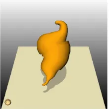
The volume visualization allows exploring the Infract itself as 3D model or with added MR image slices in all three anatomical planes. Furthermore it is possible to explore the brain from each point of view. Anatomical planes in addition to a slightly transparent Infract model provide the user with an excellent impression about the patient’s pathological state. To get an assumption about the Infract dimensions, the visualization displays the size in milli meters.The volume calculation result depends on the 3D model. Thus, to get an accurate rendering and volume calculation result, the Infract should be segmented on all three anatomical planes. Fig.8 shows the 3D visualization of the segmented region.
Fig.8 3D View of segmented Infract MRI Images
IV. CONCLUSION
Medical image segmentation is a very important technique for detecting abnormality in medical images. Segmentation is a technique which reduces the complexity in the medical images and makes the analysis easier and meaningful to understand. Region growing segmentation is a simple method which extracts the region of interest exactly. By using volume visualization technique we can easily locate the small structure of 3D volume. Through this direct volume rendering technique small structure can visible easily and extract the true information separately. Further this work can be extended for the detection, analysis and 3D visualization of tumor and haemorrhage in brain MRI Images.
REFERENCE
[1]Dzung L. Pham, ChenyangXu, and Jerry L. Princ,”Current Methods In Medical Image Segmentation,” Department of Electrical and Computer Engineering, The Johns Hopkins University,Annu. Rev. Bied. Eng. 2000. 02:315–37.
[2]Shapiro, Linda G and stockman, George C. Computer Vision, Prentice hall. ISBN 0-13-030796-3, 2002.
[3]TraneosZuva, Oludayo O, Olugbara, Sunday O. Ojo and Seleman M Ngwira,“Image Segmentation, Available Techniques, Developments and Open Issues,”Canadian Journal on Image Processing and Computer Vision Vol. 2, No. 3, March 2011
[4]KritSomkantha,Umpon,,andSansaneeAuephanwiriyakul,”Boundary Detection in Medical Images Using Edge Following Algorithm Based on Intensity Gradient and Texture Gradient Features”ieee transactions on biomedical engineering, vol. 58, no. 3, March 2011.
© 2013, IJCSMC All Rights Reserved
114
Volume Estimation in Ultrasound Images” IEEE transactions on biomedical engineering, vol. 57, no. 6, June 2010.[7] Chuan-Yu Chang and Yong-Cheng Hong Pau-Choo Chung, Chin-Hsiao “A Neural Network for Thyroid Segmentation and Volume Estimation in CT Images” IEEE computational intelligence magazine, November 2011.
[8]HodgsonR M, BaileyD G, NaylorM J ,and McNeilS J “Properties, implementations and applications of rankfilter” Butterworth & Co (Publishers) Ltd, vol3 no I February 1985.