University of Windsor University of Windsor
Scholarship at UWindsor
Scholarship at UWindsor
Electronic Theses and Dissertations Theses, Dissertations, and Major Papers
2010
Development and application of techniques for the acquisition of
Development and application of techniques for the acquisition of
ultra-wideline solid-state NMR spectra
ultra-wideline solid-state NMR spectra
Alan W. MacGregor
University of Windsor
Follow this and additional works at: https://scholar.uwindsor.ca/etd
Recommended Citation Recommended Citation
MacGregor, Alan W., "Development and application of techniques for the acquisition of ultra-wideline solid-state NMR spectra" (2010). Electronic Theses and Dissertations. 7950.
https://scholar.uwindsor.ca/etd/7950
Development and Application of Techniques for the Acquisition of Ultra-Wideline Solid-State NMR Spectra
by
Alan W. MacGregor
A Thesis
Submitted to the Faculty of Graduate Studies and Research through Chemistry and Biochemistry
in Partial Fulfillment of the Requirements for the Degree of Master of Science at the
University of Windsor
Windsor, Ontario, Canada 2010
1*1
Library and Archives CanadaPublished Heritage Branch
395 Wellington Street OttawaON K1A0N4 Canada
Bibliotheque et Archives Canada
Direction du
Patrimoine de I'edition
395, rue Wellington OttawaONK1A0N4 Canada
Your file Votre reference ISBN: 978-0-494-62748-8 Our file Notre reference ISBN: 978-0-494-62748-8
NOTICE: AVIS:
The author has granted a
non-exclusive license allowing Library and Archives Canada to reproduce, publish, archive, preserve, conserve, communicate to the public by
telecommunication or on the Internet, loan, distribute and sell theses
worldwide, for commercial or non-commercial purposes, in microform, paper, electronic and/or any other formats.
L'auteur a accorde une licence non exclusive permettant a la Bibliotheque et Archives Canada de reproduire, publier, archiver, sauvegarder, conserver, transmettre au public par telecommunication ou par I'lnternet, prefer, distribuer et vendre des theses partout dans le monde, a des fins commerciales ou autres, sur support microforme, papier, electronique et/ou autres formats.
The author retains copyright ownership and moral rights in this thesis. Neither the thesis nor substantial extracts from it may be printed or otherwise reproduced without the author's permission.
L'auteur conserve la propriete du droit d'auteur et des droits moraux qui protege cette these. Ni la these ni des extraits substantiels de celle-ci ne doivent etre imprimes ou autrement
reproduits sans son autorisation.
In compliance with the Canadian Privacy Act some supporting forms may have been removed from this thesis.
Conformement a la loi canadienne sur la protection de la vie privee, quelques
formulaires secondaires ont ete enleves de cette these.
While these forms may be included in the document page count, their removal does not represent any loss of content from the thesis.
Bien que ces formulaires aient inclus dans la pagination, il n'y aura aucun contenu manquant.
• + •
Declaration of Co-Authorship / Previous Publication
I. Co-Authorship Declaration
I acknowledge that Chapter 2 of this thesis has been submitted for publication. I
also acknowledge that my supervisor, Dr. Robert W. Schurko, has provided guidance
throughout the whole of this work and has edited this thesis.
The lead (II) thiolates discussed in Chapter 3 were synthesized and provided by
the research group of Dr. Glen Briand (Mt. Allison), and the group 13 guanidinate
compounds discussed in Chapter 4 were prepared and provided by the research group of
Dr. Sean T. Barry (Carleton). Victor Terskikh at the National Ultra-high Field NMR
facility for Solids is acknowledged for providing the data acquired at 21.1 T in Chapter 4.
Senior members our lab group trained me in the use of our NMR spectrometer,
and supervised my usage of it. The main contributors in this regard were Dr. Luke A.
O'Dell for the work discussed in Chapter 2, and Mr. Aaron J. Rossini for the work
discussed in Chapters 3 and 4. Dr. Joel A. Tang also provided supervision for the work
discussed in Chapter 4.
I am aware of the University of Windsor Senate Policy on Authorship and I
certify that I have properly acknowledged the contribution of other researchers to my
thesis, and have obtained written permission from each of the co-author(s) to include the
above material(s) in my thesis. I certify that, with the above qualification, this thesis, and
II. Declaration of Previous Publication
This thesis includes 1 original paper that has been previously published/submitted for
publication in peer reviewed journals, as follows:
Chapter 2: MacGregor, A. W.; O'Dell, L.A.; Schurko, R. W. New Acquisition Methods for
the Acquisition of Broad Solid-state NMR Spectra ofSpin-1/2 Nuclides. Phys. Chem.
Chem. Phys., Submitted for publication, April 2010.
I certify that I have obtained a written permission from the copyright owner(s) to
include the above published material(s) in my thesis. I certify that the above material
describes work completed during my registration as graduate student at the University of
Windsor.
I declare that, to the best of my knowledge, my thesis does not infringe upon
anyone's copyright nor violate any proprietary rights and that any ideas, techniques,
quotations, or any other material from the work of other people included in my thesis,
published or otherwise, are fully acknowledged in accordance with the standard
referencing practices. Furthermore, to the extent that I have included copyrighted
material that surpasses the bounds of fair dealing within the meaning of the Canada
Copyright Act, I certify that I have obtained a written permission from the copyright
owner(s) to include such material(s) in my thesis.
I declare that this is a true copy of my thesis, including any final revisions, as
Abstract
Wideline and ultra-wideline (UW) (i.e., > 250 kHz broad) solid-state NMR
(SSNMR) spectra are acquired for a wide variety of nuclei (i.e., 119Sn, 195Pt, l99Hg, 207Pb,
27A1,71Ga) using multiple acquisition techniques. The WURST-CPMG pulse sequence is
shown to acquire such patterns efficiently and without the need for piecewise acquisition.
Preliminary investigations into the use of optimal control theory (OCT) are also carried
out, and show promise for future studies. Additionally, a series of Pb(II) thiolate species,
which exhibit unique Pb bonding environments, are characterized with 207Pb SSNMR.
Density Functional Theory (DFT) calculations are performed to determine chemical
shielding (CS) tensor orientations within the molecular frame, which are then correlated
to the experimental spectra. Finally, several group 13 guanidinates (guan =
MeN-C(N'Pr2)-NMe) are studied via 27A1 and 71Ga SSNMR at 9.4 T and 21.1 T, and
experimental data is complemented with ab initio calculations, and the CS and electric
field gradient tensor parameters are found to have a great dependence on metal
Acknowledgements
First and foremost, I must thank my supervisor Dr. Rob Schurko for accepting me
into his research group, and for continually providing guidance along the way. Studying
solid-state NMR is not an easy undertaking, but Rob did his best to help me out from my
first day at the office, and for that I am grateful.
Thanks also to Dr. T. B. Carmichael and Dr. C Ezeife. for being on my M.Sc.
thesis committee, and agreeing to read through this work. Dr. P. J. Dutton is thanked for
chairing my defense.
I also must acknowledge all the members of the Schurko group: Dr. Kris Harris,
Aaron Rossini, Hiyam Hamaed, Bryan Lucier, Alex Reidel, Chris Mireault and Tatjana
Milovic, as well as former members Dr. Luke O'Dell, Dr. Joel Tang and Marcel
Hildebrand; thanks for all of your help, and for making the office an enjoyable place to
be.
Since I have arrived in Windsor, I have had the opportunity to meet lots of new
friends and foster relationships that I hope will last a long time. Thanks for all of the
great times, you know who you are. To Ben Cooper and Chris Guignion; thanks for
being a great roommate and generous landlord, respectively, and for putting up with me
over the past 2 years.
Finally, thanks to my parents, Byron and Helen, for your constant emotional (and
sometimes financial) support, and to my siblings, Andrew and Krista, for the late night
Contents
Declaration of Co-Authorship / Previous Publication iii
Abstract vi
Acknowledgements vii
List of Tables xii
List of Figures xiv
List of Abbreviations xviii
List of Symbols xx
1. Introduction 1
1.1 NMR Spectroscopy 1
1.2 NMR Interactions 1
1.2.1. General Considerations 2
1.2.2. The Zeeman Interaction 3
1.2.3. Response to Radiofrequency Pulses 4
1.2.4. Relaxation Processes 6
1.2.5. Chemical Shielding 7
1.2.6. The Quadrupolar Interaction 11
1.2.7. Euler Angles 13
1.2.8. Dipolar and Scalar coupling 15
1.3 Acquisition and Enhancement Techniques 16
1.3.1. Frequency-stepped NMR 16
1.3.2. Magic Angle Spinning 17
1.3.3. The Carr-Purcell Meiboom-Gill Pulse Sequence 18
1.3.4. Wideband Uniform-Rate Smooth Truncation
1.3.5. Cross Polarization 21
1.3.6.0ptimal Control Theory 22
1.4. Context of Research 23
Bibliography 25
2. New Methods for the Acquisition of Static CSA Patterns from
Spin-1/2 Nuclides 30
2.1. Introduction 30
2.2. Experimental Details 35
2.3. Results and Discussion 39
2.3.1. 119SnNMR 39
2.3.2. 207Pb NMR 43
2.3.3. 199HgNMR 46
2.3.4. 195PtNMR 48
2.3.5. 119Sn Ultra-Wideline SSNMR using pulses designed
with Optimal Control Theory 50
2.4. Conclusions 56
Bibliography 58
3. 207Pb SSNMR Spectroscopic Investigations of Pb(II) Thiolates 66
3.1. Introduction 66
3.2. Experimental Details 70
3.2.1. Solid-state 207Pb Spectroscopy 70
3.2.2. Density Functional Theory Calculations 71
3.3. Results and Discussion 71
3.3.1. Solid-state 207Pb Spectroscopy 71
3.3.2. Calculation of Lead Nuclear Shielding (NS) Tensors
3.4. Conclusions 93
Bibliography 95
4. Solid-state NMR Investigations of Metal Guanidinates 101
4.1. Introduction 101
4.2. Experimental Details 104
4.3. Results and Discussion 106
4.3.1. 27A1 and 7,Ga Solid-state NMR Spectroscopy 106
4.3.2. Ab initio Calculations of Nuclear Shielding and
Electric Field Gradient Tensor Parameters 119
4.3.3. Nuclear Shielding (NS) and Electric Field
Gradient (EFG) Tensor Orientations 121
4.4. Conclusions 123
Bibliography 126
5. General Conclusions and Future Work 130
Appendices
A. Supporting Information - New Methods for the Acquisition of
Static CSA Patterns from Spin-1/2 Nuclides 132
A.l. Supporting Experimental Information 132
A. 1.1. Experimental Parameters for S SNMR Experiments... 132
A. 1.2. Experimental Parameters for SSNMR Experiments
using pulses generated with optimal control theory... 134
A.1.3. Supplementary SSNMR Spectra 135 A. 1.4. Explanation for the estimated time required to
acquire the 195Pt CPMG SSNMR spectrum of
Bibliography 138
B. Supporting Information - 207Pb SSNMR Spectroscopic
Investigations of Pb(II) Thiolates 139
B.l. Supporting Experimental Information 139
B. 1.1. Experimental Parameters for 207Pb SSNMR
Experiments 139
B.l.2. DFT calculations of 3IP Shielding Parameters
for Compound 4 140
B.l.3. Supplementary SSNMR Spectra 141
B.1.4. Powder X-Ray Diffraction of Compound 4 142
C. Supporting Information - Solid-state NMR Investigations of Metal
Guanidinates 143
C.l. Supporting Experimental Information 143
C.l.l. Selected SSNMR Experimental Parameters 143
List of Tables
Table 2.1. Comparison of total experimental times for CPMG, CP/CPMG
and WURST-CPMG experiments 42
Table 2.2. Comparison of experimental chemical shift parameters with
values reported in the literature 42
Table 2.3. Comparison of selected 119Sn SSNMR spectral parameters 55
Table 3.1. Experimental 207Pb Chemical Shift Parameters 73 Table 3.2. Comparison of experimental times (hours) between CP/CPMG
and WURST-CPMG 76
Table 3.3. Theoretical and Experimental 207Pb NS and CS tensor
parameters 86
Table 3.4. Angles describing the orientation of NS tensor components with
respect to key structural and symmetry elements 89
Table 4.1. Experimental and theoretical 27A1 and 71Ga EFG and CS tensor
parameters 108
Table 4.2. 27A1 EFG Parameters of previously reported compounds 111
Table Al. Experimental parameters for WURST-CPMG NMR
experiments 132
Table A2. Experimental parameters for ' l9Sn CPMG NMR spectrum of
SnO 133
Table A3. Experimental parameters for the 207Pb CP/CPMG NMR
Table A4. Experimental details for 119Sn NMR spectra acquired with
pulses generated using OCT 134
Table Bl. CP/CPMG experimental parameters 139
Table B2. WURST-QCPMG experimental parameters 140
Table B3. Calculated 31P NS tensor components for the phosphorous
nuclei in 4 140
Table CI. 9.4 T 27A1 and 71Ga static experimental parameters 143
List Of Figures
Figure 1.1. Depiction of the distribution of resonances arising from multiple
CS tensor orientations 10
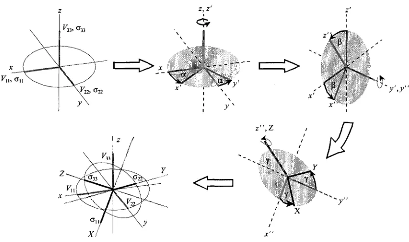
Figure 1.2. Diagram depicting the Euler angle convention used herein to describe the relative orientation of the CS and EFG tensors. Appended from
Tang (2008) 14
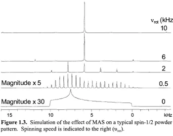
Figure 1.3. Simulation of the effect of MAS on a typical spin-1/2 powder
pattern. Spinning speed is indicated to the right (urot) 18
Figure 1.4. A block diagram of the Carr-Purcell Meiboom-Gill (CPMG)
pulse sequence 19
Figure 1.5. A free induction decay composed of multiple spin echoes 19
Figure 1.6. A comparison of the appearance of SSNMR spectra resulting from Fourier transformation of a single spin echo (top) and multiple spin
echoes (bottom) 20
Figure 1.7. A block diagram of the wideband uniform-rate smooth truncation
(WURST)-CPMG pulse sequence 21
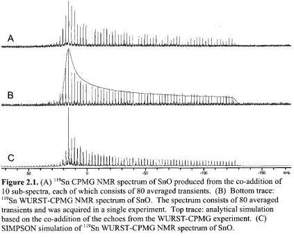
Figure 2.1. (A) ll9Sn CPMG NMR spectrum of SnO produced from the co-addition of 10 sub-spectra, each of which consists of 80 averaged
transients. (B) Bottom trace: 1,9Sn WURST-CPMG NMR spectrum of SnO. The spectrum consists of 80 averaged transients and was acquired in a single experiment. Top trace: analytical simulation based on the co-addition of the echoes from the WURST-CPMG experiment. (C) SIMPSON simulation of
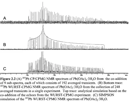
Figure 2.2. (A) 207Pb CP/CPMG NMR spectrum of Pb(OAc)2-3H20 from the co-addition of 9 sub-spectra, each of which consists of 192 averaged
transients. (B) Bottom trace: 207Pb WURST-CPMG NMR spectrum of Pb(OAc)2-3H20 from the collection of 248 averaged transients in a single experiment. Top trace: analytical simulation based on the co-addition of the echoes from the WURST-CPMG experiment. (C) SIMPSON simulation of
the 207Pb WURST-CPMG NMR spectrum of Pb(OAc)2-3H20 45
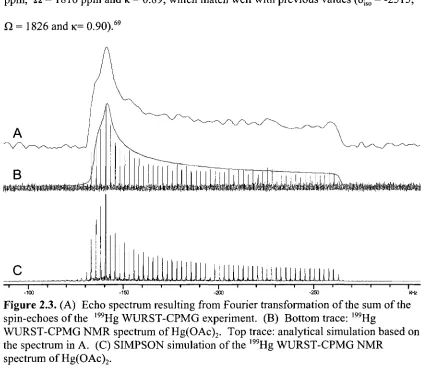
Figure 2.3. (A) Echo spectrum resulting from Fourier transformation of the sum of the spin-echoes of the l99Hg WURST-CPMG experiment. (B) Bottom trace: 199Hg WURST-CPMG NMR spectrum of Hg(OAc)2. Top trace:
analytical simulation based on the spectrum in A. (C) SIMPSON simulation
of the 199Hg WURST-CPMG NMR spectrum of Hg(OAc)2 48
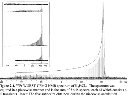
Figure 2.4.195Pt WURST-CPMG NMR spectrum of K2PtCl4. The spectrum was acquired in a piecewise manner and is the sum of 5 sub-spectra, each of which consists of 40 transients. Inset: The five subpectra obtained during the
piecewise acquisition 50
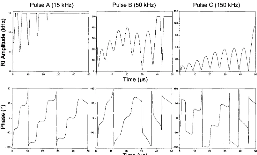
Figure 2.5. Visual descriptions of the rf amplitude (top) and phase (bottom) of
the pulses generated using OCT which were employed for this work 51
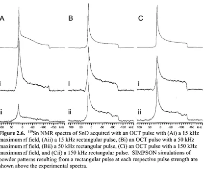
Figure 2.6. "9Sn NMR spectra of SnO acquired with an OCT pulse with (Ai) a 15 kHz maximum rf field, (Aii) a 15 kHz rectangular pulse, (Bi) an OCT pulse with a 50 kHz maximum rf field, (Bii) a 50 kHz rectangular pulse, (Ci) an OCT pulse with a 150 kHz maximum rf field, and (Cii) a 150 kHz
rectangular pulse. SIMPSON simulations of powder patterns resulting from a rectangular pulse at each respective pulse strength are shown above the
experimental spectra 54
Figure 3.1. (A) 'H - 207Pb CP/CPMG and (B) WURST-QCPMG 207Pb NMR
spectra of [(PhS)3Pb][As Ph3], 1, with WSolids simulation (top trace) 74
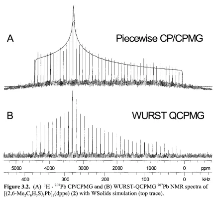
Figure 3.2. (A) 'H - 207Pb CP/CPMG and (B) WURST-QCPMG 207Pb NMR spectra of [(2,6-Me2C6H3S)2Pb]2(dppe) (2) with WSolids simulation (top
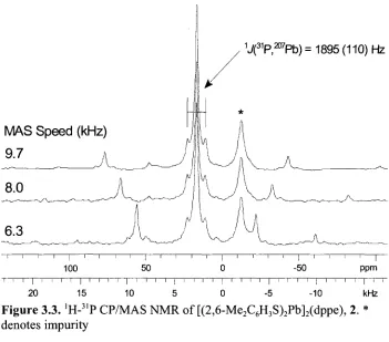
Figure 3.3. 'H-31P CP/MAS NMR of [(2,6-Me2C6H3S)2Pb]2(dppe), 2. *
denotes impurity 77
Figure 3.4. (A) 'H - 207Pb CP/CPMG and (B) WURST-QCPMG spectra of
[(2,6- Me2C6H3S)2Pb]2(tmeda), 3, with WSolids simulation (top trace) 79
Figure 3.5. (A) 'H - 207Pb CP/CPMG and (B) WURST-QCPMG spectrum of
[(2,6- Me2C6H3S)2Pb]3(dmpe), 4, with WSolids simulation 8 1
Figure 3.6. "H-31P CP/MAS NMR spectra of [(2,6-Me2C6H3S)2Pb]3(dmpe), 4.
* denotes impurity 83
Figure 3.7. (A) "Top" view and (B) "side" view of NS tensor orientation of
1, (C) NS tensor orientation of 2, and (D) 2 shown as dimer 88
Figure 3.8. NS Tensor orientations of (A) Site land (B) Site 2 of 3, and (C) Site 1 and (D) Site 2 of 4. The full molecules are shown in the insets. Note
that Site 3 of 4 has been omitted due to its similarity to Site 2 91
Figure 4.1: (A)27A1 MAS and (B) 27A1 static SSNMR spectra of Al(guan)3
acquired at 9.4 T (top) and 21.1 T (bottom), with analytical simulations 109
Figure 4.2: (A)71Ga MAS SSNMR spectrum of Ga(guan)3 acquired at 21.1 T. (B) 71Ga static SSNMR spectra of Gaguan)3 acquired at 9.4 T (top) and 21.1 T
(bottom) with analytical simulations 112
Figure 4.3: (A)27A1 MAS and (B) 27A1 static SSNMR spectra of Al(guan)2Cl
acquired at 9.4 T (top) and 21.1 T (bottom) with analytical simulations 114
Figure 4.4: Bottom trace: Al static SSNMR spectrum of Al(guan)2NMe2 acquired at 9.4 T. Middle trace: Analytical simulation including CSA. Top Trace: Analytical simulation without CSA included. Note: * denotes
impurity 116
Figure 4.5. NS (top) and EFG (bottom) tensor orientations of (A) Al(guan)3,
Figure Al. Echo spectra produced from Fourier transformation of the
time-domain sum of the spin-echoes of WURST-CPMG experiments. Shown are (A) the 1,9Sn NMR spectrum of SnO , (B) the 207Pb NMR spectrum of
Pb(OAc)2 and (C) the 195Pt NMR spectrum of K2PtCl4 135
Figure A2. (A) 207Pb CP/CPMG NMR spectrum of Pb(OAc)2-3H20 prior to recrystallization of the sample. Shown is the co-addition of 9 sub-spectra, each of which consist of 64 averaged transients. (B) 207Pb WURST-CPMG NMR spectrum of Pb(OAc)2-3H20 prior to recrystallization of the sample. The spectrum consists of 198 averaged transients and was acquired in a single experiment. The large "lump" at the high-frequency end of the spectra is not present after recrystallization, indicating that the sample used initially was
partially dehydrated 136
Figure Bl. SIMPSON simulations (top traces) and experimental 207Pb CP/MAS spectra (bottom traces) of 1 at spinning speeds of (A) 9.7 kHz, (B)
8.0 kHz and (C) 6.3 kHz 141
List of Abbreviations
ADF B3LYP CP CPMG CS CSA CT DFT DMPE DPPE EFG Et FID FT guan 'Pr MAS Me NMR NS OCT o.d. ppmAmsterdam Density Functional
Becke's three parameter hybrid fimcti functional of Lee, Yang and Parr
cross-polarization
Carr-Purcell Meiboom-Gill
chemical shielding
chemical shielding anisotropy
central transition
density functional theory
dimethylphosphinoethane
diphenylphosphinoethane
electric field gradient
ethyl group, CH2CH3~
free induction decay
Fourier transform
guanidinate group = (Me2N-C(N'Pr)2)
isopropyl group, -CH2(CH3)2
magic angle spinning
methyl group, CH3~
nuclear magnetic resonance
nuclear shielding
optimal control theory
Outer diameter
QCPMG
rf
RHF
S/N
SSNMR
TMEDA
WURST
ZORA
quadrupolar Carr-Purcell Meiboom-Gill
radiofrequency
restricted Hartree-Fock
signal to noise
solid state nuclear magnetic resonance
tetramethylethylenediamine, C6H16N2
wideband uniform-rate smooth truncation pulse sequence
List of Symbols
a, P, y Euler angles
a, P nuclear spin state labels
y gyromagnetic ratio
8U, 522, 533 principal components of the chemical shift tensor
5iso isotropic chemical shift
r\Q asymmetry of the electric field gradient tensor
K skew of the chemical shielding tensor
on, a22, a3 3 principal components of the chemical shielding tensor
9 angle describing the orientation of spin magnetization from the external magnetic field
xp length of applied pulse
x,, x2, x3, T4 inter-pulse delays
ji nuclear spin magnetic m o m e n t
co0 Larmor frequency (rad s1)
coj nutation frequency (rad s"1)
©Q quadrupolar frequency (rads1)
t>0 Larmor frequency (Hz)
u, nutation frequency (Hz)
urf frequency of a radiofrequency pulse (Hz)
t»Q quadrupolar frequency (Hz)
Q. span of the chemical shielding tensor
B0 external magnetic field strength
B, applied magnetic field strength
Planck's constant
plack's constant, divided by 2n
Quadrupolar Hamiltonian
nuclear spin
vector describing net magnetization
quadrupole moment
spin-lattice relaxation time constant
transverse relaxation time constant
Chapter 1
Introduction
1.1 NMR Spectroscopy
Nuclear magnetic resonance was first reported simultaneously by the research
groups of Bloch and Purcell in 1946.12 In the decades that have passed since then, NMR
has become an indispensable tool for the characterization of structure and dynamics at
the molecular level in virtually every area of chemical research. The advent of large,
superconducting magnets, and advancements in hardware and software, have made it
possible to conduct experiments which would have been considered virtually impossible
in the past. The field of solid-state nuclear magnetic resonance (SSNMR) has greatly
benefited from these advancements and has become an area of intense research over the
past several decades.3"8 SSNMR has been employed to study a wide variety of molecules
and materials, including proteins,9 polymers,10 inorganic materials"12 and clays and
minerals.13 In this section, the interactions that give rise to SSNMR spectra are
examined, along with a variety of techniques employed in their acquisition.
1.2 NMR Interactions
In this section, we discuss the physical interactions which give rise to the NMR
phenomenon, and the informative fine structure observed in NMR spectra, as well as the
section meant to be considered a complete treatment of these interactions; rather, the
reader is presented with a rudimentary background of the basis for NMR. More
comprehensive descriptions of the interactions which govern NMR are elegantly
described elsewhere.14"18
1.2.1 General Considerations
The nuclear spin, I, is an intrinsic property of the nucleus and gives rise to spin
angular momentum, described by a vector I, and a nuclear magnetic moment, p, which
are related through an inherent nuclear property called the gyromagnetic ratio:18
H = yl (1)
The spin can interact with a static magnetic field, B0, or an oscillating magnetic field
induced by a radiofrequency (rf) pulse, B,. In the presence of a magnetic field, \i will
begin to precess about the axis of the field, B0, which is conventionally taken to be along
the z-axis.
Spin is quantized such that the z-component of I, Iz = mh, where m= I, I - 1, ...,
-7, and represents the spin state of a given spin. The energy of interaction between |u, and
B0 is given as:
E = ~f* ' Bo = -V-A = -mfiyB0 (2)
The rate of precession, co0, is known as the Larmor frequency and is dependent upon the
co0=-y£0 (3)
Note, that Levitt's convention for the sign of the gyromagnetic ratio is applied here, and
that:
co0 = 2TW0 (4)
1.2.2 The Zeeman Interaction
The Zeeman interaction is the fundamental interaction that gives rise to the NMR
phenomenon. Outside of a magnetic field, the spin states of a nucleus are degenerate.
This degeneracy is removed when a magnetic field is introduced, resulting in 2(1+ 1/2)
quantized spin states, which differ in energy by AE = Ao0.18 The simplest case to
consider is that of a nucleus with / = 1/2, which has two possible spin states, m, = +1/2
and m, = -1/2, or a and P, respectively.20 An individual spin can be visualized as a
magnetic moment which precesses about the external magnetic field, B0 (z-axis) at the
Larmor frequency. The quantization of spin along B0 dictates that there are a limited
number of possible states for precession; in the case of the spin-1/2 nucleus, there are two
possibilities: a and p. Note that for a nucleus with a positive gyromagnetic ratio, the a
spin state is lower in energy than the p spin state.
In treating the NMR experiment classically, a large number of spins (i.e., an
ensemble) is normally considered. The spin states are populated according to the
-^ = e kT (5)
where Na and TVp represent the populations of the a and P spin states, k is the Boltzmann
constant and T'\s the temperature (in K). The Boltzmann distribution dictates that there
are more spins in the lower energy a state; therefore, the net magnetization of the
ensemble is a vector, M, that is parallel to B0 The population difference between the a
and P states can be increased by increasing B0 or lowering the temperature. Both of these
actions result in increased sensitivity, as the NMR signal can be shown to be proportional
to:
Nfh2B% ital
AkT
where N describes the natural abundance of the isotope under study, or the number of
spins present.
1.2.3 Response to radiofrequency pulses
In an NMR experiment, the sample is placed in a coil (normally on a probe head)
which is inserted into the large external magnetic field. When an oscillating (AC) current
is passed through the coil, an oscillating magnetic field, B,, is produced along the axis of
the coil, which is directed perpendicular to B0 (note, that this is the case for a solenoid
magnetic moments precess about B0 at the Larmor frequency, co0. If B, is applied such
that it oscillates at a transmitter frequency of corf, and corf ~ co0 then it is possible to "tip"
the net magnetization away from B0. The reason for this is best considered using the
rotating frame, which is a frame rotating at corf (corf is the transmitter frequency in this
case). If corf = co0, corf is said to be "on resonance," and the individual magnetic moments
appear to be stationary in the rotating frame. Since the magnetic moments are stationary,
it is as if B0 is absent, and the stationary B, field is present. The magnetization vector,
M, begins to precess about the stationary B, field at a frequency of co, = -yfi,. which
known as the nutation frequency, and depends on the magnitude of Bx produced by the rf
coil.
The introduction of a pulse with a field magnitude of 5, for a time period xp "tips"
M, by an angle 9 away from the z-axis:
0 = X p • yB } = -T p • CO, (7)
Hence, to tip the magnetization into the xy-plane of the rotating frame, xp is set such that
0 = JI/2. The phase of the pulse dictates the orientation of the magnetization vector
within this plane, following the right hand rule. For example, a pulse applied along the
x-axis will direct the magnetization vector to the -y-x-axis. Once the pulse is turned off, B,
is removed and the magnetic moments once again precess about B0 at the Larmor
frequency. As M precesses about B0, it induces a current in the sample coil, which is
recorded and digitized as the free induction decay (FID), which in turn is used to generate
If corf * co0, then the magnetic moments no longer appear stationary in the rotating
frame, and B0 no longer appears to be absent, but rather, is attenuated by a factor of
(1 -cOrt/co,)). The result is an effective magnetic field, Beff:
Beff = (B0 - <*Ji)-Z + BAX (8)
If (B0 - (Brf/y) » Beff, then corf is considered to be "off resonance", and Beffis ineffective at
tipping the magnetization into the xy-plane (where the signal is detected). In the case of
ultra-wideline (UW, i.e., > 250 kHz broad) SSNMR spectra, resonances may be
separated by hundreds of kHz, or more often, may spread over hundreds of kHz or tens of
MHz, which makes it difficult (if not impossible) to acquire the entire powder pattern in a
single experiment with traditional acquisition methods. Hence, specialized techniques
are employed in such cases (vide infra).
1.2.4 Relaxation Processes
There are two types of nuclear spin relaxation that are essential for the NMR
phenomenon. Spin-lattice relaxation (or longitudinal relaxation) is described by the Tx
relaxation time constant, and provides a measure of the time required for the
magnetization to return to equilibrium along B0. The Tx is measured experimentally with
the inversion recovery pulse sequence.21 Spin-spin relaxation (or transverse relaxation) is
described by the T2 relaxation time constant, and is a measure of the time required for
complete dephasing of the magnetization in the xy-plane. The T2 time constant is most
infra). All of the NMR interactions that are discussed in the next few sections can
contribute to mechanisms which induce nuclear spin relaxation; a detailed discussion of
relaxation mechanisms is beyond the scope of this introduction, and is well covered
elsewhere.24
1.2.5 Chemical Shielding
The local magnetic field at a nucleus is dependent upon the external magnetic
field, B0, and the induced magnetic field at the nucleus Bind:
Bloc = Bind + B0 (9)
The Bind varies between different nuclei, due to the induced circulation of electrons
within the surrounding molecular orbitals. The effect that Bind has on B,oc is referred to as
the chemical shielding or nuclear magnetic shielding of the nucleus; if Bjnd is aligned in
the same direction as B0, causing Bloca| to increase, it is considered deshielding. If Bind is
aligned in the opposite direction to B0, causing B|oca] to decrease, it is considered
shielding. The chemical shielding is reported in ppm with respect to the bare nucleus,
which is assigned a value of 0 ppm. In practice, one cannot experimentally measure the
chemical shielding of a bare nucleus for comparison to the chemical shielding of a
nucleus in a sample of interest. In experimental NMR spectra, the chemical shift (CS) is
measured which is the shielding of the nucleus, a, with respect to that of a reference
5 = °ref ~ °
1 " O. ref " Cref ~ ° (10)
An arbitrary standard reference compound is chosen for each nucleus, and all chemical
shifts are reported with respect to this reference.
The chemical shielding or nuclear shielding (NS) interaction can be described by
a second rank tensor (3 x 3 matrix):
a =
CT a a
xx xy xz
a a a
yx yy yz
. a a a , \ zx zy zz )
(11)
This tensor is not traceless and is anti-symmetric. It can, however, be broken down into
symmetric and anti-symmetric components. Only the former contributes to the
observable chemical shifts, and it is represented by a symmetric second rank tensor
which, in its own principal axis system (PAS), is written as:
o„ 0 0
0 G22 0
0 0 o 33
(12)
a,,, a22, and a33 are referred to as the principal components of the NS tensor, and are
arranged such that a,, < a22 £ c33 {i.e., a,, is the least shielded component, and a33 is the
as:
8 =
f
s
uo o
x0 522 0
0 0 5
(13)
' 3 3 ;
where 8,, > 522 > 833.
The CS interaction is orientation dependent or anisotropic, meaning the values of
the tensor components can change depending upon the orientation of the tensor with
respect to B0. There are several conventions available for describing the chemical shift
anisotropy (CSA); herein, the Herzfeld-Berger convention26'27 is used to describe the
CSA. The isotropic shift, 8jso, describes the isotropic or average chemical shift of the
nucleus, and is found at the centre of gravity of the NMR powder patterns of spin-1/2
nuclides:
(511 + 82 2 + 533>
(14)
The span, CI, describes the breadth of the pattern in ppm (often referred to as the
magnitude of the CSA),
^ = 511 " 53 3 (15)
and the skew, K, gives the position of 822 with respect to 8iso, and describes the axial
K = -3 (522 " §iso)
Q 1 < K < -1 (16)
In solution NMR experiments, the CSA is averaged to zero due to the rapid
tumbling of the molecules, and as a result, only the isotropic chemical shift is observed.
In the solid state, where such rapid isotropic motion is absent, the orientation dependence
of the CS tensor can be observed. Most solid state samples, including microcrystalline
and amorphous or disordered solids have a vast number of tensor orientations which are
represented within the bulk sample. The bulk of early SSNMR experiments were
conducted upon microcrystalline powder samples, and as a result, the patterns arising
from the distributions of these shifts are commonly referred to as powder patterns.
Powder patterns represent the weighted sum of all of the resonances arising from
individual CS tensor orientations (Figure 1.1), and its shape can be directly correlated to
the principal components of the CS tensor.
1.2.6 The Quadrupolar Interaction
Nuclei with / > V2 are quadrupolar nuclei, and have an asymmetric distribution of
the charge in the nucleus. This asymmetry in charge distribution is described by a scalar
parameter known as the nuclear quadrupole moment (NQM or Q) which has dimensions
of m2 (or barn = 1028 m2). The nuclear quadrupole moment interacts with the electric
field gradients (EFGs) in the molecule. The EFG at a nuclear site is described by a
symmetric, traceless tensor, with principal components arranged such that \vu I < \V2 2 ' 2:
slK, 3 3 '
V EFG
V
n0 0
x0 v22 0
O O F . 33
(17)
The quadrupolar interaction (QI) is typically described by the quadrupolar
coupling constant, CQ,
C eQK 33 (18)
and the asymmetry parameter, r|Q,
%
V -V
y 11 *22 0 < r,Q < 1 (19)
33
ground-state electronic environment at the quadrupolar nucleus, and it increases in
magnitude as the degree of spherical symmetry decreases.13 nQ, which is a dimensionless
parameter, is a measure of the axial symmetry of the EFG tensor; for an EFG tensor with
a high degree of axial symmetry, r|Qis near zero or one. The magnitude of the QI is also
sometimes reported as the quadrupolar frequency, coQ:
co = 3eQ F33 = ^— (20)
Q 21(21-I)A 33 21(21-I)A y '
The QI is described by the quadrupolar Hamiltonian, which is comprised of first
and second order terms:
Under the high-field approximation, where co0 » coQ, it is possible to treat the QI as a
perturbation on the Zeeman Hamiltonian. To first order, the perturbation is given by:28
° W i = - ~ 0 ~ 2w)(3cos29 - 1 + r\Qsin2Qcos2q>) (22)
where 8 and 9 are polar angles which describe the relative orientation of B0 in the PAS of
the EFG.
AE does not change for the +1/2 « -1/2 transition (the central transition, CT)
since the shift in energy is the same for both spin states. The remaining transitions
signal of the satellite transitions to be broadened, typically over many MHz, so they are
often not observed. The first order perturbation is also orientation dependent, and can be
removed through the use of magic angle spinning (MAS) (vide infra). However, the
second order term:28
2
<»+i/2-i/2 = - ^ _ { | s i n26 [ ( , 4 + £)cos2e - B]
12co0 2
rucos2(psin29[(,4 + 5)cos20 + B] . . . .
+ ^Q-[A - (A + 45)cos2e
- (A + 5)cos2(p(cos20 - 1)22]}
where:
A = 24m(m - 1) - 4/(7+1) + 9
and B = -[6m(m - 1) - 21(1 + 1) + 3] <24>
4
The orientation dependence of the second-order QI is such that MAS cannot
completely average the pattern to a single sharp peak; rather, averaging of this interaction
must be accomplished by rotating the sample about two axes simultaneously (e.g., the
double-rotation or DOR technique),29,30 or by selecting multiple-quantum (MQ)
coherences in two-dimensional experiments for which the second-order QI can be
averaged (e.g., dynamic-angle spinning, DAS31"35 and MQ-MAS).36"38
1.2.7 Euler Angles
the appearance of a SSNMR spectrum. To best describe how these two tensors are
aligned with respect to each other, three Euler angles, a, p, and y, are employed. In this
work the Euler angles are defined as follows: the two tensors are aligned so that, in a
standard xyz coordinate system, F33 and a33 are aligned along the z-axis, V22 and o22 are
aligned along thejy-axis, and Vn and a,, are aligned along the jc-axis. First, the CS tensor
is rotated about z by an angle, a. The CS tensor orientation is then taken as the reference
frame, with a33, a22 and a,, becoming x\ y' and z\ respectively. A rotation of the new
frame by an angle p is then made about the /-axis, yielding a third frame of reference
with axes of x", y" and z". A rotation by an angle of y is then made about the z" axis,
resulting in the final orientations of the tensors (Figure 1.2).
C = ^ >
4r
< :
/ X
1.2.8 Dipolar and Scalar Coupling
Dipolar direct spin-spin coupling (or just dipolar coupling) and indirect spin-spin
coupling (or scalar or J) coupling are two-spin NMR interactions. The dipolar
interaction is a through space interaction, described by a traceless second rank tensor.
Secular effects (i.e., frequency shifts) arising from the dipolar interaction are observed
only in the solid state (and some oriented samples); in solution, the rapid isotropic motion
of molecules average dipolar effects to zero.40 For the purposes of this thesis, dipolar
coupling will not be discussed further, as its effects have largely been removed through
the use of decoupling schemes where applicable. It is also noted that the dipolar
interaction is extremely important for the application of cross-polarization NMR
experiments (vide infra). The interested reader may find a thorough description of the
dipolar interaction elsewhere.17
./-coupling occurs between two nuclei, as for dipolar coupling; however,
J-coupling is a through-bond interaction, mediated by electrons within the molecule.41
J-coupling is described by a anti-symmetric tensor with a non-zero trace. The symmetric
portion of the tensor makes both isotropic and anisotropic secular contributions, defined
as the isotropic ./-coupling, J or Jiso, and the J-anisotropy, AJ. While the former is very
commonly observed in both solution- and solid-state NMR spectra, the latter is only
observed in solid-state NMR spectra of nuclei which are ./-coupled to heavy nuclei (or
vice versa).42 In this thesis, we are only concerned with isotropic contributions to
J-couplings. The ./-coupling of a nucleus A of spin / to a nucleus X of spin S results in a
the spectrum of X, with the spacing between the peaks in each spectrum equal to the
value of the Jiso in Hz. Since ./-coupling is a through-bond interaction, it provides
valuable structural information about the bonding/connectivity within a given molecule.41
Except for a few select cases, scalar couplings are not visible in the SSNMR spectra
presented herein, as their relatively small magnitude (hundreds of Hz) are dwarfed by the
broad (tens to hundreds of kHz) powder patterns.
1.3 Acquisition and enhancement techniques
There are a wide variety of techniques that can be employed to enhance the NMR
signal and/or spectral resolution. Some involve specialized hardware, though many are
simply pulse sequences that manipulate the spins in such a way as to increase the
efficiency of the experiment. In this thesis, we are concerned with rapidly acquiring
broad powder patterns with high S/N, and hence, will focus on techniques employed to
that end.
1.3.1 Frequency-stepped NMR
The majority of the spectra in this work are either wideline NMR spectra (i.e.,
broad patterns ranging from ca. 20 to 250 kHz in breadth), or much broader spectra we
designate as ultra-wideline (UW) NMR spectra (i.e. > 250 kHz in breadth).43 UW spectra
exceed the excitation bandwidths of standard, high-power, rectangular pulses; as such,
specialized hardware, pulse sequences or acquisition methodologies must be applied.
done with the "point-by-point" method, where the echo intensity of the free induction
decay was plotted as a function of transmitter frequency44'45 or magnetic field strength.46'4
More recently, it has become common practice to co-add a series of Fourier transformed
spectra, which have been acquired at evenly-spaced transmitter frequencies.48'49 The
acquisition of spectra in much larger frequency increments, combined with spectral
processing prior to co-addition, greatly reduces experimental times and yields spectra
with considerably higher S/N than conventional point-by-point acquisitions. This
technique is known as the variable offset cumulative spectrum (VOCS) method,48 or
simply as a piecewise spectral acquisition. There are also some specialized pulse
sequences which can be utilized to acquire UW NMR spectra in a single experiment, as
well as to acquire extremely broad UW NMR spectra in a piecewise fashion (vide infra).
1.3.2 Magic-Angle Spinning
Magic-angle spinning (MAS) NMR spectroscopy50 is a technique that involves
rotating the sample holder (i.e., rotor) about an axis at an angle of 54.74° from B0, which
averages the CSA to zero (or at least enough that the effects of CSA are negligible, vide
infra). The result is observation of solution-like NMR signals from spin-1/2 nuclei, and
classic second-order quadrupolar lineshapes from quadrupolar nuclei. Hence, MAS
allows for the isotropic shift and EFG tensor parameters to be determined with great
accuracy.
In order for MAS to be most effective, a general rule is that the sample must be
For narrow patterns (i.e., < 15 kHz), this is readily achieved with what have become
standard NMR probes. However, for broad SSNMR spectra (like most of those
contained within this work) limitations on NMR hardware make it difficult to spin the
sample at a high enough rate to average the CSA to zero. Currently, the upper limit on
spinning speed is ca. 70 kHz, which is attainable on specialized SSNMR probes.
Vrot ( ^ Z )
10
Magnitude x 5
Magnitude x 30
~i 1 r
15 10 5 0 kHz
Figure 1.3. Simulation of the effect of MAS on a typical spin-1/2 powder
pattern. Spinning speed is indicated to the right (urot).
1.3.3 The Carr-Purcell Meiboom-Gill (CPMG) Pulse Sequence
The CPMG (Carr-Purcell Meiboom-Gill) pulse sequence,22'23 also called QCPMG
when applied to quadrupolar nuclei,5'"54 has become widely used in the acquisition of
(*/2)x
00y (7l)y
N Figure 1.4. A block diagram of the Carr-Purcell Meiboom-Gill (CPMG) pulse sequence.
The sequence takes advantage of the large T2 relaxation constants exhibited by some
nuclei by continually refocusing the magnetization with a series of n pulses. As a result,
a series of spin-echoes (Figure 1.5) is acquired in a single scan, significantly enhancing
the signal intensity when compared to standard echo experiment,55 in which a single
refocusing pulse is made.
M
r^:;'';il,
:r
l^i
,f)^l;!'if
lti-V!'ifV*:-l;:
;t>
0.005 0.010 0.015 s
Figure 1.5. A free induction decay composed of multiple spin echoes.
This "echo train" can be directly processed by FT, results in a series of spikelets,
spikelets (in Hz) are inversely proportional to the distance between the echoes (in s).
Equivalently, the spin-echoes can be summed to produce an FID comprised of a single
echo, which upon Fourier transformation, takes the form of a normal SSNMR powder
pattern (Figure 1.6).
Figure 1.6. A comparison of a SSNMR spectrum acquired with a conventional echo seqeunce (top) and the CPMG sequence (bottom).
1.3.4. Wideband Uniform-Rate Smooth Truncation (WURST) QCPMG
In 2009, O'Dell et al. developed a new pulse sequence, wideband uniform-rate
smooth truncation (WURST) QCPMG.56 This sequence is a combination of the
(WURST) pulse sequence57'58 and a CPMG-type train of WURST pulses (Figure 1.7).
The use of WURST pulses57 in the acquisition of UW SSNMR spectra was proposed by
Bhattacharyya and Frydman.58 These pulses perform a frequency sweep which generates
a Beff field that sweeps from the +z direction to the -z (or to the xy-plane), which results
in broadband excitation.43 The CPMG-type train of WURST pulses then serves to
compared to a standard echo experiment. This combination of broadband excitation and
signal enhancement has made the WURST-CPMG sequence a valuable tool in the
acquisition of UW SSNMR spectra, permitting acquisition of some spectra in a single
experiment, and increasing the capacity to do UW NMR experiments on extremely broad
patterns. 56,59
WURST
Pulse 1
WURST
Pulse 2
N
Figure 1.7. A block diagram of the wideband uniform-rate smooth truncation (WURST)-CPMG pulse sequence.
1.3.5. Cross Polarization
Cross polarization (CP) is employed in the acquisition of SSNMR spectra of
nuclides which are considered unfavourable for NMR experiments due to a number of
factors, such as low natural abundance and/or large 71, relaxation constants.60"62 Use of
CP involves transferring magnetization from an abundant nucleus, I (i.e., "H or 19F), to
the "dilute" nucleus of interest, S, which can significantly enhance the observed signal,
by a maximum theoretical factor of y/ys.63
A (7i/2)x pulse is first made on the / channel, which is then followed by a lower
power contact pulse that is made on both the / and S channels. To allow the transfer of
magnetization to occur under static conditions, the contact pulses have the same rf field
TiBn = TsBis (25)
The FID is then recorded by observing on the X channel. When MAS is employed, CP
the H-H match is dependent upon the spinning speed. In addition, the efficiency of CP is
decreased as spinning speed increases, because the I-S and I-I dipolar couplings are
averaged when the spinning speed is of comparative magnitude.63 If the combination of
fast MAS and CP is required, it is possible to alleviate these averaging effects with pulse
sequences such as variable-amplitude CP (VACP),64'65 ramped-amplitude CP
(RAMPCP),66 and numerous others.
1.3.6 Optimal Control Theory
The most recent version of SIMPSON (Ver. 2.0)67'68 NMR simulation software
package includes an optimal control theory (OCT) functionality. OCT is capable of
numerically optimizing a large number of variables, which makes it ideal for generating
NMR pulses; one can input the spectral parameters of a given spectrum and generate a
pulse designed specifically for the excitation of that spectrum. OCT studies on the
optimization of NMR pulses have largely focussed on high-resolution solution
experiments,69"71 though recently our group examined the effectiveness of OCT at
generating pulses for use in acquiring the solid-state spectra of quadrupolar nuclei.72
Currently, the development of SSNMR pulses with OCT is still in its infancy and an
in-depth treatment of the theory behind OCT is beyond the scope of this thesis, however it is
1.4. Context of Research
The first research chapter of this thesis, Chapter 2, describes the application of the
WURST-CPMG pulse sequence to various spin-1/2 nuclides (ll9Sn, 207Pb, 199Hg and 195Pt)
in order to examine its effectiveness for acquiring wideline SSNMR patterns.
WURST-CPMG is compared to the CP/WURST-CPMG and WURST-CPMG pulse sequences, and its usefulness in
acquiring ultra-wideline (UW) SSNMR spectra in a piecewise fashion is explored.
Additionally, the potential of pulses generated with SIMPSON 2.067'68 using optimal
control theory (OCT) in acquiring wideline SSNMR spectra is investigated.; pulses
generated with OCT are compared with standard, rectangular pulses at several pulse
powers.
In Chapter 3,207Pb CP/CPMG and WURST-CPMG SSNMR experiments are
carried out in the characterization of a series of (2,6-Me2C6H3S)2Pb adducts which exhibit
unique Pb(II) coordination environments. The 207Pb SSNMR spectra are found to exhibit
extremely large lead CSAs which span several hundred kHz, requiring piecewise
acquisition with CP/CPMG. WURST-CPMG, however, is capable of exciting the entire
powder pattern, eliminating the need for multiple subspectra to be acquired. 207Pb NS
tensor orientations are determined via Amsterdam Density Functional (ADF)
calculations to examine the molecular origins of the experimentally determined CS tensor
parameters.
In Chapter 4, 27A1 and 7lGa solid-state NMR (SSNMR) are employed to examine
a series of metal guanidinate (guan = MeN-C(N'Pr2)-NMe) complexes at 9.4 T and 21.1
associated NMR interaction tensor parameters. The 27A1 CS and EFG tensor parameters
are found to be greatly influenced by site symmetry at the metal centre; in particular, the
magnitude of CQ is determined to be strongly influenced by the nature of the bonding
ligands, in addition to the aluminum coordination number. 7lGa SSNMR experiments
indicate that Ga(guan)3 is less spherically symmetric and exhibits a much broader CSA
than the similar Al analogue.
Initially, the focus of the study presented in Chapter 4 was to develop a
methodology for the analysis of oriented surface-bound aluminum materials, which
would require the application of signal-enhancing UW experiments. However, due to
low sample loading levels and the inability to establish a lower detection limit, we chose
to focus on a fundamental structural characterization of these compounds via 27Al
Bibliography
(1) Bloch, F.; Hansen, W. W.; Packard, M. Phys. Rev. 1946, 69, 127.
(2) Purcell, E. M.; Torrey, H. C ; Pound, R. V. Phys. Rev. 1946, 69, 37-38.
(3) Ashbrook, S. E. Phys. Chem. Chem. Phys. 2009, 11, 6892-6905.
(4) Lesage, A. Phys. Chem. Chem. Phys. 2009, 11, 6876-6891.
(5) Geppi, M.; Borsacchi, S.; Mollica, G.; Veracini, C. A. 2009, 44, 1-89.
(6) Singhal, A. 2009, 49-51, 149-192.
(7) Tishmack, P. A. 2009, 192, 381-435.
(8) Widdifield, C. M.; Chapman, R. P.; Bryce, D. L. 2009, 66, 195-326.
(9) Ketchem, R. R.; Hu, W.; Cross, T. A. Science 1993, 261, 1457-1460.
(10) Schmidt-Rohr, K.; Speiss, H. W. Multidimentsional solid-state NMR and Polymers;
Academic Press: San Diego, 1994.
(11) Fitzgerald, J. J., Ed. Solid-State NMR Spectroscopy of Inorganic Materials; Oxford
University Press: Washington, 1999.
(12) MacKenzie, K. J. D.; Smith, M. E. Multinuclear Solid-state NMR of Inorganic
Materials; Pergamon: New York, 2002; Vol. 6.
(13) Kentgens, A. P. M. Geoderma 1997, 80, 271-306.
(14) Harris, R. K. Nuclear Magnetic Resonance Spectroscopy; Longman Scientific &
Technical: New York, 1986.
(15) Mason, J., Ed. Multinuclear NMR; Plenum Press: New York, 1987.
(16) Slichter, C. P. Principles of Magnetic Resonance; 3rd ed. New York, 1990.
Oxford, 2004.
(18) Levitt, M. Spin Dynamics: Basics of Nuclear Magnetic Resonance; 2nd ed.; John
Wiley and Sons: Chichester, 2008.
(19) Levitt, M. J. Magn. Reson. 1997, 126, 164-182.
(20) Dirac, P. A. M. The Principles of Quantum Mechanics; 4 ed.; Oxford University
Press: Oxford, 1958.
(21) Keeler, J. Understanding NMR Spectroscopy; John Wiley and Sons: Chichester,
2005.
(22) Carr, H. Y.; Purcell, E. M. Physical Review 1954, 94, 630.
(23) Meiboom, S.; Gill, D. Rev. Sci. Inst. 1958, 29, 688-691.
(24) Wasylishen, R. E. In NMR Spectroscopy Techniques; Dybowski, C , Lichter, R. L.,
Eds.; Marcel Dekker, Inc.: New York, 1996, pp 45-85.
(25) Tang, J. A.; Kogut, E.; Norton, D.; Lough, A. J.; McGarvey, B. R.; Fekl, U.;
Schurko, R. W. J. Phys. Chem. B 2009, 113, 3298-3313.
(26) Herzfeld, J.; Berger, A. E. J. Chem. Phys. 1980, 73, 6021-6030.
(27) Harris, R. K.; Becker, E. D.; De Menezes, S. M. C ; Granger, P.; Hoffman, R. E.;
Zilm, K. W. Magn. Reson. Chem. 2008, 46, 582-598.
(28) Autschbach, J.; Zheng, S.; Schurko, R. W. Cone. Magn. Reson 2010, In Press.
(29) Samoson, A.; Lippmaa, E.; Pines, A. Mol. Phys. 1988, 65, 1013-1018.
(30) Wu, Y.; Sun, B. Q.; Pines, A.; Samoson, A.; Lippmaa, E. 1990, 89, 297-309.
(31) Llor, A.; Virlet, J. Chem. Phys. Lett. 1988, 152, 248-253.
1990,86,470-487.
(33) Wu, Y.; Chmelka, B. F.; Pines, A.; Davis, M. E.; Grobet, P. J.; Jacobs, P. A. Nature
1990, 346, 550-552.
(34) Mueller, K. T.; Wooten, E. W.; Pines, A. 1991, 92, 620-627.
(35) Mueller, K. T.; Chingas, G. C ; Pines, A. Rev. Sci. lustrum. 1991, 62, 1445-1452.
(36) Frydman, L.; Harwood, J. S. J. Am. Chem. Soc. 1995, 117, 5367-5368.
(37) Medek, A.; Harwood, J. S.; Frydman, L. J. Am. Chem. Soc. 1995, 117, 12779-12787.
(38) Medek, A.; Frydman, L. J. Braz. Chem. Soc. 1999, 10, 263-277.
(39) Tang, J. A. PhD Thesis; University of Windsor: Windsor, 2008.
(40) Jameson, C. J.; Mason, J. In Multinuclear NMR; Mason, J., Ed.; Plenum Press: New
York, 1987, pp 8-9.
(41) Jameson, C. J.; Mason, J. In Multinuclear NMR; Mason, J., Ed.; Plenum Press: New
York, 1987, pp 9-11.
(42) Wasylishen, R. E. In Encyclopedia of Nuclear Magnetic Resonance; Grant, D. M.,
Harris, R. K., Eds.; Wiley Inc.: Chichester, UK, 1996; Vol. 3, pp 274-282.
(43) O'Dell, L. A.; Schurko, R. W. Chem. Phys. Lett. 2008, 464, 97-102.
(44) Rhodes, H. E.; Wang, P. K.; Stokes, H. T.; Slichter, C. P.; Sinfelt, J. H. Phys. Rev. B
1982,26,3559-3568.
(45) Bastow, T. J.; Smith, M. E. Solid State Nucl. Magn. Reson. 1992, 1, 165-174.
(46) Bryant, P. L.; Harwell, C. R.; Mrse, A. A.; Emery, E. F.; Gan, Z. H.; Caldwell, T.;
Reyes, A. P.; Kuhns, P.; Hoyt, D. W.; Simeral, L. S.; Hall, R. W.; Butler, L. G. J. Am.
(47) Sampathkumaran, E. V.; Fujiwara, N.; Rayaprol, S.; Madhu, P. K.; Uwatoko, Y.
Phys. Rev. B 2004, 70.
(48) Massiot, D.; Farnan, I.; Gautier, N.; Trumeau, D.; Trokiner, A.; Coutures, J. P. Solid
State Nucl. Magn. Reson. 1995, 4, 241-248.
(49) Medek, A.; Frydman, V.; Frydman, L. J. Phys. Chem. A 1999, 103, 4830-4835.
(50) Andrew, E. R. Philos. Trans. R. Soc. Lond. Ser. A-Math. Phys. Eng. Sci. 1981, 299,
505-520.
(51) Larsen, F. H.; Jakobsen, H. J.; Ellis, P. D.; Nielsen, N. C. J. Phys. Chem. A 1997,
101,8597-8606.
(52) Larsen, F. H.; Jakobsen, H. J.; Ellis, P. D.; Nielsen, N. C. Mol. Phys. 1998, 95,
1185-1195.
(53) Larsen, F. H.; Jakobsen, H. J.; Ellis, P. D.; Nielsen, N. C. Chem. Phys. Lett. 1998,
292,467-473.
(54) Larsen, F. H.; Jakobsen, H. J.; Ellis, P. D.; Nielsen, N. C. J. Magn. Reson. 1998,
131, 144-147.
(55) Hahn, E. L. Phys. Rev. 1950, 80, 580-594.
(56) O'Dell, L. A.; Rossini, A. J.; Schurko, R. W. Chem. Phys. Lett. 2009, 468, 330-335.
(57) Kupce, E.; Freeman, R. J. Magn. Reson. Ser. A 1995, 115, 273-276.
(58) Bhattacharyya, R.; Frydman, L. J. Chem. Phys. 2007, 127.
(59) Tang, J. A.; O'Dell, L. A.; Aguiar, P. M.; Lucier, B. E. G.; Sakellariou, D.; Schurko,
R. W. Chem. Phys. Lett. 2008, 466, 227-234.
2004,108,2218-2226.
(61) Hung, I.; Rossini, A. J.; Schurko, R. W. Journal of Physical Chemistry A 2004, 108,
7112-7120.
(62) Briand, G. G.; Smith, A. D.; Schatte, G.; Rossini, A. J.; Schurko, R. W. Inorg.
Chem. 2007, 46, 8625-8637.
(63) MacKenzie, K. J. D.; Smith, M. E. In Pergamon Materials Series; Pergamon: New
York, 2002; Vol. 6, pp 85-90.
(64) Peersen, O. B.; Wu, X. L.; Kustanovich, I.; Smith, S. O. J. Magn. Reson. Ser. A
1993, 104,334-339.
(65) Peersen, O. B.; Wu, X. L.; Smith, S. O. J. Magn. Reson. Ser. A 1994, 106, 127-131.
(66) Metz, G.; Wu, X. L.; Smith, S. O. J. Magn. Reson. Ser. A 1994, 110, 219-227.
(67) Bak, M.; Rasmussen, J. T.; Nielsen, N. C. J. Magn. Reson. 2000, 147, 296-330.
(68) Tosner, Z.; Vosegaard, T.; Kehlet, C ; Khaneja, N.; Glaser, S. J.; Nielsen, N. C. J.
Magn. Reson. 2009, 197, 120-134.
(69) Skinner, T. E.; Reiss, T. O.; Luy, B.; Khaneja, N.; Glaser, S. J. J. Magn. Reson.
2003, 163,8-15.
(70) Skinner, T. E.; Reiss, T. O.; Luy, B.; Khaneja, N.; Glaser, S. J. J. Magn. Reson.
2004, 167,68-74.
(71) Kobzar, K.; Skinner, T. E.; Khaneja, N.; Glaser, S. J.; Luy, B. J. Magn. Reson. 2004,
170,236-243.
Chapter 2
New Methods for the Acquisition of Static CSA
Patterns from Spin-1/2 Nuclides
2.1 Introduction
Solid-state NMR (SSNMR) powder patterns can vary in breadth from a few Hz to
several MHz. In the former extreme, the widths of the spectral lines are on the order of
those encountered in many solution-state NMR spectra. Such narrow lines occur in
SSNMR spectra due to the absence of large anisotropic interactions or dipolar couplings,
low magnetic susceptibility broadening, averaging via mechanical rotation and/or
specialized pulse sequences, or combinations of these factors. However, the latter
extreme describes the situation for many nuclides across the periodic table, where large
anisotropic interactions dominate the appearance of the NMR powder patterns, and
techniques for averaging (or partially averaging) these interactions are generally
ineffective. Nonetheless, there is much information to be garnered from the acquisition
of such patterns; in particular, analysis of the anisotropic NMR interaction tensors which
give rise to these broad patterns can provide detailed information on structure and
dynamics at the molecular level.
Wideline NMR spectroscopy is a term that has been in use since the 1950's to refer
to NMR experiments conducted on nuclei with broad patterns arising from anisotropic
dipole-dipole interactions (e.g., 'H, 19F), quadrupolar interactions {e.g., 2H) and large
breadth from tens of kHz to ca. 300 kHz. However, there are numerous nuclei, both
spin-1/2 and quadrupolar {i.e., spin > 1/2), which can yield NMR patterns with breadths
of hundreds of kHz to tens of MHz. Acquisition of such NMR spectra can be
challenging, since (i) the signal intensity is spread over a wide spectral range, thereby
decreasing the inherent signal to noise ratio (S/N), (ii) standard, high-power rectangular
pulses are insufficient for uniform excitation of these broad patterns' and (iii) the probe
detection bandwidths are often very limited. Such spectra cannot be acquired with
routine NMR experiments, but require specialized methodologies, pulse sequences and/or
hardware. We have previously suggested the term ultra-wideline (UW) NMR
spectroscopy to describe the set of techniques designed to ensure uniform excitation of
such extremely broad patterns.2 Improvements in NMR hardware, the availability of
ultra-high magnetic fields and the introduction of an array of different pulse sequences
and experimental schemes have made the acquisition of UW NMR spectra feasible, and
opened up the periodic table of NMR-active nuclides to investigation via SSNMR.3"9
Early UW NMR spectra were acquired using a "point-by-point" method, where
the transmitter is stepped in even increments across the entire spectral range at constant
magnetic field, and the echo intensity is plotted as a function of the transmitter frequency
to obtain the total powder pattern.10'1' There are recent reports that feature this
acquisition technique, or describe experiments in which the magnetic field is
incrementally stepped while holding the transmitter frequency constant.3'12 This basic
technique is very time-consuming, since a large number of experiments must be
later suggested that the total UW NMR experiment can be conducted more efficiently by
acquiring echoes at evenly spaced transmitter offsets, Fourier transforming the individual
echoes, and then co-adding13,14 or skyline projecting15 the resulting sub-spectra to produce
the final pattern. This reduces the number of experiments required to obtain the complete
powder pattern, and provides spectral resolution more closely associated with the dwell
time of the echo experiment than the transmitter spacing.
There have been several modifications to the aforementioned UW NMR
techniques that involve specialized pulse sequences or hardware. The quadrupolar
Carr-Purcell Meiboom-Gill (QCPMG) pulse sequence, which was reintroduced for the
acquisition of wideline NMR spectra of half-integer quadrupolar nuclei,16 has been
particularly useful in enhancing the S/N of individual sub-spectra and reducing total
acquisition times in UW NMR experiments.15'17"22 Another recently explored method for
acquiring UW NMR spectra involves the use of microcoils,2'23'24 which typically have a
1.5 mm inner diameter or less. Microcoils are capable of producing large, homogeneous
Bx fields from very modest power inputs. This feature partially offsets the loss in S/N
from reduced sample size and allows for excitation pulses with correspondingly wider
excitation bandwidths. In addition, the small coil size may also be advantageous in cases
where only a limited amount of sample is available.
A recent development in the acquisition of UW spectra was initiated by
Bhattacharyya and Frydman,25 who proposed the use of WURST (Wideband Uniform
Rate Smooth Truncation) broadband excitation pulses,26 which greatly increase excitation
upon this work with the WURST-QCPMG pulse sequence,22 which utilizes a train of
WURST pulses and acquisition windows in a fashion similar to the QCPMG sequence.
The combination of the broad excitation bandwidths of WURST pulses with the signal
enhancements of the QCPMG sequence has proven useful in the acquisition of NMR
spectra of both half-integer and integer quadrupolar nuclei.27
Another potential method for acquiring UW NMR spectra lies in using optimal
control theory (OCT), which was recently implemented in the SIMPSON (ver. 2.0)
software package.28'29 OCT can be used to numerically optimize a system composed of a
very large number of variables, and can therefore be utilized to adjust pulse amplitudes,
phases, transmitter offsets, etc. in order to obtain an "optimal NMR pulse sequence" in
which the experimental efficiency is maximized. OCT has been used to generate
broadband excitation schemes for high-resolution 13C NMR at high fields, with excitation
profiles of approximately 50 kHz,30"32 as well as spectra of half-integer quadrupolar
nuclei.33 However, to the best of our knowledge, OCT has not yet been employed to
optimize broadband excitation pulses for the acquisition of static UW powder patterns.
UW NMR studies to date have largely focussed on half-integer quadrupolar
nuclei with broad central-transition NMR spectra resulting from large nuclear quadrupole
moments and/or low gyromagnetic ratios coupled with sizeable electric field gradients
(EFGs).8'9'15'19'34"36 However, there are several heavy spin-1/2 nuclei such as ll9Sn, 195Pt,
'99Hg and 207Pb, which often have large chemical shift anisotropics (CSAs) and
correspondingly broad patterns that could potentially be acquired efficiently using UW