ABSTRACT
HERMAN, KIMBERLY NICOLE. Evaluation of the Role of Human DNA Polymerase η on Mutagenesis in a Cell-Based Model. (Under the direction of Scott D. McCulloch).
DNA damage occurs constantly throughout the cell cycle, and can occur intrinsically from DNA synthesis errors and as a side effect of oxidative respiration or extrinsically from DNA damaging agents such as chemical and dietary exposures or exposure to ultraviolet light (UV) from the sun. The main DNA damage caused by UV is cyclobutane pyrimidine dimers (CPDs). Additionally, DNA can be damaged by chemical exposure. An example is the use of mustard gas in military applications, which were later modified and utilized as chemotherapeutic agents. Two chemicals that we are studying include one of these modified mustard gases, cis-diamminedichloroplatinum (cisplatin), as well as another chemical called
N-methyl-N’-nitro-N-nitrosoguanidine (MNNG). These treatments can cause lesions which block the replication fork and have to be bypassed by a special tolerance mechanism called translesion synthesis (TLS). TLS is performed by special polymerases which have wide open active sites to accommodate bulky lesions. These open active sites are great for
accommodating lesions, but also do not have the tight fit of replicative polymerases which can lead to errors and mutagenesis.
sequences. However, we do note additional types of mutations with UV-B that are not previously reported after UV-C exposure.
Reactive oxygen species (ROS) can also cause DNA damage. One of the main
damaging oxidative lesions is 7,8-dihydro-8-oxoguanine (8-oxoG). This lesion can inhibit the replication fork and has been linked to mutagenesis, cancer and aging. In vitro studies have shown that the translesion synthesis polymerase, DNA polymerase η (pol η), is able to efficiently bypass 8-oxoG in DNA. In this study we wanted to investigate the mutagenic effects of oxidative stress, and in particular 8-oxoG, in the presence and absence of pol η. We quantified levels of oxidative stress, 8-oxoG levels in DNA, and nuclear mutation rates. We found that most of the 8-oxoG detected were localized to the mitochondrial DNA, opposed to the nuclear DNA. We also saw a corresponding lack of mutations in a nuclear encoded gene. This suggests that oxidative stress’ primary mutagenic effects are not predominantly on genomic DNA.
Evaluation of the Role of Human DNA polymerase η on Mutagenesis in a Cell-Based Model
by
Kimberly N. Herman
A dissertation submitted to the Graduate Faculty of North Carolina State University
in partial fulfillment of the requirements for the degree of
Doctor of Philosophy
Toxicology Raleigh, North Carolina
2015
APPROVED BY:
_______________________________ ______________________________
Scott D. McCulloch Yoshiaki Tsuji
Committee Chair Committee Vice-Chair
________________________________ ________________________________
John Cavanagh Robert Smart
DEDICATION
BIOGRAPHY
Kimberly N. Herman attended the University of Rochester, in Rochester, NY. After
ACKNOWLEDGMENTS
I would like to thank my mentor, Dr. Scott D. McCulloch. I couldn’t have asked for a better boss and advisor; without you this journey to a Ph.D. would have been much more difficult. You are a great teacher, and have imparted a vast amount of wisdom my way. Thank you for making such a rough road a little easier to travel.
I would like to thank my colleagues; Dr. Sam Suarez, Renee Beardslee and Shannon Toffton for all of their support, scientific advice and life advice.
TABLE OF CONTENTS
LIST OF TABLES ... viii
LIST OF FIGURES ... ix
GENERAL INTRODUCTION ...1
DNA Replication ...2
DNA Damage ...6
8-oxo-guanine ...11
Mutagenesis ...13
Translesion Synthesis ...15
Regulation and Availability of the Polymerases ...20
Repair Mechanisms: Nucleotide Excision Repair ...23
Repair Mechanisms: Base Excision Repair ...24
Cancer ...25
Xeroderma Pigmentosum ...27
RATIONALE ...46
CHAPTER 1 – Detrimental Effects of UV-B Radiation in an XP-Variant Cell Line ...48
Abstract ...49
Introduction ...50
Material and Methods ...52
Cell lines, growth conditions, and treatment protocols ...52
Mutation frequency ...53
Analysis of HPRT mutation spectra ...54
Results ...55
Verification of cell lines and doses ...55
Cell proliferation and effects of caffeine ...56
Mutagenesis of UV-B ...58
Mutation spectra evaluation ...59
Discussion...61
CHAPTER 2 – Minimal Detection of Nuclear Mutations in XP-V and Normal Cells Treated with Oxidative Stress Inducing Agents ...79
Abstract ...80
Introduction ...81
Material and Methods ...83
Cell lines, growth conditions, and treatment protocols ...83
Total cellular ROS detection ...84
Protein ROS detection ...85
8-oxoG detection by alkaline gel ...86
8-oxo-dG detection by two-dimensional mass spectrometry ...86
Mutation frequency ...87
Analysis of mutations at the ura3-29 locus after MBL dosing...87
Results ...88
Production of cellular oxidative stress ...88
Effects of oxidative stress: protein oxidation...89
Evaluation of nuclear mutations using the HPRT locus ...91
Oxidative stress dependent Ura3-29 mutations in yeast cells ...93
Discussion...94
CHAPTER 3 – DNA Polymerase mRNA Expression Changes Following DNA Damaging Agents ...107
Abstract ...108
Introduction ...109
Material and Methods ...111
Cell lines ...111
DNA damaging agent treatments ...111
RNA collection and processing ...112
qPCR primer verification...113
qPCR ...113
Statistical analysis ...114
Results ...115
General information ...115
One-way anova and T-test analysis ...116
Model statistical evaluation ...118
Discussion...121
GENERAL DISCUSSION ...134
LIST OF TABLES
GENERAL INTRODUCTION
Table I.1 Categorization of XP subtypes by gene and
complementation group ...45
CHAPTER 1
Table 1.1 Mutation Rates ...71
Table 1.2 Mutations observed in the HPRT gene of UV-B irradiated XP-V cells after 6-thioguanine selection ...72
Table 1.3 Mutations observed in the HPRT gene of UV-B irradiated NHF cells after 6-thioguanine selection ...74 CHAPTER 2
Table 2.1 Oxidative stress flow cytometry ...98
Table 2.2 Analysis of 8-oxoG damage in nuclear and mitochondrial DNA ...101 CHAPTER 3
LIST OF FIGURES
GENERAL INTRODUCTION
Figure I.1 Example of a double strand DNA sequence ...30
Figure I.2 Depiction of DNA organization with chromatin ...31
Figure I.3 Assembly of the proteins at the replication origin ...33
Figure I.4 Replication fidelity...34
Figure I.5 Replication fork model ...35
Figure I.6 Main biochemical mechanisms for producing ROS ...36
Figure I.7 Varying models of TLS ...37
Figure I.8 Structure of human pol η ...38
Figure I.9 Structure Domains of the Y-family polymerases ...40
Figure I.10 Nucleotide excision repair ...41
Figure I.11 Base excision repair ...43
CHAPTER 1 Figure 1.1 Sequence verification and cell viability ...67
Figure 1.2 Schematic diagram of HPRT mutation assay ...69
Supplementary
Figure 1.1 Raw data for luminescence values 1 and 2 days
post treatment ...77
Supplementary
Figure 1.2 Dot blot of genomic DNA collected 1 and 24 hours after
UV-B irradiation ...78
CHAPTER 2
Figure 2.1 ROS and oxidative stress analysis ...96 Figure 2.2 Analysis of 8-oxoG damage in nuclear and
mitochondrial DNA ...99 Figure 2.3 Oxidative stress induced mutation frequency at the URA3-29
Locus is dependent on OGG1 and RAD30 activity ...102 Supplementary
Figure 2.1 Gated regions of forward scatter (FSC: Y-axis) and side
Scatter (SSC: X-axis) analysis in NHF cell lines...104 Supplementary
Figure 2.2 Gated regions of FSC and SSC analysis in XP-V cell lines ..105
Supplementary
CHAPTER 3
Figure 3.1 Individual gene changes by cell and treatment ...127
Figure 3.2 Additional individual gene changes by cell and treatment ....129 Figure 3.3 Linear model analysis of UV-B treatments ...131
Introduction
DNA is the essential genetic material that guides and controls the growth of organisms—it determines our individuality and defines our family history. It is why one sibling might be tall and have blue eyes while another is short with brown eyes; or why some individuals are more prone to certain diseases. DNA was discovered and described in the late nineteenth century, but that knowledge was limited to its chemical components
diseases, aging and cancer. The cellular response in the prevention and repair of these genetic changes is vitally important to cellular well-being.
DNA Replication
The human genome, or the entirety of genetic information in the form of DNA in each cell, is enormous. A normal human cell has approximately 2-3 meters worth of DNA (if measured linearly), which makes storing and replicating this genetic material a difficult task.4 In order to package DNA in a way that fits into cells, it is organized into chromosomes. Humans have 46 chromosomes within a somatic cell and 23 within reproductive cells. Within the chromosomes DNA is tightly packed around proteins called histones, and the
DNA/protein complex is then called chromatin.5 In order for DNA replication to occur, the chromatin and helices both need to be unwound to make the individual strands of DNA accessible. A depiction of DNA organization can be seen in Figure I.2. DNA replication occurs within the S (synthesis) phase of the cell cycle. The cell cycle is divided into G1 in which the cell is growing, S phase; in which cellular DNA is duplicated, G2 when the cell is growing and preparing for division, and M (mitosis) phase, in which chromosomal
condensation and nuclear division occur. There are checkpoints during the cell cycle which are used to control the cell cycle, and either causes it to continue or to pause. G1/S is the first checkpoint, if the cell continues through this checkpoint then it is committing to proceeding through the cell cycle. The G2/M checkpoint stops after synthesis and before mitosis to check for accuracy of the DNA replication and can pause to repair the DNA if it has been
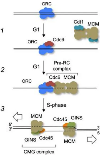
When the cell is getting prepared to enter S phase, proteins must be assembled in preparation of replication. The origin replication complex (ORC), a multi-subunit protein serves as the initiator protein for replication; meaning that it binds to a recognition start site in DNA in an ATP dependent manner. ORC is also responsible for the start of protein assembly for the pre-replicative complex (pre-RC) by interacting with CDC6.7 CDC6 is one of the most important proteins for assembling proteins onto the DNA for replication. CDC6 protein levels are regulated by the cell cycle and are at high expression in M-G1 transition and the G1-S transition. Without CDC6, cells are unable to replicate their DNA, and with too much CDC6 they replicate it overly abundantly without stopping for mitosis.7 From this it appears that ORC when chromatin bound recruits CDC6, and then the ORC/CDC6 complex in turn recruits minichromosome maintenance (MCM) proteins in this case MCM2. MCM2-7 in eukaryotes is likely the helicase responsible for unwinding the DNA helix.7-8 The pre-replicative complex of the ORC, CDC6 and MCM2 are assembled during G1 phase, however replication does not begin until the cells have entered S phase. The control of DNA
replication appears to be initiated by cyclin-CDKs which phosphorylates and causes the disassembly of the pre-RC. In order for replication to begin, the pre-RC must be
disassembled and the pre-IC must become assembled.7, 9
and GINS.9 The CMG complex encircles the DNA and is the active replicative helicase responsible for unwinding the double stranded DNA helix, and is the core of the replisome progression complex (RPC).10-11 A summary of the basic steps to initiate replication as described above is depicted in Figure I.3. Additional components of the RPC include Tof1-Csm3 complex which is used to allow the complex to pause at protein-DNA barriers, along with FACT, a histone chaperone; Mrc1, a checkpoint mediator, Top1 a type I topoisomerase, Mcm10 and Ctf4 which are proteins bound to DNA polymerase α-primase (pol α).10
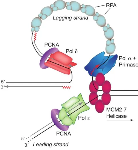
The GINS complex is also able to associate with DNA polymerases α, δ and ε. DNA is only replicated in a 5' to 3' manner; therefore one strand is replicated continuously—known as the leading strand, while the other is done in a non-continuous manner—known as the lagging strand, this is termed semi-conservative replication.1, 12 DNA polymerase (pol) α primase is utilized to prime the leading and lagging strands with RNA making a DNA-RNA primer hybrid to use as template for replication by creating a 30-40 nucleotide sequence of DNA-RNA with a 3'-OH group.8, 10 Once the template is made, replication factor C (RFC) facilitates polymerase switching from pol α to proliferating cell nuclear antigen (PCNA); PCNA is the sliding clamp that holds the replicative polymerase onto the DNA template.8, 10
There are three major stages to DNA replication: initiation, elongation and
proofreading activity and mismatch repair (MMR) lowers this error rate to ~1 x 10⁻¹⁰ for any given replication cycle, as shown in Figure I.4.8, 14 MMR is a repair process which removes normal bases that are not in the correct pairing, helping correct DNA replication errors. This process uses strand discrimination to remove the base from the newly synthesized strand instead of the parental strand.15-16 One of the reasons for this low replicative error rate is the differences in free energy between a correct base pair and an incorrect base pair, as well as the proofreading exonuclease activity associated with some of the replicative polymerases all help ensure the correct base is inserted lending to the low spontaneous mutation rate of replication. The estimated rate of spontaneous replication error is about ~1 x 10⁻¹⁰, or 1 in every 10,000,000,000 per nucleotide per cell replication.8, 14, 17-19 While this rate (i.e. < 1 mutation per genome per cell generation) may seem like a small number, the number of cells that are replicating and the number of times cells replicate over the human life span add up, leading to consequences such as aging and cancer.
inherently more complicated due to its fragmented replication. Replication protein A (RPA) coats the single stranded DNA region of the lagging strand to prevent reannealing while it waits to be replicated by pol δ.8, 21 The DNA polymerases add the 5'-phosphate group of the incoming nucleotide to the 3'-OH group on the existing DNA, thus migrating in a 5' to 3' direction.22 A simplified cartoon model of the process of eukaryotic replication can be visualized in Figure I.5.
After initiation and elongation, termination is when replication has been completed and the replisome is disassembled.13 Termination is promoted from converging replication forks, and it is facilitated by 71 chromosomal termination regions (TERs) which influence how fast the replication forks progress and therefore merge. Then Rrm3 which is a DNA helicase and DNA topoisomerase 2 (Top2) help facilitate the fusion of the replication forks at the TERs.23 DNA must then be rewound and packaged back into chromatin. This occurs in a replication-coupled (RC) nucleosome assembly, where new and parental H3-H4 are
deposited followed by the H2A-H2B dimer onto the DNA, and this is all regulated and assisted by multiple histone chaperone proteins.24 After DNA replication is complete, the cell cycle can move onto mitosis and cytokinesis.
DNA Damage
site.17, 25 Depurination is very mutagenic as it can occur in physiologic conditions and happens with a high frequency, with an estimate of 10,000 depurinations per cell per day.17 One of the reasons depurination is so mutagenic is the seeming preference for the insertion of deoxyadenosine across from the abasic site.17 Abasic sites can also occur at pyrimidines (apyrimidinic sites) but they do so with less frequency than apurinic sites. Abasic sites can also be caused by base excision repair (a repair mechanism detailed later). Abasic sites can cause fork stalling leading to either fork collapse or mutations when they are bypassed by translesion synthesis (TLS), a tolerance mechanism.25 Base deamination is the loss of an exocyclic amino group which can occur spontaneously on cytosine, adenine or guanine but occurs with greatest frequency on cytosine. Deamination of cytosine leads to a uracil paired with adenine—this can result in a C to T mutation, but cells have a robust repair mechanism to prevent this error.15
double-term “TT dimer”, however they can also occur as TC, CT, or CC which are influenced by the DNA sequence of the irradiated region. CPDs are unable to be bypassed by replicative polymerases and cause a replication fork block. CPDs can also be bypassed by translesion synthesis (TLS), a tolerance mechanism that is thought to exist to allow a cell to potentially have an incorrect base inserted across from a lesion rather than end up with a prolonged replication fork blockage which would likely cause a strand break, almost assuredly leading to either cell death or extensive mutagenesis. TT dimers have been visualized by the help of crystal structures which shows that the DNA helix is distorted, bending approximately 30° towards the major groove and also unwound by 9°.15 C containing pyrimidine dimers are much less stable, often deaminating to uracil.15 Another major UV related lesion is a
pyrimidine-pyrimidone (6-4) photoproduct (6-4PP); which links the C6 of a 5' pyrimidine to the C4 of the neighboring pyrimidine. 6-4PP usually occurs in TC or CC and occasionally in TT. This 6-4PP lesion only is formed in one stereoisomer and it causes a major distortion in the DNA helix due to the 3' pyrimidine ring being almost parallel to the DNA helix axis and being perpendicular to the 5’ pyrimidine ring, this lesion is usually repaired by nucleotide excision repair although if TLS is performed on this lesion, it usually inserts a G.15-16 A correctly paired TT (6-4) photoproduct has been analyzed by NMR and shown that it causes the DNA to bend towards the major groove by 44° and become unwound by 32°.
called an intrastrand crosslink, and if it occurs on two separate strands then it is called a interstrand DNA cross-link. Interstrand crosslinks are caused by cisplatin, and are important because they prevent the DNA from separating and therefore blocks DNA replication and transcription. Additionally, cisplatin also causes 1,2-intrastrand linkages between adjacent guanines on N7.16 Cisplatin causes mutations mainly in the form of G:T and A:T
transversions. Pol η can help suppress mutagenesis across from cisplatin-GG intrastrand crosslinks by inserting the correct C pair.15N-methyl-N’-nitro-N-nitrosoguanidine (MNNG) is another chemical agent. MNNG causes methylation of the DNA, and its major lesion is O6-methylguanine (O6-MeG). MNNG is an SN1 alkylating agent, and defects in MMR can lead to increased resistance of such agents. MNNG normally leads to S phase checkpoint followed by MMR-dependent G2/M arrest and apoptosis. O6-MeG inhibits DNA replication and triggers repair through MMR. O6-MeG is recognized by MSH2-MSH6 heterodimer and then signals downstream checkpoints.16
Imbalance of oxidative stress, and accumulation of oxidative damage can lead to mutations and increase in certain cancers.16
In addition to naturally made ROS from cellular processes, there are many chemicals that can interact with these cellular processes causing an increase in the amount of ROS produced, two examples are menadione (MD) and methylene blue plus light (MBL). Methylene blue (MB) is a redox dye which carries a positive charge and is reduced on the cell surface before being transported into the cell membrane. Once within the cell membrane, MB is re-oxidized and therefore it cannot escape the cell. MB within the cell can then
interact directly with oxygen or with heme-containing proteins.28 MB can also activate the pentose phosphate pathway within the cell which is the pathway utilized to make
nicotinamide adenine dinucleotide hydride (NADH) and some precursors of nucleic acid. One way this pathway might be activated is by the reduction of NADPH to NADP+ caused by MB.28 MB is utilized as a way to generate oxidative stress in the laboratory, specifically a lesion called 8-oxo-guanine (8-oxoG), but it is also utilized in the clinic for numerous
reasons including a treatment for methemoglobinemia and as an antidote for paraquat poisoning.28-29 Menadione (MD) is a chemotherapeutic agent which can be reduced at the Complex I of the mitochondrial respiratory chain and lead to the production of O2-. MD can activate apoptosis through a Ca2+ -dependent mitochondrial pathway.30-31
8-oxo-guanine
base pair with cytosine. When 8-oxoG is in the syn conformation it can base pair in a 8-oxoG:A pair which looks very similar to a normal T:A base pair making it hard to detect and repair and can lead to a transversion mutation.16, 32 Eukaryotic replicative polymerases can replicate 8-oxoG by inserting an A across from it, causing a G:C to T:A transversion.33 This is a very common place lesion and bypass error, however eukaryotes have multiple
mechanisms to try and prevent and repair 8-oxoG from being mutagenic.
In addition to damage to guanine after the incorporation into DNA, damaged guanines can be directly incorporated into DNA. 8-oxoG also denoted in some of the
literature as dGTP, can come from oxidation of dGTP, or from the oxidation of 8-oxo-dGDP and then nucleoside diphosphate kinase can convert the nucleoside diphosphate to a triphosphate. In order to try and prevent the incorporation of 8-oxoG into DNA, cells utilize 8-oxo-dGTPase which are used to hydrolyze 8-oxo-dGTP to 8-oxo-dGMP which helps to reduce the 8-oxo-dGTP pool. This GTPase is encoded by MTH1. Once the pool is reduced to 8-oxo-dGMP, it is unable to convert back to dGDP or dGTP as human guanylate kinase is not able to phosphorylate dGMP. This leads to the further dephosphorylation of dGMP to 8-oxo-deoxyguanosine which can then be excreted in the urine.15-16
Since it has been established that 8-oxoG can occur either by oxidative damage onto the dGTP pool or to already utilized dGTP within the DNA strand, there are multiple pathways to try and repair the damage once it is within the DNA. One protein within E.coli
another glycosylase within E.coli that can help remove 8-oxoG if it is mispaired to an A, it is called MutY. MutY does not directly remove 8-oxoG but rather removes the incorrect A from the undamaged strand, BER then usually inserts a C leading to a GO: C pair which can then be acted on by MutM which can in turn remove the 8-oxoG. One more enzyme, termed MutT helps to prevent the incorporation of 8-oxoG by hydrolyzing 8-oxodGTP as previously described. Humans are known to have a homolog to MutY called MYH.15-16
Mutagenesis
DNA damage does not inherently mean mutations. In order for DNA damage to turn into a mutation, the damage must occur, must be unsuccessfully repaired and must be replicated incorrectly, therefore imprinting it into the DNA. Many chemicals and
vice versa (e.g. G to T).16 Base substitution mutations can be further categorized by their effect. If a base substitution results in a change from one amino acid to another amino acid, then the mutation is called a missense mutation. This type of mutation is frequently
responsible for changes in gain or loss of function mutations which drive carcinogenesis. A nonsense mutation occurs when a point mutation changes the original amino acid to a stop codon, which usually leads to early termination and a non-functional protein. There are also silent mutations, in which the nucleotide sequence changed due to the mutation but the codon still reads for the same amino acid therefore the mutation goes unnoticed physiologically.16 Mutations do not only occur at a single point, or single base; often there are insertions or deletions of more than one base in a row, as well as tandem base substitutions and complex base substitutions. When one of these types of mutations occurs, specifically insertions and deletions of greater than one base, then it often leads to (but not always) a frameshift. A frameshift occurs when there is an insertion or deletion of 3n +/- 1bp, resulting in the amino acid sequence which is used for translation is shifted, this can lead to alterations in the function of the protein or lead to a non-functional protein.16
Translesion Synthesis (TLS)
Translesion synthesis is a tolerance pathway used to bypass lesions that block the replication fork. Once the replication fork is stalled, specialized polymerases called TLS polymerases are brought in, they insert a nucleotide across from the damage, and extend past the damage—thus allowing replicative polymerases to return and continue replicating DNA, giving the term “lesion bypass”. TLS can insert the right base and be high fidelity, but often times it inserts the wrong base which leads to mutagenesis. TLS is a tolerance pathway and not a repair pathway because it does not take out the lesion, but rather inserts a base whether correct or incorrect across from the lesion and extends past the damage in order to allow DNA replication to continue. This occurs when replication is occurring and replicative polymerases stall.8, 15-16, 34-43
The TLS polymerases include DNA polymerase η, encoded by Rad30A, DNA polymerase ι (pol ι) encoded by Rad30B, DNA polymerase κ (pol κ) encoded by DINB1, and dCMP transferase Rev1 encoded by UmuC all of the Y family of polymerases as well as DNA polymerase ζ, of the B family of polymerases. Each of these polymerases has varied cognate lesions which they are able to bypass with differing fidelities based on which polymerase is bypassing which lesion.15-16, 19
TLS polymerases lack the 3' – 5' exonuclease activity associated with normal
active sites which allow for the accommodation of large bulky lesions. This structural difference allows for variability in what nucleotide is incorporated, as it is not a
geometrically constrained fit like in a replicative polymerase.15, 38 All polymerases contain a finger, palm and thumb domain.44 The A-, B- and Y-family polymerases all share highly conserved palm domain, but the secondary structures of the thumb and finger domains are vastly different between these families. The Y-family polymerases thumb and finger regions are considerably smaller than that of the A- and B- families. Y-family polymerases all contain four domains, a palm, finger, thumb and little finger (LF), with the thumb and LF constituting the active site (see the structure in Figure I.7).36 Within these regions, the Y-family polymerases all have a conserved catalytic active site within the N-terminal
polymerase which is approximately 350-450 residues, of which about 100 of the residues are only in the Y-family and make up the little finger domain. The little finger, in combination with the thumb domain is used to hold the DNA substrate, and this is where the flexibility lies allowing for the flexibility to accommodate varying lesions. On the opposite side, the C-terminal side contains an appendage which is used for regulatory protein-protein interactions which varies in size depending on the polymerase. Human and mouse Rev1 contains an extra N-terminal BRCT domain not found in the other Y-family polymerases.45
lesion but a second polymerase then is used to extend past the lesion. Whether the system is either one or two polymerases is based on the type of lesion and which polymerase(s) are used.15 An example of each mechanism is: the bypass of a TT dimer by pol η is likely a one polymerase system, but bypass of AP sites by Rev1 and pol ζ is likely a two polymerase mechanism.15 Due to the low fidelity of these polymerases they are highly regulated and kept at low levels during normal replication in order to keep mutagenesis to a minimum and are recruited when needed to damaged DNA during fork stalling. These polymerases are likely regulated transcriptionally and post-transcriptionally in order to keep them at low levels until needed.15
POLH, also known as hRAD30A, is the gene that encodes for human DNA
polymerase η, one of the TLS polymerases. It was first discovered in S. cerevsiae in 1999 and is found in mice, drosophila and humans.38 POLH contains 11 exons over 40 kilobases and is located on human chromosome 6p21.1-6p12, with one of the 11 exons is
untranslated.34, 38 Human pol η is 713 amino acids long and 78 kDa. Protein complementation studies found that the addition of pol η into a xeroderma pigmentosum variant (XP-V) cell line, discussed below, was able to restore the cells ability to bypass CPDs.38 Based on further
in vitro replication and biochemical studies of pol η, it is known that pol η bypasses CPDs well.34 Bypass of CPDs by pol η is more accurate than other polymerases, and less mutagenic than double strand breaks (DSBs) which would occur if the lesion was unable to by
shown that dATP is inserted across from both the 3' and 5' T in the TT dimer. When TT dimers were studied biochemically in a nucleotide competitive environment it showed that dimer bypass fidelity was relatively low. This study by McCulloch et al. showed that pol η bypasses the 5' T of the dimer 4.4 times more accurately than on undamaged T (32x10-4 versus 140x10-4). The most frequent insertion was of G across from the 5'T with an error rate of 32x10-4 which created a T to C transversion, whereas the 3'T has an error rate of 390x10-4. This experiment shows that there is a bias on the lesion position for the fidelity and it was determined that this was due to the lesion and not the sequence content as the experiment analyzed multiple sequence contexts. The difference in fidelity by placement is likely due to base pair energetics and base pairing stability.46 In order to better understand how the fidelity of pol η works, the crystal structure of the catalytic domain (amino acids 1-432) was solved in 2010. See Figure I.9 for the structure of pol η. The structure of pol η acts as a molecular splint in which it keeps the damaged DNA section straight due to the high positive charge on the DNA-binding surface, this allows it to interact with four upstream template nucleotides above the active site. The LF domain (aa 316-324) has a β-strand which is almost parallel to the template strand and this LF domain also interacts with the template and primer in the minor groove.16, 36 Based on the interaction of pol η with the template strand, it leads to the ability of pol η to extend past the lesion 3 base pairs, after which the major-groove
damage by UV, pol η is recruited into foci and it was determined that the N-terminal zinc finger motif is required for this foci formation16.
Rev1 is considered a dCMP transferase because it preferentially inserts a C no matter what the template is. Therefore, if Rev1 is replicating on a run of G’s it puts in the correct base of C, however it continues to put in C’s even across the other bases and lesions. The reasoning for its choice of base was discovered to be due to the use of Arg324 within its own protein as the template instead of using the DNA template.15, 45, 48 Pol κ is the TLS
polymerase known for its bypass ability of (-)-trans-anti-BPDE-N2-dG adduct, although it can also bypass the (+)-trans-anti-BPDE-N2-dG adduct which are both adducts formed by benzo[a]pyrene, and pol κ is able to insert the correct C nucleotide across from these adducts.16 Pol κ also can bypass AP sites but does so in a potentially error prone manner, often inserting an A opposite the AP site. Pol ι preferentially inserts G across from an
One of the other uses for TLS in addition to damage bypass is somatic hypermutation. Somatic hypermutation is the production of antibodies by B cells which is accomplished by base substitutions within the DNA of the V regions of Ig genes. This process is started by cytosine deamination followed by replication using TLS polymerases, specifically Rev1, and pol ζ with some implications of pol η and ι also might be involved.15, 44
This process creates around one billion antibody variants for the immune system.48
Regulation and availability of the polymerases
One of the main regulatory mechanisms known for TLS is the ubiquitination of PCNA. After DNA damage occurs, a DNA damage response is triggered including the monoubiquitination of PCNA by Rad6-Rad18 on Lys164, which leads to the recruitment of TLS polymerases.19, 49 Rad18 binds to single stranded DNA, which may be the trigger to cause its ligase activity for PCNA. When replication is blocked, Rad18 binds to ssDNA, once bound it recruits Rad6 and together they monoubiquitinate PCNA.50 Once
monoubiquitination and deubiquitination of this region. The UBZ is important due to the fact that it binds to the K164 ubiquitin on PCNA. Polyubiquitination of K63 on PCNA is thought to be involved in restarting the stalled replication fork by recruiting ZRANB3/AH2.19, 49
Pol η after UVC damage localizes into nuclear foci. This was investigated also in pol ι and was found that after UV-C treatment, pol ι also localizes into nuclear foci but only in the presence of pol η; in cells lacking pol η, pol ι was unable to localize. The interaction of pol η and pol ι is through a direct interaction using the residues 492-715 of the C-terminal end, which is not the catalytic region, of pol ι.53
The region of pol η necessary for the interaction with pol ι is likely between aa 352-595, although the experiment could not rule out the residues 595-713 also being necessary. This direct interaction is only a fragment of the overall pol η within the cell, but is likely the reason for the co-localization within the nuclear foci.53 The C-terminal region of pol η, amino acids 595-713 is region responsible for pol η’s localization to replication foci.54
While it is well known that the two interact, this leads to questions as a lot of the mutagenesis data in the field suggests that pol ι is a backup mechanism for pol η.55-56
The human relevance of DNA polymerases is in their involvement in cancer; whether by creation of mutations, or their involvement in possible chemotherapeutic resistance. Although the levels of the various polymerases appear to vary widely depending on the patient and the tumor, there is the possibility of predicting the outcome of treatment if the investigator would analyze for the level of the polymerases before and after treatment. Previous research has shown TLS polymerases ι and η are often over expressed in cancers, however pol κ is usually down regulated, except for its increase in lung cancer.61-63
Additionally, a few other polymerases including pol β, which is involved in NER are also overexpressed in many cancers.63 The overexpression of these polymerases can lead to increased mutagenesis and help fuel cancer progression, as well as cause resistance to chemotherapy. A very pertinent example is the presence of pol η during treatment with cisplatin. Based on a structural analysis, as well as a retrospective analysis of tumors
analyzed for pol η mRNA levels determined that higher levels of mRNA was correlated with response to platinum based therapy: with low levels of pol η showing a greater response to platinum based therapy, and higher levels of pol η more likely failing, or having
recurrence.62, 64-65 This leads to the potential to either check for expression before
Repair Mechanisms: Nucleotide Excision Repair
Nucleotide excision repair (NER) is used for the removal of many types of DNA damage including bulky adducts. NER recognizes damage by looking for distortions within the helix instead of looking at the lesion itself.15-16 NER is one of the most complex repair mechanisms but it can be broken down into six main steps. First it must find the damage, and in humans it is believed that the protein complex NPC-RAD23B is responsible for
recognition. Second it must be able to get access to the lesion by unwinding the DNA in the local region, which is done by TFIIH. Third, it must make incisions on both sides of the lesions which are performed by XPF on the 3' and ERCC1-XPF on the 5'. The fourth step is the release of the DNA region containing the lesion, fifth is DNA synthesis by pol ε or δ in the excised region and sixth is ligating the newly synthesized region to the previously synthesized region by DNA ligase. These steps are summarized in Figure I.10. NER is usually a post-replication repair model, but there is also transcription-coupled NER (TC-NER) which is triggered when the RNA polymerase complex is blocked at a damage site. TC-NER is useful in that it helps transcription continue and not stop, therefore it helps prevent cell death. TC-NER is a fast and efficient repair model. One other type of NER is global genome repair (GGR) which is responsible for the repair of nontranscribed strands and unexpressed regions of the genome. GGR is therefore helps prevent mutagenesis by
Repair Mechanisms: Base Excision Repair
Base excision repair (BER), is one of the major repair pathways to get rid of damaged DNA bases. It uses enzymes called DNA glycosylases which recognize the damaged base and makes an incision/cleaves the N-glycosidic bond which is what connects the base to the deoxyribose-sugar backbone. Once the base is removed, it creates an AP site, next whether using an AP site caused by BER or from spontaneous base loss, a protein called an AP endonuclease hydrolyzes the phosphodiester bond on the 5' side of the AP site. DNA
synthesis then occurs in either a one base, short-patch BER, or multiple bases which displace some of the previously synthesized strand, long-patch BER. For short-patch BER dRpase modify the ends and DNA ligase ligates the bases together. In long-patch BER, FEN1 is used to cleave the flap that was created from the displacement of some of the previously
synthesized DNA, and then it is ligated by DNA ligase.15-16, 44, 67 See Figure I.11 for a
rendition of BER. The polymerase used to fill in the patch varies depending on the length, for short-patch, pol β is used, where in long-patch usually pol δ or ε synthesizes the patch. There are many different glycosylases which are utilized within the first step of detecting the damaged bases. Of interest to this research would be Fpg also named MutM which removes 8-oxoG in addition to other oxidized and ring-opened purines, as well as MutY which removes adenine when it is paired with 8-oxoG. MutY and MutM are both isolated from
Cancer
The study of mutations is valuable because mutations are the start of what can turn into cancer. Cancer is a disease that needs a mutation which has a growth advantage over normal cells, leading to proliferation of the mutated cell. The first step towards cancer termed initiation or mutagenesis is the focus of this research. Initiation is the generation of a “stable, heritable change”, and is usually the result of unsuccessfully repaired DNA damage.15-16, 18, 68 Once initiated, the mutated cell might die if the mutation makes it unviable, it could also stay in a static state based on the conditions and normal cells around it, or it could contain a selective advantage resulting in cell proliferation. The second stage, promotion, can be brought on either by the selective advantage growth of the mutated cell on its own, or by the use of a tumor promoter.68 Two endogenous mechanisms for the promotion of growth are either by the activation of a proto-oncogene or by the loss of function of a tumor suppressor gene. Proto-oncogenes are usually involved in positively regulating cell proliferation and help to ensure cell survival so mutations within this type of gene lead to a gain in function oncogene which often block apoptosis and stimulates cell survival. Tumor suppressor genes are usually negative regulators of proliferation and positive regulators of apoptosis, so this gene tends to have a loss of function mutation which leads to unregulated cell growth69 In either case, the initiated cell begins to clonally expand which can create a preneoplastic lesion. Progression is the next and final step leading to carcinogenesis, and this is when the preneoplastic lesion which was benign, now converts into neoplastic cancer. During
timing are crucially important in the model of initiation through promotion; without initiation there will be no mutagenesis even if there is promotion, and without promotion even the mutation from the initiation event will not turn into cancer.15, 69
DNA damage as described previously is hard to avoid in life due to exposure to chemicals, radiation and oxidative stress. The main categories of DNA damaging agents are (1) direct-acting carcinogens, which are chemicals that do not need metabolically activated such as N-methyl-N’-nitro-N-nitrosoguanidine (MNNG). (2) There are also indirect-acting carcinogens which do need metabolically activated before they can react with DNA such as benzo[a]pyrene. (3) There is radiation and oxidative damage which can occur both directly and indirectly and (4) inorganic agents which have unknown mechanisms such as arsenic. UV light which is studied here, is recorded to cause around one million new cases of skin cancer each year, not including melanomas.15, 69
Carcinomas make up the majority of human cancers. These are tumors derived from epithelial cells in the various organs. The other types of cancers which are slightly less common are sarcomas which are derived from mesenchymal tissues, leukemia which are derived from blood forming stem cells and lymphomas which come from B and T
Xeroderma Pigmentosum
Xeroderma pigmentosum (XP) is a sun sensitive, cancer prone disease phenotype that can be comprised of 8 varying subsets of the overall disease which are labeled XP-A through XP-G and XP-V. XP-A through XP-G all have varying deficiencies within NER, while XP-V is deficient in functional pol η. XP is an autosomal recessive disorder which is more
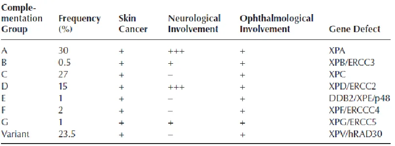
prevalent in some populations: for example cases are rare in Caucasians approximately 1 in 250,000 people, however populations such as Japan and Egypt have increased rates at about 1 in 40,000 people.34 Often patients with XP have parents who are consanguineous. There are a number of clinical hallmarks for XP including severe photosensitivity which is presented as especially painful sunburns in early childhood, skin dryness, premature skin aging, and malignant tumors including squamous cell carcinoma, basal cell carcinomas and melanoma on the face, head and neck. Some subsets of XP also have a neurological component.34 Table I.1 illustrates the breakdown of the various subtypes by their effects and gene affected. The risk of tumors in XP is 1000 times greater than in the normal population and it starts much earlier in life.
The neurological involvement is thought to be due to neuronal death from oxidative stress induced DNA damage—leading to irreversible progressive symptoms. Some
neurological symptoms include lowered IQ, spasticity, peripheral neuropathy and ataxia.34 Additionally, approximately 40% of XP patients have ophthalmological symptoms including blepharitis, ectropion, and corneal abnormalities. They also have increased risk of
metabolic and genetic tests, unfortunately there is no cure for the disease and treatment includes treating the symptoms and preventing sun exposure.34 XP-A through G patients often develop their tumors in their very early years with an average around age 8, and have a life expectancy into their 20s. XP-V is a more mild phenotype that does not have a
neurological component, and they develop their tumors slightly later, in the mid-teens, and therefore can sometimes live into their 40s.34
XP-A through G constitutes 80% of XP patients, however XP-V is approximately 20% just on its own. XP-A patients are deficient in the NER protein XPA which is also utilized in transcription coupled repair (TCR), and therefore have some of the most severe clinical manifestations of the disease. XPB and XPD are helicases which are utilized in both NER and TCR. XPC is the largest subgroup of XP, and XPC is only deficient in global genome repair and not in TCR, they are free of neurological symptoms and only manifest in the severe UV sensitivity and skin tumors. XP-E, F and G are more rare then the other subsets. XP-E is deficient in the DNA damaging binding gene (DDB2), which is associated with helping repair CPDs. XP-F and XP-G are proteins used in NER, but at a lesser capacity than some of the other proteins and therefore present with a mild phenotype of XP and these patients have a slightly longer life expectancy. XP-V patients have functional NER but are deficient in functional pol η which is necessary for the TLS of CPDs.
carcinoma (BCC) is most prevalent in the normal population, there seems to be a higher proportion of squamous cell carcinomas (SCC) within the XP population. Part of the
Figure I.2 – Depiction of DNA organization within chromatin. A. This is a simplified depiction of DNA within the chromatin structure. With the lowest level of organization being the nucleosome which has two superhelical turns of DNA around a histone octamer. These nucleosomes are then connected by short linker DNA regions. At the next level these
Figure I.3 – Assembly of the proteins at the replication origin. A. A simplified depiction of the steps leading to replication origin unwinding in yeast which has the same steps as in humans. Replication origins are bound by ORC, and during G1 phase, ORC recruits Cdc6 (step 1). This in turn recruits Cdt1/MCM complex forming the pre-RC complex (step 2). MCM is loaded onto double-stranded DNA. S phase beings when MCM is activated by GINS and Cdc45 (step 3). One CMG complex functions at each replication fork. Figure reprinted and legend adapted from Onesti, MacNeill 2013.9
Figure I.4 – Replication fidelity. This is a depiction showing the relative contribution of the three main factors of replication fidelity. The ranges are overlapping due to the fact that there are multiple mechanisms to increase the level of fidelity, and within each family of
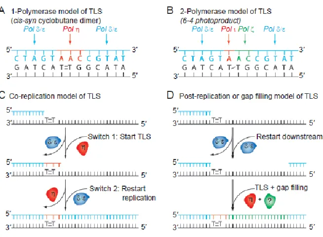
Figure I.7 – Varying models of TLS. (A) A one-polymerase model of TLS illustrated over a TT dimer. This shows a single polymerase being responsible for both the insertion and extension past the lesion. (B) Illustrated here is a two-polymerase model of TLS over a 6-4PP showing that different polymerases are responsible for the insertion versus extension step. In this model it shows that pol ι inserts across from the 5’ lesion, and pol ζ inserts across from the 3’ lesion and extends past the lesion. Both A and B are completing TLS within a
Figure I.9 – Structure Domains of the Y-family polymerases. The polymerase domains are labeled in the following colors: red (palm), blue (finger), green (thumb), purple (LF) and yellow (N-terminal addition for pol κ and Rev1). The regulatory units are listed in a legend within the figure. Abbreviations include: UBM for ubiquitin-binding motif, UBZ for
ubiquitin-binding zinc finger and BRCT for Brca1 C-terminal domain. Figure reprinted and legend adapted from Yang and Woodgate.45
Figure I.10 – Nucleotide Excision Repair. This figure illustrates the sub pathways of GG-NER versus TC-GG-NER using UV damage as a model. In GG-GG-NER on the left the damage sensor XPC works with the UV excision repair protein RAD23B and centrin 2 (CETN2) which constantly probes the DNA helix for helix distorting lesions which are recognized by the help of UV-DNA damage-binding protein (UV-DDB). Once the XPC complex binds to the damage, RAD23B dissociates. In TC-NER on the right, damage is recognized indirectly due to stalling of RNA polymerase II (RNA pol II). During transcript elongation
Figure I.11 – Base Excision Repair. This figure is showing the effects of oxidative stress which causes 8-oxoG or an abasic site. DNA glycosylase OGG1 removes 8-oxoG and leaves an AP site. APE1 incises at the 5’ side of the AP site sugar leaving a 5’-sugar phosphate which is either native (non-oxidized) or oxidized. A native 5’-sugar phosphate can be repaired by short patch (single nucleotide) BER (SN-BER) but the oxidized sugar phosphate group must be repaired by long-patch BER (LP-BER). SN-BER proceeds after the incision by APE1, pol β dRP lyase removes the 5’-sugar phosphate. Pol β then fills in the gap and the nick is ligated by ligase. In LP-BER when there is an oxidized phosphate group, the
Research Hypothesis and Rationale
DNA damage can lead to stalling of the replication fork, and if not immediately repaired it can lead to prolonged fork stalling and eventually fork collapse and either cell death or gross chromosomal changes caused by double strand breaks.16 In order to prevent this, cells have developed a process, termed translesion synthesis (TLS), which utilizes specialized DNA polymerases with wide open active spaces able to accommodate the large bulky lesions created by damaging events and agents. This allows the polymerases to insert a base, either correct or incorrect across from the lesion and allows replication to continue.35, 39, 44
In this process the correct base can help prevent mutagenesis, whereas an incorrect base can lead to mutagenesis; however by utilizing TLS the cell accepting the potential base substitution as compared to gross chromosomal changes.16, 72 One of the main polymerases involved in TLS is pol η which is characteristically known to bypass CPD dimers created by UV-C. In this study we wanted to expand on the literature which describes the effects of the presence and absence of pol η when bypassing dimers induced by UV-C. Therefore, we evaluated the effects of UV-B on mutagenesis in the presence and absence of pol η. We also wanted to investigate another cognate lesion that pol η is able to bypass, 8-oxoG. We did by this by measuring ROS levels in cells, the effect of oxidative stress on proteins and
mutagenesis. We then evaluated the mRNA expression levels of the TLS polymerases in a time course following treatment with DNA damaging agents. Our hypotheses are:
2. That pol η will help suppress oxidative stress induced mutagenesis.
3. That pol η expression will increase after DNA damaging agents that cause CPDs, or platinum based cross-links, and that in the absence of pol η we will see an increase in a backup polymerase expression, most likely by pol ι.
CHAPTER 1
Detrimental effects of UV-B radiation in an XP-Variant cell line* Kimberly N. Herman1, Shannon Toffton1, Scott D. McCulloch 1,2 1
Department of Biological Sciences, Environmental and Molecular Toxicology Program 2
Center for Human Health and the Environment North Carolina State University, Raleigh, NC 27695 *
Running Title: Effects of UV-B radiation on XP-V cells
Corresponding Author: Scott McCulloch
850 Main Campus Drive Campus Box 7633 Raleigh, NC [email protected]
Key Words: DNA damage, DNA polymerase η, mutagenesis, translesion synthesis, ultraviolet radiation
Published In:
Abstract
DNA polymerase η (pol η), of the Y-family, is well known for its in vitro DNA lesion bypass ability. The most characterized bypass event for this polymerase is that of
Introduction
Human exposure to solar radiation is constant due to its large array of wavelengths, varying from the infrared down to short wave ultraviolet and beyond. Exposure to ultraviolet radiation has been shown to cause detrimental effects to human skin; including sunburns and skin cancer.73 Interestingly, some clinical and epidemiological studies have shown a
correlation between sunscreen use and an increase in some forms of skin cancer.74 While this may seem paradoxical, increased sunblock use tends to correlate with increased sun
exposure. For decades, commercially available sunblock has primarily contained ingredients that block both UV-C and UV-B radiation, even though nearly all UV-C is filtered by the stratospheric ozone layer and only ~5% of ultraviolet exposure coming from UV-B
wavelengths.75 Despite this, UV-C has most often been used for research due to its potency and ability to generate cyclobutane pyrimidine dimers (CPD) and other photoproducts.76 There is ample evidence that both UV-B and UV-A radiation can also generate CPDs;75, 77 however they also make other secondary lesions through their ability to generate reactive oxygen species (ROS).16, 77-80 The ability to generate multiple types of lesions adds relevance to the study of UV-B and UV-A radiation with respect to mutagenesis.
The photoproducts caused by UV light include 6-4 photoproducts (6-4 PP) and CPDs. Both lesions occur between adjacent pyrimidines, but the 6-4 PP is structurally more bulky and is readily repaired by the nucleotide excision repair (NER) pathway.81 CPDs are
encountered during replication and therefore require translesion synthesis (TLS), making them primarily responsible for the mutagenesis that occurs after UV light exposure.82 There are multiple types of CPD dimers based on the identity of the adjacent bases (i.e. CC, TC, CT, or TT). DNA polymerase η (pol η) is able to readily bypass at least the thymine-thymine dimer (TT dimer) with high efficiency and moderate fidelity.83 It is assumed that pol η is able to bypass all of the dimer combinations (and also the corresponding uracil containing lesions created by deamination of the cytosine base) with similar properties. There is in vitro
experimental support for bypass of a TU (thymine-uracil) dimer84-85 and genetic evidence that in yeast the CC and TC lesions are preferentially processed by pol η.86
The potency of UV-C has led to its popularity as an investigative tool for UV photoproduct mutagenesis. However, UV-C is not the only wavelength spectra that can cause photoproduct lesions. UV-B and UV-A, although less energetic, are much more abundant and
environmentally relevant than UV-C,75-76, 80, 87 and they can produce photoproduct lesions as well as generate significant levels of reactive oxygen species (ROS).84, 88 ROS can lead to a wide variety of DNA lesions,89-90 at least one of which, 7-8-dihydro-7,8-oxoguanine (8-oxoG) is also bypassed by pol η.33, 42-43, 91 Therefore, investigating the types of mutations generated after exposure to UV-B and UV-A radiation may provide clues as to what types of lesions are commonly generated and which of the multiple polymerases available in human cells are involved in their bypass.44 In this study, we sought to understand the role of CPDs compared to other potential lesions caused by environmentally relevant exposures of
bypass polymerase, pol η. We have evaluated the cytotoxicity in the presence and absence of caffeine, as well as mutation rates and spectra after single dose of primarily UV-B exposure. We compare our results to previously published work using UV-C and simulated sunlight sources of radiation.
Materials and Methods
Cell lines, growth conditions and treatments protocols
GM02359-hTERT (XP-V strain XP115LO) Clone 1B, denoted here as XP-V (containing a non-functional DNA polymerase η due to a truncating mutation at ORF
position 1117) has been previously described.92-93 We use as a control, an apparently normal neonatal foreskin fibroblast line (NHF1-hTERT), denoted here as NHF, which has been previously characterized.93-94 These cell lines were a generous gift from Dr. Marila Cordiero-Stone (University of North Carolina). All media and supplements were obtained from Sigma-Aldrich (Saint Louis, MO) or Gibco-Life Technologies (Grand Island, NY). XPV cells were maintained in Dulbecco’s MEM with 2mM L-glutamine, 10% FBS and 2x MEM Non-Essential amino acids. NHF cells were maintained in Eagle’s MEM with 10% FBS and 2 mM L-glutamine. Some expansion cultures for HPRT sequence analysis were maintained with the addition of 100 U penicillin/100 µg streptomycin. All cultures were maintained in 37ᵒC with 5% CO2. UV-B treatments were performed by removal of growth media and addition of HBSS to cells. P100 plates used 3 ml HBSS and 96-well plates were dosed using 100 l of HBSS per well. The lamp used has been described previously.95
a 5 or 10 mJ/cm2 final dose was performed with no lid and took ~10 and ~20 seconds, respectively.
Cell Viability
Cell viability after UV-B treatment was determined using the CellTiter-Glo Luminescent Cell Viability Assay Kit (Promega, Madison, WI). Cells were plated into 96 well, flat bottom plates (655083; Greiner Bio-one, Austria) at 5,000 cells per well based on preliminary plating studies as suggested by the manufacturer. 24 hours after plating, cells were treated with UV-B, UV-B plus caffeine or no treatments. UV-B dosing was as described above. For treatments using caffeine (1 mM final), a 1 hour pre-treatment was used, it was removed with the media for UV-B treatment and replaced after treatment; then it was left in the media until viability was measured. At 24 and 48 hours, cells were lysed and luminescence was measured using a FLUOstar Omega plate reader (BMG Labtech,
Offenburg, Germany) using the protocol recommended by the manufacturer.
Mutation Frequency
The HPRT mutation assay was conducted essentially as previously described.94 Briefly, cells were preselected for functional HPRT by growth in 1x HAT (hypoxanthine, aminopterin, thymidine) media (H0262; Sigma-Aldrich, Saint Louis, MO) for 10 days. Cells were then expanded and plated at 15-20% confluence and grown for 24 hours, followed by treatment of 10 mJ/cm2 UV-B, or no treatment. These cells were then maintained in
re-Aldrich, Saint Louis, MO). Cells were given fresh media/6-TG on day 7 of selective growth day. Mutants were counted and collected on the 14th day of selective growth. Colony forming efficiency was determined at the time of mutant selection by plating 750 cells per P100 plate in non-selective media (normal growth media). Colony forming efficiency plates were counted on the same day as mutant collection. Mutation frequencies were calculated as previously described.94 For colonies not destined for mutation spectra analysis, colonies were fixed with methanol/acetic acid (vol:vol 3:1) and stained with crystal violet prior to counting. Statistics for HPRT mutation frequencies were conducted using a one-way ANOVA with the Tukey post test, performed using GraphPad Prism version 5.00 for Windows, GraphPad Software, San Diego California USA, www.graphpad.com.
Analysis of HPRT mutation spectra
cDNA production, PCR amplification of the HPRT gene was done using LongAmp Taq DNA Polymerase (New England Biolabs, Ipswich, MA), and PCR primers
5′-CTGCTCCGCCACCGGCTTCC and 5′-GATAATTTTACTGGCGATGT (Primers 1 and 2)96 with the cycling parameters: 95C for 5 min, 35 cycles of 95C for 1 minute, 48C for 1 minute, 65C for 2 min, with a final extension of 65C for 2 minutes. PCR cleanup was performed using a QIAquick PCR Purification Kit (Qiagen, Louisville, KY), followed by a second round of PCR using 1:100 dilution of PCR product, and primers
5′-CCTGAGCAGTCAGCCCGCGC and 5′-CAATAGGACTCCAGATGTTT (primers 3 and 4).96 Sequence analysis was performed using Geneious Pro Version 5.4.4(Biomatters, Auckland, New Zealand). Analysis of the POLH gene in both cell lines was performed by PCR amplification using primers CCTCACCTCTCCAGACCTGC and
5′-GAGGAGACCATTCTGTCTGGA and the cycling parameters: 95C for 1 min, 30 cycles of 95C for 30 sec, 45C for 1 minute, 68C for 1.5 min, with a final extension of 68C for 5 minutes using Quick-load Taq 2X Master Mix (New England Biolabs, Ipswitch, MA). This amplifies a product of ~750 base pairs that flanks the altered position. Sequencing was performed by Genewiz (Research Triangle Park, NC) using the same primers as the PCR reaction.
Results
Verification of Cell Lines and Dosing
human foreskin fibroblast and a dermal fibroblast line deficient in DNA polymerase η activity, in which the deficient cell line contains a point mutation leading in the POLH gene at codon 373, leading to a truncation in the protein that results in a complete loss of DNA polymerase η activity.92-93
First we verified the genotypes of both cell lines and found the expected mutation in the XP-V cell line (Figure 1.1A). We then exposed exponentially growing cells to doses of biologically relevant UV-B radiation.95 These doses can be achieved by relatively short exposure to direct sunlight (Dr. Jonathon Hall, personal communication) and with the lamp used and a monolayer of cells in culture, required only ~21 seconds for the highest dose. While the peak output of this lamp occurs at 312 nm, it likely does have a minor output in the UV-C range as it emits between 280-350 nm
wavelengths (according to manufacturer literature). Based on preliminary studies, we chose 5 and 10 mJ/cm2 as doses which were not overly cytotoxic to cells (data not shown). The goal of these experiments was to examine if relatively low levels of primarily UV-B radiation were able to affect cells in a manner similar to the potent UV-C exposures that characterize many studies involving pol η.55, 93, 98
Cell Proliferation and Effects of Caffeine
The CellTiter Glo™ assay was used to measure ATP levels in whole cell lysates. This correlates with cell number and preliminary studies verified that we were in the linear
luminescence values are shown in Figure S1.1). In each experiment, multiple replicate wells were used and the values averaged. Each value plotted is the average of multiple independent experiments. As can be seen in Figure 1.1C and 1.1D, exposure to both 5 and 10 mJ/cm2 UV-B radiation was not overly cytotoxic to NHF cells in the presence or absence of caffeine (solid lines). At 1 day and 2 days post radiation exposure, cell numbers were at least 90% those of un-irradiated cells with no striking difference noted. Figure S1.1 shows that
luminescence values increased with time, indicating that cells were indeed proliferating. This demonstrates that the dose is not excessively cytotoxic. That caffeine does not have any noticeable effect suggests that whatever damage is occurring is not enough to activate the ATM/ATR pathway in response to stalled replication forks. In contrast, we do see a noticeable effect of adding caffeine to the XP-V cells. At day 1, there is no apparent
difference in cell number, but by day 2 we see a decrease in the number of cells exposed to combination treatment of both caffeine and UV-B radiation. The drop in total luminescence over time seen in Figure S1.1 also supports the conclusion that in XP-V cells, the
Mutagenesis of UV-B
To investigate this, we employed the hypoxanthine-guanine
magnifies this phenotype. We acknowledge that these cell lines are not isogenic and therefore the difference could be due to factors other than pol η. However, at 1 and 24 hour post
irradiation, we saw no evidence of different levels of TT dimer lesions by dot blot analysis (Figure S1.2). This suggests that both initial levels of and overall repair rates for this lesion are comparable in these two lines.
Mutation Spectra Evaluation
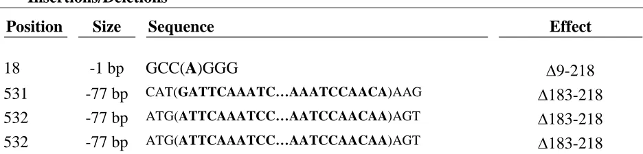
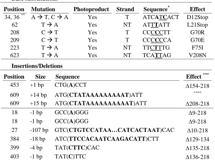
Since UV-B and UV-C radiation can produce multiple different types of lesions,80, 87 we were interested in how the spectra of mutations caused by UV-B radiation compared to those observed after UV-C exposure. To investigate this, we generated spectra of mutations caused by UV-B light in pol η proficient and deficient cells by isolating RNA from 6-TG resistant clones, PCR amplifying the HPRT open reading from cDNA and then sequencing these products. Tables 1.2, 1.3 and Figure 1.3 show a marked difference in the types of mutations observed in NHF and XP-V cells. The most obvious difference is the relative levels of base substitutions to insertion/deletions. In NHF cells, insertions/deletions account for 67% of the total mutations, compared to only 26% for XP-V cells. We also observe more variability in the types of insertion/deletion mutations in NHF cells. In NHF cells, we
detected 3 different insertions at 2 different locations and 6 different types of deletions ranging from a single base up to over 100 bases deleted. These occurred at 7 separate
the deletions were of a much more limited scope (a single 1 base deletion and 3 separate instances of a 77 base deletion) (Table 1.2).
The most striking difference was observed in the numbers and distribution of base substitutions mutations. In NHF cells, all of the base substitutions can be attributed to dipyrimidine sites in one of the two strands (Table 1.3). These mutations account for a minority of total changes in the NHF cells, though. Four of the 6 sites are either TT or CC sequences while only 1 is of a mixed base sequence (the last could be either TT or CT). Of the photoproduct associated mutations, there were 3 changes each at C and T bases. In XP-V cells, there is a similar trend of most base substitutions occurring in dipyrimidine
photoproduct sites, but we also detected 2 changes that cannot be attributed to such
Discussion
It is generally believed that most non-melanoma skin cancers are caused by UV-B and/or UV-A radiation. Most of UV-C radiation is filtered by stratospheric ozone before reaching the ground level, leading to the composition of UV irradiation we are normally exposed to being ~95% UV-A and UV-B.75, 102 We were interested in how these less energetic but more environmentally relevant wavelengths of solar radiation affect human cells with respect to cytotoxicity and mutagenesis. A study of yeast strains proficient or deficient for either the
rad30 (pol η) or rev3 (pol ) genes was conducted comparing UV-C to simulated sunlight
(SSL; UV-B and UV-A in proportions that mimic solar radiation).103 Their results suggested that the lesions created by UV-C are more mutagenic. Similarly, we found that UV-B did indeed cause an increase in mutagenesis in normal cells, it was less than the reported values for this line using UV-C.93 Interestingly, they saw little to no effect on mutation rate when pol η was missing using only SSL. This differs somewhat from our results in that our XP-V line showed a further increase in mutation rate compared to normal cells (Figure 1.2). One explanation could be the different types of radiation used. Another explanation could be the lack of pol in yeast, as it is known to be involved in UV light dependent mutagenesis in the absence of pol η.55
cytotoxicity both in the presence and absence of caffeine, as a specific enhancement of cytotoxicity by caffeine is a well described phenotype of XP-V cells.100, 104 We also were interested in whether pol η deficiency would cause an increase in mutations after UV-B radiation and if so, whether the types of mutations found was similar to or different from that reported after UV-C radiation. To this end, we performed the HPRT mutagenesis assay and sequenced the coding sequence of 6-TG resistant mutants.