University of Mississippi
eGrove
Faculty and Student Publications
Communication Sciences and Disorders,
Department of
6-3-2017
Disrupted Olfactory Integration in Schizophrenia:
Functional Connectivity Study
Sara Kiparizoska
University of Mississippi Medical Center
Toshikazu Ikuta
University of Mississippi
, [email protected]
Follow this and additional works at:
https://egrove.olemiss.edu/csd-facpubs
Part of the
Neuroscience and Neurobiology Commons
This Article is brought to you for free and open access by the Communication Sciences and Disorders, Department of at eGrove. It has been accepted for inclusion in Faculty and Student Publications by an authorized administrator of eGrove. For more information, please [email protected].
Recommended Citation
Sara Kiparizoska, Toshikazu Ikuta, Disrupted Olfactory Integration in Schizophrenia: Functional Connectivity Study,
International
Journal of Neuropsychopharmacology
, Volume 20, Issue 9, September 2017, Pages 740–746, https://doi.org/10.1093/ijnp/pyx045
Received: January 13, 2017; Revised: May 5, 2017; Accepted: June 3, 2017 © The Author 2017. Published by Oxford University Press on behalf of CINP.
doi:10.1093/ijnp/pyx045
Advance Access Publication: June 3, 2017 Regular Research Article
740
This is an Open Access article distributed under the terms of the Creative Commons Attribution Non-Commercial License (http:// creativecommons.org/licenses/by-nc/4.0/), which permits non-commercial re-use, distribution, and reproduction in any medium, provided the original work is properly cited. For commercial re-use, please contact [email protected]
regular research article
Disrupted Olfactory Integration in Schizophrenia:
Functional Connectivity Study
Sara Kiparizoska, Toshikazu Ikuta
School of Medicine, University of Mississippi Medical Center, Jackson, Mississippi (Ms Kiparizoska); Department
of Communication Sciences and Disorders, University of Mississippi, University, Mississippi (Dr Ikuta).
Correspondence: Toshikazu Ikuta, PhD, 311 George Hall, 352 Rebel Drive, University of Mississippi, University, MS 38672 ([email protected]).
Abstract
Background: Evidence for olfactory dysfunction in schizophrenia has been firmly established. However, in the typical understanding of schizophrenia, olfaction is not recognized to contribute to or interact with the illness. Despite the solid presence of olfactory dysfunction in schizophrenia, its relation to the rest of the illness remains largely unclear. Here, we aimed to examine functional connectivity of the olfactory bulb, olfactory tract, and piriform cortices and isolate the network that would account for the altered olfaction in schizophrenia.
Methods: We examined the functional connectivity of these specific olfactory regions in order to isolate other brain regions associated with olfactory processing in schizophrenia. Using the resting state functional MRI data from the Center for Biomedical Research Excellence in Brain Function and Mental Illness, we compared 84 patients of schizophrenia and 90 individuals without schizophrenia.
Results: The schizophrenia group showed disconnectivity between the anterior piriform cortex and the nucleus accumbens, between the posterior piriform cortex and the middle frontal gyrus, and between the olfactory tract and the visual cortices. Conclusions: The current results suggest functional disconnectivity of olfactory regions in schizophrenia, which may account for olfactory dysfunction and disrupted integration with other sensory modalities in schizophrenia.
Keywords: olfaction, schizophrenia, functional connectivity, resting state fMRI, sensory integration
Introduction
There is established evidence supporting olfactory dysfunction in schizophrenia. Olfactory disturbances have been reported since the 1960s (Hoffer and Osmond, 1962; Hurwitz et al., 1988; Kopala et al., 1992; Wu et al., 1993; Brewer et al., 1996,
2003, 2007; Turetsky et al., 2009; Kästner et al., 2013). A previ-ous and recent meta-analysis showed robust olfactory deficits in schizophrenia and also at-risk youths (Moberg et al., 1999,
2014). Although olfaction may not be the central pathology in the typical dopaminergic understanding of schizophrenia, it has been shown that olfactory dysfunction can be induced by NMDA antagonists (Javitt and Zukin, 1991) and that olfac-tory performance is associated with clinical measurements (Kästner et al., 2013).
In olfactory sensation, the olfactory nerve sends afferent pro-jections from the olfactory epithelium to the forebrain structure, the olfactory bulb. Unlike other sensory pathways, the olfactory pathway bypasses the brain stem and directly enters to the olfactory bulb, further projecting to the lateral olfactory tract and the anterior and posterior piriform cortices. In humans, the posterior piriform cortex in the temporal lobe has been isolated to encode categorical perception of odors (Howard et al., 2009), while the anterior piriform cortex in the frontal lobe is sensitive to higher order attentional control (Zelano et al., 2005).
There is evidence suggesting that the pathology of olfac-tory dysfunction in schizophrenia may lie in the olfacolfac-tory bulb. Olfactory bulb volume has been found to be smaller
Kiparizoska and Ikuta | 741
International Journal of Neuropsychopharmacology (2017) 20(9): 740–746
doi:10.1093/ijnp/pyx045
Advance Access Publication: June 3, 2017 Regular Research Article
in schizophrenia (Turetsky et al., 2003). A postmortem study showed decreased synaptic efficacy in the olfactory bulb of schizophrenia patients (Egbujo et al., 2015). Smaller olfactory bulb volume has also been seen in first-degree relatives of schiz-ophrenic patients (Kamath et al., 2011).
However, a limited number of neuroimaging studies of the olfactory system in schizophrenia have ever been conducted. A structural MRI study showed that association with olfactory sensation was found in the region that receives direct affer-ents from the olfactory bulb (Turetsky et al., 2003). Cortical volume of the anterior ventromedial temporal lobe is smaller in schizophrenia and shows association with olfactory thresh-old sensitivity. In a PET study, limbic and paralimbic regions showed lesser activation while experiencing unpleasant odors compared with the healthy cohorts. Patients of schizophrenia showed an impairment in detecting pleasant odors but not unpleasant ones (Crespo-Facorro, B et al., 2001). In another PET study, the left anterior cortex adjacent to the putamen, and the left inferior frontal gyrus showed association to dysfunction in olfactory sensation in schizophrenia (Plailly et al., 2006).
It remains largely unclear how and/or whether olfactory dys-function relates and contributes to other dysdys-functions in schiz-ophrenia. To clarify the brain regions that are associated with olfactory dysfunction in schizophrenia, this study aimed to isolate olfactory connectivity disrupted in schizophrenia by examining resting state functional connectivity from the olfactory bulb, olfac-tory tract, anterior piriform cortex, and posterior piriform cortex.
Materials and Methods
Data AcquisitionThe MRI images, the clinical data, and the demographic data from the Center for Biomedical Research Excellence in Brain Function and Mental Illness (Çetin et al., 2014) were obtained from Collaborative Informatics and Neuroimaging Suite (http:// coins.mrn.org/). This data subset consisted of 183 individuals for whom both resting state and structural data were available. Among these individuals, 84 had a diagnosis of schizophrenia or schizoaffective disorder (hereafter SZ group, 36.85 ± 14.09 years old) based on the Structural Clinical Interview for DSM-IV for Axis I DSM-IV Disorders (First et al., 1998), and 90 were nonpsy-chiatric age-matched controls (control group, 37.5 ± 11.40 years old). Nine individuals who had a diagnosis of bipolar disorder were excluded from further analysis.
Resting state echo planner image (EPI) volumes had 32 slices of 4 mm 64 x 64 matrix with 4-mm thickness (voxel size = 3 x 3 x 4 mm), with repetition time (TR) of 2000 milliseconds and echo time (TE) of 29 milliseconds. A total of 150 volumes (5 minutes) were used in the analysis. High-resolution struc-tural T1 volume was acquired as 176 sagittal slices of 256 mm x 256 mm with 1-mm thickness (voxel size = 1 x 1 x 1 mm, TR = 2530 milliseconds and TE = 3.25 milliseconds).
Data Processing
Data preprocessing and statistical analyses were conducted using FMRIB Software Library (FSL) as well as Analysis of Functional NeuroImages. The anatomical volume for each sub-ject was skull stripped, segmented (gray matter, white matter, and CSF), and registered to the MNI 2-mm standard brain. The first 4 EPI volumes were removed. Transient signal spikes were removed by despiking interpolation. To correct head motion, the volumes were linearly registered to the then-first volume, through which 6 motion parameters and displacement dis-tance between 2 consecutive volumes were estimated. Each of the resting state volumes was regressed by white matter and cerebrospinal fluid signal fluctuations as well as the 6 motion parameters. After smoothing with a 6-mm FWHM Gaussian ker-nel, the volumes were resampled, spatially transformed, and aligned to the MNI 2-mm standard brain space. Through this registration, 12 affine parameters were created between rs-fMRI volume and MNI152 2-mm space, so that a seed ROI can later be registered to each individual rs-fMRI space. To perform scrub-bing where the volumes with excess motion are removed, as a displacement distance between 2 EPI volumes, the root mean square deviation was calculated from motion correction param-eters, at an r =40-mm spherical surface using FSL’s rmsdiff tool (Power et al., 2012, 2015). Volumes whose displacement distance exceeded the threshold (0.3 mm) were removed (scrubbed) from further statistical analyses (Siegel et al., 2014).
The olfactory bulb, olfactory tract, anterior piriform cortex, and posterior piriform cortex were manually segmented in the MNI 2-mm space (Figure 1) following anatomical descriptions in the literature (Howard et al., 2009; Gottfried, 2010; Scherfler et al., 2013). For each ROI, voxel-wise connectivity analysis was conducted. The time course was spatially averaged within the ROI that was registered to the EPI space so that correlations can be tested between the ROI and each individual voxel across the brain. The Z-scores representing the correlations between the ROI and a voxel were used for group-level analysis after registra-tion to the MNI 2-mm brain space.
The SZ and control groups were compared by randomise
script in FSL. Z-statistic images were estimated where clusters were determined by the statistical threshold of Z > 2.326 with a family-wise error-corrected cluster significance threshold, assuming a Gaussian random field for the Z-statistics. The peak voxels within a cluster were calculated by using voxelwise fam-ily-wise error corrected t test images.
Results
Voxel-wise statistical analysis of functional connectivity from the olfactory tract, and anterior and posterior piriform cor-tices showed regions that have lesser connectivity in the SZ group, compared with the control group (Table 1; Figures 2–4). Specifically, the olfactory tract in the SZ group showed less
Significance Statement
Olfactory dysfunction has been repeatedly and consistently found in schizophrenia. However, it is unclear how olfactory dys-function relates to other dysdys-functions in schizophrenia and their pathophysiology. We examined dys-functional connectivity of olfactory regions in schizophrenia using resting state fMRI data. Olfactory regions in schizophrenia showed less functional con-nectivity to other sensory regions and, multisensory integrative regions, and the prefrontal cortex compared with the healthy control group. Our results suggest that olfactory integration to other sensory modalities is disrupted in schizophrenia.
connectivity with the left occipital and parietal region, the ante-rior piriform cortex with the nucleus accumbens and prefrontal cortex, and the posterior piriform cortex with the left frontal cortex. There was no group difference found with the olfactory bulb. None of the 4 ROIs indicated any greater connectivity in the SZ group than the control group.
Discussion
To the best of our knowledge, this is the first study that exam-ined functional connectivity from the olfactory regions in schizophrenia. The results indicate the presence of disrupted connectivity between the olfactory tract and visual cortices, between the anterior piriform cortex and the nucleus accum-bens, and between the posterior piriform cortex and the mid-dle frontal gyrus. The findings here are new but coherent with previous literature suggesting disrupted olfactory integration in schizophrenia.
The anterior piriform cortex (APC) showed disconnectivity to the nucleus accumbens (NAcc). While the NAcc and olfac-tory regions have been known to have similar projections (Heimer and Wilson, 1975; Brog et al., 1993), the association between the piriform cortex and NAcc has been shown as a
part of a feeding-related circuitry for which odors play criti-cal roles (Truong et al., 2002; Stratford, 2005). In schizophre-nia, stronger functional connectivities of the NAcc have been found to be associated with hallucination (Rolland et al., 2015). In patients with auditory hallucination, NAcc functional con-nectivity to the superior temporal gyrus is increased, as well as to the cingulate gyrus and ventral tegmental area, suggesting the enhanced connectivity between the NAcc and auditory cor-tices that may underlie the auditory hallucinations. The NAcc has been implicated in audio visual integration of speech in schizophrenia (Szycik et al., 2009), suggesting that the NAcc is involved in multi-modal sensory integration. Our finding of decreased connectivity between the APC and NAcc may sug-gest that olfactory input to the NAcc is disrupted and may contribute to multi-sensory disintegration whose outcome includes auditory hallucinations.
The piriform regions in schizophrenia showed disrupted connectivities to the prefrontal regions. Disconnectivity in schizophrenia was found between the APC and right superior frontal gyrus extending to the orbitofrontal cortex (Figure 3) and between the posterior piriform cortex (PPC) and middle frontal gyrus (Figure 4). There has been known anatomical projections between prefrontal cortex and olfactory regions
Figure 1. Four regions of interest (ROIs) specified in the MNI 152 2-mm brain space. Axial images shown included regions anterior to the brain stem (y ≧ -12).
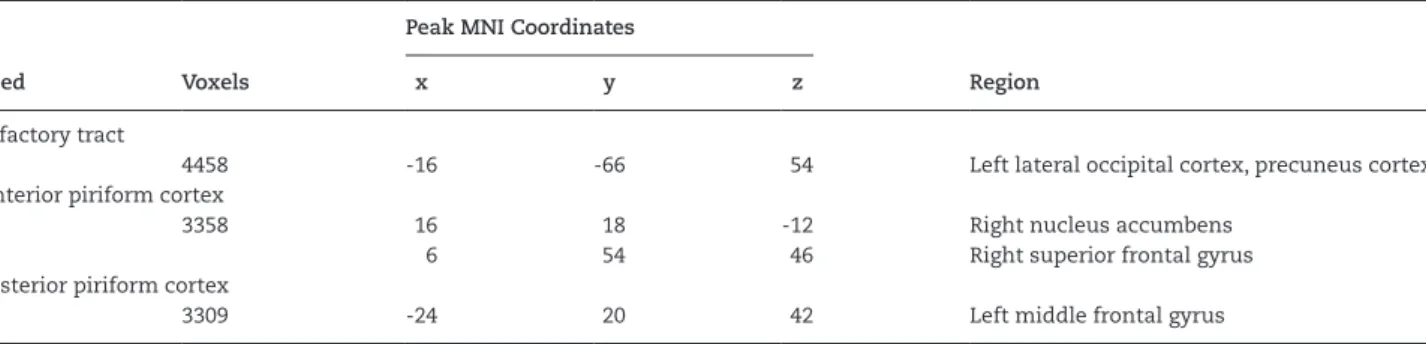
Table 1. Regions of Lesser Connectivity in the SZ Group (Cluster Z > 2.326)
Seed Voxels
Peak MNI Coordinates
Region
x y z
Olfactory tract
4458 -16 -66 54 Left lateral occipital cortex, precuneus cortex
Anterior piriform cortex
3358 16 18 -12 Right nucleus accumbens
6 54 46 Right superior frontal gyrus
Posterior piriform cortex
3309 -24 20 42 Left middle frontal gyrus
Kiparizoska and Ikuta | 743
in rats and primates (Carmichael et al., 1994; Carmichael and Price, 1995). In human fMRI, the orbitofrontal cortex has been shown to be coding olfactory valence, independent from inten-sity (Anderson et al., 2003), suggesting that the connectivity between olfactory regions to orbitofrontal cortex accommo-dates odor valence. The piriform-prefrontal disconnectivity in schizophrenia in our results may account for the disrupted olfactory discrimination and identification in schizophrenia (Brewer et al, 1996; Moberg et al., 2014).
The PPC showed disconnectivity to the middle frontal gyrus. The PPC has been shown to furnish categorical perception (Howard et al., 2009). At the same time, the middle frontal gyrus has been implicated in detecting mixtures of odors (Boyle et al., 2009) compared with simplex odors, suggesting the associa-tive role of categorical perception processed in the PPC. Indeed, tasks used in the previous studies employed complex odors where disruptions in performance were found in the schizo-phrenia group (Hoffer and Osmond, 1962; Hurwitz et al., 1988;
Figure 3. Anterior piriform connectivity. Regions that showed significantly lesser connectivity in the schizophrenia (SZ) group than the control group.
Figure 2. Olfactory tract connectivity. Regions that showed significantly lesser connectivity in the schizophrenia (SZ) group than the control group.
Kopala et al., 1992; Wu et al., 1993; Kästner et al., 2013). Although it remains unclear whether schizophrenia influences more selectively to sensing mixtures of odors, our finding suggests that the PPC-middle frontal gyrus disconnectivity in schizo-phrenia may account for the olfactory dysfunctions found in the literature.
Disconnectivity between the olfactory tract and visual corti-ces including the lateral occipital cortex and precuneus corticorti-ces suggests that olfactory information may not be synchronized with visual information in schizophrenia. This may be consist-ent with disrupted multisensory integration of olfactory input. At the same time, abnormalities in the occipital lobe have been consistently reported (Meador-Woodruff et al., 1997; Narr et al., 2005; Onitsuka et al., 2007). The current methods do not provide evidence to account for the relations between sensory integra-tion and occipital disrupintegra-tions.
While olfactory malfunction has been more widely known in Alzheimer’s disease (Murphy et al., 1990; Devanand et al., 2015) and Parkinson’s disease (Ross et al., 2008), olfactory malfunction is not usually considered to be one of the central symptoms in either disorder. Since it has been suggested that the pathology is in the central nervous system and not in the olfactory epithe-lium, we aimed to detect olfactory disruption in the olfactory bulb and its system (Minovi et al., 2015). The pathology, how-ever, may originate further in the periphery. A reduced quantity of chondroitin sulfate proteoglycans is found in the olfactory epithelium tissues of patients of schizophrenia (Pantazopoulos et al., 2013), which may be accounted for by the pathology of olfactory dysfunction in schizophrenia. A reduction of protein synthesis in olfactory cells in schizophrenia has been reported (English et al., 2015). Olfactory neural epithelium has been recently suggested to indicate signatures of schizophrenia (Horiuchi et al., 2016). The design used in our study does not per-mit any judgment on whether the pathology in the epithelium affects the disconnectivity of the central olfactory regions found in this study. Although our study did not detect any disconnec-tivity in the olfactory bulb, our finding in the other olfactory
regions may be the consequences of dysfunctions in the olfac-tory epithelium.
It has to be noted that the SZ groups are not treatment naïve. Therefore, our finding may be due to antipsychotic medications or other complications from the illness. Although olfactory dysfunctions have been reported in first relatives (Kamath et al., 2011), the current methodology does not distinguish schizophrenia pathology and consequences of medications.
This study suggests the presence of functional disconnectiv-ity of olfactory regions in schizophrenia. The functional discon-nectivity may account for olfactory dysfunction and disrupted integration with other sensory modalities in schizophrenia.
Acknowledgments
Data was downloaded from the Collaborative Informatics and Neuroimaging Suite Data Exchange tool (COINS; http://coins. mrn.org/dx) and data collection was performed at the Mind Research Network, and funded by a Center of Biomedical Research Excellence (COBRE) grant 5P20RR021938/P20GM103472 from the NIH to Dr. Vince Calhoun.
Interest Statement
Dr. Ikuta has received speaker’s honoraria from Eli Lilly, Daiichi Sankyo, and Dainippon Sumitomo.
References
Anderson AK, Christoff K, Stappen I, Panitz D, Ghaheremani DG, Glover G, Gabrieli JDE, Sobel N (2003) Dissociated neural rep-resentations of intensity and valence in human olfaction. Nat Neurosci 6:196–202.
Boyle JA, Djordjevic J, Olsson MJ, Lundström JN, Jones-Gotman M (2009) The human brain distinguishes between single odor-ants and binary mixtures. Cereb Cortex 19:66–71.
Figure 4. Posterior piriform connectivity. Regions that showed significantly lesser connectivity in the schizophrenia (SZ) group than the control group.
Kiparizoska and Ikuta | 745
Brewer WJ, Edwards J, Anderson V, Robinson T, Pantelis C (1996) Neuropsychological, olfactory, and hygiene deficits in men with negative symptom schizophrenia. Biol Psychiatry 40:1021–1031. Brewer WJ, Wood SJ, McGorry PD, Francey SM, Phillips LJ, Yung
AR, Anderson V, Copolov DL, Singh B, Velakoulis D, Pantelis C (2003) Impairment of olfactory identification ability in indi-viduals at ultra-high risk for psychosis who later develop schizophrenia. Am J Psychiatry 160:1790–1794.
Brewer WJ, Wood SJ, Pantelis C, Berger GE, Copolov DL, McGorry PD (2007) Olfactory sensitivity through the course of psycho-sis: Relationships to olfactory identification, symptomatol-ogy and the schizophrenia odour. Psychiatry Res 149:97–104. Brog JS, Salyapongse A, Deutch AY, Zahm DS (1993) The patterns
of afferent innervation of the core and shell in the “Accum-bens” part of the rat ventral striatum: immunohistochemi-cal detection of retrogradely transported fluoro-gold. J Comp Neurol 338:255–278.
Carmichael ST, Clugnet M- C, Price JL (1994) Central olfactory connections in the macaque monkey. J Comp Neurol 346: 403–434.
Carmichael ST, Price JL (1995) Sensory and premotor connec-tions of the orbital and medial prefrontal cortex of macaque monkeys. J Comp Neurol 363:642–664.
Çetin MS, Christensen F, Abbott CC, Stephen JM, Mayer AR, Cañive JM, Bustillo JR, Pearlson GD, Calhoun VD (2014) Thala-mus and posterior temporal lobe show greater inter-network connectivity at rest and across sensory paradigms in schizo-phrenia. NeuroImage 97:117–126.
Crespo-Facorro B, Paradiso S, Andreasen NC, et al (2001) Neural mechanisms of anhedonia in schizophrenia: a pet study of response to unpleasant and pleasant odors. JAMA 286:427–435. Devanand DP, Lee S, Manly J, Andrews H, Schupf N, Doty RL,
Stern Y, Zahodne LB, Louis ED, Mayeux R (2015) Olfactory defi-cits predict cognitive decline and Alzheimer dementia in an urban community. Neurology 84:182–189.
Egbujo CN, Sinclair D, Borgmann-Winter KE, Arnold SE, Turetsky BI, Hahn C-G (2015) Molecular evidence for decreased synap-tic efficacy in the postmortem olfactory bulb of individuals with schizophrenia. Schizophr Res 168:554–562.
English JA, Fan Y, Focking M, Lopez LM, Hryniewiecka M, Wynne K, Dicker P, Matigian N, Cagney G, Mackay-Sim A, Cotter DR (2015) Reduced protein synthesis in schizophrenia patient-derived olfactory cells. Transl Psychiatry 5:e663.
First MB, Spitzer RL, Miriam G, Williams JBW (1998) Structured clinical interview for DSM-IV-TR axis I disorders, patient edi-tion. (SCID-I/P). New York: Biometrics Research, New York State Psychiatric Institute.
Gottfried JA (2010) Central mechanisms of odour object percep-tion. Nat Rev Neurosci 11:628–641.
Heimer L, Wilson RiD (1975) The subcortical projections ofthe allocortex: similarities in the neural associations of the hip-pocampus, the piriform cortex, and the neocortex. In: Golgi Centennial Symposium. New York: Raven.
Hoffer A, Osmond H (1962) Olfactory changes in schizophrenia. Am J Psychiatry 119:72–75.
Horiuchi Y, Kondo MA, Okada K, Takayanagi Y, Tanaka T, Ho T, Varvaris M, Tajinda K, Hiyama H, Ni K, Colantuoni C, Schretlen D, Cascella NG, Pevsner J, Ishizuka K, Sawa A (2016) Molecular signatures associated with cognitive deficits in schizophre-nia: a study of biopsied olfactory neural epithelium. Transl Psychiatry 6:e915.
Howard JD, Plailly J, Grueschow M, Haynes J- D, Gottfried JA (2009) Odor quality coding and categorization in human posterior piriform cortex. Nat Neurosci 12:932–938.
Hurwitz T, Kopala L, Clark C, Jones B (1988) Olfactory deficits in schizophrenia. Biol Psychiatry 23:123–128.
Javitt DC, Zukin SR (1991) Recent advances in the phencyclidine model of schizophrenia. Am J Psychiatry 148:1301–1308. Kamath V, Moberg PJ, Kohler CG, Gur RE, Turetsky BI (2011) Odor
hedonic capacity and anhedonia in schizophrenia and unaf-fected first-degree relatives of schizophrenia patients. Schiz-ophr Bull 187.
Kästner A, Malzahn D, Begemann M, Hilmes C, Bickeböller H, Ehrenreich H (2013) Odor naming and interpretation perfor-mance in 881 schizophrenia subjects: association with clinical parameters. BMC Psychiatry 13:1–12.
Kopala LC, Clark C, Hurwitz T (1992) Olfactory deficits in neu-roleptic naive patients with schizophrenia. Schizophr Res 8:245–250.
Meador-Woodruff JH, Haroutunian V, Powchik P, Davidson M, Davis KL, Watson SJ (1997) Dopamine receptor transcript expression in striatum and prefrontal and occipital cortex: focal abnormalities in orbitofrontal cortex in schizophrenia. Arch Gen Psychiatry 54:1089–1095.
Minovi A, Dombrowski T, Brüne M, Dazert S, Juckel G (2015) Olfactory function and morphology of olfactory epithelium in an adult population with schizophrenia. Schizophr Res 161:513–514. Moberg PJ, Agrin R, Gur RE, Gur RC, Turetsky BI, Doty RL (1999)
Olfactory dysfunction in schizophrenia: a qualitative and quantitative review. Neuropsychopharmacology 21:325–340. Moberg PJ, Kamath V, Marchetto DM, Calkins ME, Doty RL, Hahn
C-G, Borgmann-Winter KE, Kohler CG, Gur RE, Turetsky BI (2014) Meta-analysis of olfactory function in schizophrenia, first-degree family members, and youths at-risk for psycho-sis. Schizophr Bull 40:50–59.
Murphy C, Gilmore MM, Seery CS, Salmon DP, Lasker BR (1990) Olfactory thresholds are associated with degree of dementia in Alzheimer’s disease. Neurobiol Aging 11:465–469.
Narr KL, Toga AW, Szeszko P, Thompson PM, Woods RP, Robin-son D, Sevy S, Wang Y, Schrock K, Bilder RM (2005) Cortical Thinning in cingulate and occipital cortices in first episode schizophrenia. Biol Psychiatry 58:32–40.
Onitsuka T, McCarley RW, Kuroki N, Dickey CC, Kubicki M, Demeo SS, Frumin M, Kikinis R, Jolesz FA, Shenton ME (2007) Occipital lobe gray matter volume in male patients with chronic schizophrenia: a quantitative MRI study. Schizophr Res 92:197–206.
Pantazopoulos H, Boyer-Boiteau A, Holbrook EH, Jang W, Hahn C-G, Arnold SE, Berretta S (2013) Proteoglycan abnormalities in olfactory epithelium tissue from subjects diagnosed with schizophrenia. Spec Sect Negat Symptoms 150:366–372. Plailly J, d’Amato T, Saoud M, Royet J-P (2006) Left temporo-limbic
and orbital dysfunction in schizophrenia during odor famili-arity and hedonicity judgments. NeuroImage 29:302–313. Power JD, Barnes KA, Snyder AZ, Schlaggar BL, Petersen SE (2012)
Spurious but systematic correlations in functional connec-tivity MRI networks arise from subject motion. NeuroImage 59:2142–2154.
Power JD, Schlaggar BL, Petersen SE (2015) Recent progress and outstanding issues in motion correction in resting state fMRI. NeuroImage 105:536–551.
Rolland B, Amad A, Poulet E, Bordet R, Vignaud A, Bation R, Del-maire C, Thomas P, Cottencin O, Jardri R (2015) Resting-state functional connectivity of the nucleus accumbens in audi-tory and visual hallucinations in schizophrenia. Schizophr Bull 41:291–299.
Ross GW, Petrovitch H, Abbott RD, Tanner CM, Popper J, Masaki K, Launer L, White LR (2008) Association of olfactory
function with risk for future Parkinson’s disease. Ann Neurol 63:167–173.
Scherfler C, Esterhammer R, Nocker M, Mahlknecht P, Stockner H, Warwitz B, Spielberger S, Pinter B, Donnemiller E, Decristoforo C, Virgolini I, Schocke M, Poewe W, Seppi K (2013) Correlation of dopaminergic terminal dysfunction and microstructural abnormalities of the basal ganglia and the olfactory tract in Parkinson’s disease. Brain 136:3028–3037.
Siegel JS, Power JD, Dubis JW, Vogel AC, Church JA, Schlaggar BL, Petersen SE (2014) Statistical improvements in functional magnetic resonance imaging analyses produced by censor-ing high-motion data points. Hum Brain Mapp 35:1981–1996. Stratford TR (2005) Activation of feeding-related neural circuitry
after unilateral injections of muscimol into the nucleus accumbens shell. Brain Res 1048:241–250.
Szycik GR, Münte TF, Dillo W, Mohammadi B, Samii A, Emrich HM, Dietrich DE (2009) Audiovisual integration of speech is disturbed in schizophrenia: an fMRI study. Schizophr Res 110:111–118.
Truong BG, Magrum LJ, Gietzen DW (2002) GABAA and GABAB receptors in the anterior piriform cortex modulate feeding in rats. Brain Res 924:1–9.
Turetsky BI, Moberg PJ, Roalf DR, Arnold SE, Gur RE (2003) Decre-ments in volume of anterior ventromedial temporal lobe and olfactory dysfunction in schizophrenia. Arch Gen Psychiatry 60:1193–1200.
Turetsky BI, Hahn CG, Borgmann-Winter K, Moberg PJ (2009) Scents and nonsense: olfactory dysfunction in schizophre-nia. Schizophr Bull 35 Available at: http://dx.doi.org/10.1093/ schbul/sbp111.
Wu J, Buchsbaum MS, Moy K, Denlea N, Kesslak P, Tseng H, Plos-naj D, Hetu M, Potkin S, Bracha S, Cotman C (1993) Olfactory memory in unmedicated schizophrenics. Schizophr Res 9: 41–47.
Zelano C, Bensafi M, Porter J, Mainland J, Johnson B, Bremner E, Telles C, Khan R, Sobel N (2005) Attentional modulation in human primary olfactory cortex. Nat Neurosci 8:114–120.