Oncologist
2015-03-03
Cervical Cancer
Cervical Cancer
Anne Garrison
University of Massachusetts Medical School Et al.
Let us know how access to this document benefits you.
Follow this and additional works at: https://escholarship.umassmed.edu/cancer_concepts Part of the Female Urogenital Diseases and Pregnancy Complications Commons, Neoplasms Commons, Obstetrics and Gynecology Commons, Oncology Commons, Radiology Commons, and the Women's Health Commons
Repository Citation Repository Citation
Garrison A, Fischer AH, Karam AR, Leary A, Pieters RS. (2015). Cervical Cancer. Cancer Concepts: A Guidebook for the Non-Oncologist. https://doi.org/10.7191/cancer_concepts.1014. Retrieved from https://escholarship.umassmed.edu/cancer_concepts/13
Creative Commons License
This work is licensed under a Creative Commons Attribution-Noncommercial-Share Alike 4.0 License.
This material is brought to you by eScholarship@UMMS. It has been accepted for inclusion in Cancer Concepts: A Guidebook for the Non-Oncologist by an authorized administrator of eScholarship@UMMS. For more information, please contact [email protected].
Adib R. Karam, MD Antonella Leary, MD Richard S. Pieters, MD
Citation: Garrison A, Fischer A, Leary A, Pieters RS. Cervical Cancer. In: Pieters RS, Liebmann J, eds. Cancer Concepts: A Guidebook for the Non-Oncologist. Worcester, MA: University of Massachusetts Medical School; 2015. Doi: http://dx.doi.org/10.7191/cancer_concepts.1014.
Copyright: All content in Cancer Concepts: A Guidebook for the Non-Oncologist, unless otherwise noted, is licensed under a Creative Commons Attribution-Noncommercial-Share Alike License,
http://creativecommons.org/licenses/by-nc-sa/4.0/
Notes & Additional Reading
Summary and Key Points
1. Development of cervical cancer can be tracked as it progresses from cervical intraepithelial neoplasia to cervical carcinoma in situ to invasive cervical cancer.
2. Human papillomavirus infection is central to the development of all cervical cancers.
3. Most cervical cancers are squamous cell carcinomas.
4. Treatment of cervical cancer can involve surgery, radiation, and/or chemotherapy.
5. Close cooperation between gynecologic oncologists and radiation oncologists is mandatory for successful treatment of these cancers.
Introduction
Cervical cancer is an invasive cancer of the uterine cervix. It spreads either by direct extension, growing to involve the uterus, vagina, parametria (tissue just lateral to the cervix and uterus) and/or the pelvic side walls or through the lymphatic and less often vascular system.
Cervical cancer is the 3rd most common gynecologic malignancy and
the 6th most common solid malignant neoplasm among women in the
United States (US). In 2010, approximately 12,200 cases were
diagnosed in the US with 4,210 deaths.1 However; worldwide,
cervical cancer is the 2nd most common cause of all cancer related
deaths in women. In 2002, ~500,000 new cases were diagnosed in developing countries and there were ~ 275,000 deaths observed, corresponding to a ~50% mortality rate.
Through careful study of the cervical epithelium next to invasive cancers, it became clear that changes within the cervical squamous epithelium precede the development of invasive cervical cancer. Recognition of these initial intraepithelial changes enabled researchers and clinicians to develop effective screening modalities aimed at early detection. Furthermore the concept of a step-wise development of a cancer provided important insights into basic carcinogenic mechanisms applicable not only to cervical cancer but certain other human cancers. It is for these reasons that this disease provides an excellent model to learn prevention, progression, epidemiology and possible eradication of cancer.
This chapter provides an overview of cervical cancer. The etiology, pathology, staging, and principles of treatment will be reviewed.
Screening and Pre-Invasive Disease
The cervical cytology known as Pap smear is a first screening tool that detects cervical precancerous changes. Treatment of these changes prevents progression to invasive or advanced stage of the disease. The Pap smear was named after George Papanicolaou, who developed it in the 1940’s. It was widely implemented in 1950’s in the USA. It works by evaluating scrapings of cervical cells under a microscope. This microscopic evaluation allows the pathologist to categorize cells as either normal or with varying degrees of precancerous changes (Figure 1).
Figure 1. The Pap test is designed to sample only cells at the surface of the epithelium. In this image, there are several normal mature squamous cells (pink or blue depending on the presence of particular keratins—a feature that has no importance for students at this level). An arrow points to a
Adib R. Karam, MD Antonella Leary, MD Richard S. Pieters, MD
“Koilocyte” diagnostic of HPV infection. The koilocyte has a collapse of its keratins peripherally, leaving an empty space around the nucleus. The nucleus also shows increased amounts of hematoxylin staining, reflecting increased DNA content due to unscheduled DNA synthesis from the HPV. University of Massachusetts Medical School, Department of Pathology.
The word “precancerous” has acquired a large number of synonyms. In part the large number of similar terms reflects medical-legal pressure to avoid implying that precancerous changes inevitably progress to invasive cancer. Synonyms include “pre-invasive”, “dysplastic”, “cervical intraepithelial neoplasia” (a term used to define histologic features in biopsies), or “squamous intraepithelial lesion” (a term used to define the cellular-level features seen in Pap smears Cervical cancer was one of the first human cancers to illustrate the now generally accepted concept that there is a step-wise progression from normal cells, to dysplastic cells, and finally to invasive disease. Cervical intraepithelial neoplasia (CIN) refers to pre-invasive dysplasia of cervical epithelial cells. It is the antecedent of malignant disease. CIN is categorized primarily according to the extent to which squamous cells fail to mature as they migrate from the basal zone next to the basement membrane to the surface of epithelium. In CIN I, (also known as “mild dysplasia”) cells begin to “mature” within the lower third of the epithelium (Figure 2).
Figure 2. Cervical biopsy showing normal ectocervical squamous epithelium on the left and CIN 1 (low grade squamous epithelial lesion, mild dysplasia) on the right. CIN 1 is distinguished from normal squamous epithelium on the basis of irregular halos around enlarged nuclei, especially toward the upper half of the epithelium. These halos are caused by collapse of the keratin intermediate filaments due to their disassembly by the E4 protein of HPV. Such cells are called “koilocytes”, and the disassembly of the keratin probably causes the infected epithelial cells to slough off more readily,
thereby spreading virus.
Normal squamous epithelium, under the influence of physiologic amounts of estrogen, accumulates glycogen as cell mature upward away from the basal layer. Glycogen is extracted in tissue processing and therefore also appears as a white-colored area around the nucleus in the maturing squamous cells. Compared to CIN 1, the normal glycogenated clear zone around the nuclei is more regular, giving rise to a “basket-weave” pattern, whereas the HPV infected cells show a disordered basket weave pattern.
In CIN 1, multiple rounds of unscheduled DNA synthesis induced by HPV E6 and E7 genes cause exponential production of viral genomes. The unscheduled DNA synthesis also causes the nuclei to acquire polyploid amounts of DNA (2, 4, 8 or more times normal), which is reflected in the nuclear enlargement and hyperchromasia.
The epithelium in both sides of the figure has the same thickness (about 400 microns in this 200X magnification image). CIN 1 shows a relatively thin zone of basal cells, not too different from the normal squamous mucosa. It is this basal zone, at the lowest level of the epithelium adjacent to the underlying stroma, where cell division takes place. University of Massachusetts Medical School Department of Pathology.
In CIN II (also known as “moderate” dysplasia), cells begin to mature in the middle third of the epithelium (Figure 3). In CIN III (also known as “severe dysplasia” or “squamous cell carcinoma in situ”) cells do not mature until the upper third of the epithelium (Figure 4). In Pap testing, a “low-grade squamous intraepithelial lesion” is synonymous with CIN I, whereas a “high grade squamous intraepithelial lesion” is synonymous with CIN II or CIN III (Figure 5 & Figure 6).
Figure 3. This is a different cervical biopsy stained for Ki67 antigen, an antigen expressed in cells that have retained replication competence. The location of Ki67 staining cells is indicated by the brown color. The surface of the epithelium is toward the top and arrowheads show the location of the basal layer of the epithelium. In this biopsy, there is CIN 2 on the left half of the image, and normal squamous epithelium on the right half.
Note that there is an increase in the thickness of the zone in which replication competence is maintained in the CIN 2. In CIN 2, replication competence is maintained for about half of the thickness of the epithelium. University of Massachusetts Medical School Department of Pathology.
Adib R. Karam, MD Antonella Leary, MD Richard S. Pieters, MD
Figure 4. A cervical biopsy is shown with CIN III, also known as “severe dysplasia”. Short arrows point to the location of the basal aspect of the epithelium; the underlying stromal tissue has been ripped away artifactually. Note the lack of change of the appearance of the cells as they grow away from the basal zone: it is as if the cells retained basal-like features, or lost the ability to differentiate as they reach the surface. Note also the mitotic cell that is approximately 2/3 of the way from the basal area to the surface (long thin arrow). University of Massachusetts Medical School Department of Pathology.
Figure 5. A Pap test cytology sample is shown with a high grade squamous intraepithelial lesion, corresponding to a cervical biopsy with CIN III. A diagnostic feature of high grade dysplasia is the cell to cell variation in total amount of hematoxylin, corresponding to a cell by cell variation in total DNA content. In other words, there is “genetic instability” characterized by frequent gains and losses of chromosomes at each cell division. As an internal control for normal DNA content, look at the normal squamous cells (the two cells with the most cytoplasm) and compare their DNA content to the cluster of dysplastic squamous cells toward the left. High grade dysplastic cells do not acquire much cytoplasm, so the “nuclear to cytoplasmic ratio” is much higher (about 1:1.5) compared to the normal squamous cell (with an N:C ratio of about 1:7).University of Massachusetts Medical School Department of Pathology.
Figure 6. Which of these two cervical biopsies shows normal squamous mucosa, and which one shows CIN? University of Massachusetts Medical School Department of Pathology.
Expert Answer
By definition, carcinoma in situ is limited to the epithelium and invasive carcinoma only occurs when the cells achieve the ability to grow in the extracellular matrix deep to the basement membrane. The slow progress of this transformation from dysplastic to invasive disease leads to a long latency period. Estimates of the time from the earliest changes to invasive cancer range from several years to decades.
The great majority of cervical lesions have the potential to regress spontaneously, probably through immune mechanisms, allowing abnormal epithelial cells to become replaced with normal epithelium. This occurs commonly in adolescents, often leading to regression of their cervical dysplasia. Up to 90% of adolescents with HPV infection will have regression of their disease in 3 years and up to 75% of adults will have regression in this same time period. As a result of the high rate of spontaneous regression and the long latency, current recommendations are to start screening women with Pap smears at age 21.
Etiology and Risk Factors
The human papilloma virus (HPV) is known to be the major etiologic factor in development of cervical dysplasia and cancer. And it was recognized to be caused by specific HPV types. HPV is a sexually transmitted infection. There are many different strains of this virus that can infect the cervix, but only about 15 strains are associated with risk of cancer. Testing directly for these “high risk” HPV types is playing an increasingly large role in screening for cervical cancer, with the potential to replace Pap testing within the decade. To date, the most common strains associated with cervical intraepithelial
neoplasia and cancer are HPV 16 and 18 (Figure 1).
Adib R. Karam, MD Antonella Leary, MD Richard S. Pieters, MD
50% of all women will acquire HPV at some time in their life. Most HPV infections are cleared spontaneously by the body’s immune system, and infection with the virus alone is not sufficient to cause the development of cervical cancer. In fact, it is when HPV infection persists over time due to local or generalized humoral milieu that the development of intraepithelial neoplasia or invasive cancer, takes place. Time from initial infection to the development of invasive cervical cancer is estimated to take an average of 15-30 years depending on HPV strain involved (Figure 7).
Expert Answer
Figure 7. Which one of the two images shows HPV infection? University of Massachusetts Medical School Department of Pathology.
There are several other risk factors associated with the development of cervical cancer. These include, early onset of sexual activity, a
history of sexually transmitted diseases, smoking,
immunosuppression, low socioeconomic status, and diethylstilbestrol (DES) exposure in utero. For many of these, except prenatal DES exposure, the increased risk is likely to be directly related to the risk of acquiring HPV infections. DES is associated with a different histologic type of cervical cancer, beyond the scope of this chapter. In the United States, the incidence of cervical cancer is 50 percent higher in African American women compared to their Caucasian counterparts and 66 percent higher in Hispanics compared to non-Hispanic women.
Pathology
Most cervical cancers arise from the transformation zone of the cervix where the squamous and columnar cells join, often referred to as the squamo-columnar junction (Figure 8).
Figure 8. The squamo-columnar junction of the cervix is the zone between the squamous epithelium and the endocervical epithelium. This is the area in which cervical cancers start. In this image, a CIN III lesion is pushing from the left toward the normal columnar endocervical cells. University of Massachusetts Medical School Department of Pathology.
In the US about 70% of cervical cancers are squamous cell carcinomas, arising from the squamous epithelial lining. Adenocarcinoma (arising from the endocervical mucous-producing glandular epithelium) accounts for approximately 25% (Figure 9) and a mixed adenosquamous carcinoma in about 3-5 percent. Other more infrequent malignant histologies include neuroendocrine or
small cell carcinomas, rhabdomyosarcomas, lymphomas, and
cervical sarcomas. These latter four forms of cervical cancer have no apparent relation to HPV.
Figure 9. An endocervical adenocarcinoma, associated with HPV infection, is present at the right. A normal endocervical gland is on the left. Compared to the normal endocervical glandular cells, the endocervical adenocarcinoma shows nuclei that show a subtle variation (typically increased) in total hematoxylin content, and the ability of the malignant endocervical cells to stratify and survive in spite of the absence of a connection to the basal lamina. These features (DNA content variation reflecting genetic instability and ability to survive without a basal lamina
Adib R. Karam, MD Antonella Leary, MD Richard S. Pieters, MD
connection) are common diagnostic features of adenocarcinomas arising in many different sites. University of Massachusetts Medical School Department of Pathology.
In developing nations, the vast majority of cervical cancers diagnosed each year are squamous cell carcinomas, with adenocarcinomas occurring much less commonly. The relative under representation of endocervical adenocarcinoma in the United States is likely to be due to the difficulty in detecting endocervical glandular lesions in Pap tests compared to the relative ease of detecting squamous precursors, although this trend is changing, either due to better detection technique with Thin Prep Cytology or increasing incidence of endocervical adenocarcinoma.
Symptoms and Signs
Cervical neoplasia has no symptoms until it becomes invasive in which time abnormal vaginal discharge that is unresponsive to treatment, post coital bleeding, irregular and out of phase menstrual bleeding and in advanced stage pelvic pain occur. The signs of invasive cervical cancer include an irregular firm feeling of the cervix. A knobby feeling can sometimes be appreciated when using a spatula to sample the cervix for a pap test. The cervical ulceration is seen in some cases on cervical inspection (Figure 10).
Figure 10. This hysterectomy was performed for treatment of invasive cervical cancer. The cervical cancer can be seen at the arrow to be a “friable” (easily torn and prone to bleed) mass that projects out of the cervix. Compare the smooth tan texture of the normal squamous epithelium at 12:00 in this image to the pinkish mass with a slightly irregular surface of the cancer. This patient presented with post-coital bleeding as a symptom. The cancer showed mixed squamous and endocervical histologic features—an “adenosquamous carcinoma”. University of Massachusetts Medical School Department of Pathology.
Staging
Staging of cervical cancers differs from other cancers of the gynecologic tract. The majority of gynecologic malignancies
are staged surgically; however cervical cancer is clinically staged.
Since the extent of local disease is critical to staging, a good pelvic exam is mandatory. A thorough examination of the external genitalia and internal vagina needs to be performed in order to look for lesions since multifocal disease is not uncommon. The cervix itself is evaluated and may appear grossly normal if there is only microinvasive disease. Visible cervical lesions may appear
exophytic, endophytic, polypoid, papillary, ulcerative or necrotic (Figure 10 and Figure 11). A bimanual rectovaginal examination to assess uterine size, vaginal wall, the rectovaginal septum, the parametrium, the uterosacral ligaments and the pelvic sidewall should be performed.
Figure 11. 41-year-old female patient with biopsy proven cervical cancer. A sagittal image from an MRI of the pelvis demonstrates an endophytic lesion involving the posterior aspect of the cervix, consistent with the biopsy proven cervical cancer. Note the preserved low signal intensity (dark color) of the normal anterior aspect of the cervix. University of Massachusetts Medical School, Department of Radiology, Abdominal Imaging Division, Department of Radiology.
Adib R. Karam, MD Antonella Leary, MD Richard S. Pieters, MD
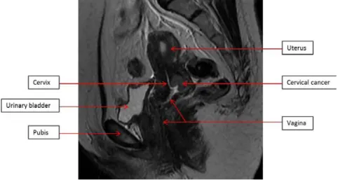
Due to discrepancies between the clinical staging and findings at surgery, the revised FIGO staging system now recommends obtaining pelvic MRI when available to better stage locally advanced disease. MRI is able to define the local extent of the disease including parametrium and pelvic sidewall invasion, urinary bladder and rectal invasion (Figure 12). Chest radiograph, CT scan, and PET scan can be utilized to evaluate the distant disease spread including
lymphadenopathy and distant organ metastasis2. CT scan has
replaced the traditional intravenous pyelography (IVP) to evaluate hydronephrosis (Figure 13).
Figure 12. 45-year-old female patient with biopsy proven cervical cancer. Axial (a) and Sagittal (b) images from an MRI of the pelvis demonstrate abnormal signal intensity involving the entire cervix (loss of the normal dark color), consistent with the patient’s cervical cancer. The cervical cancer is extending into the myometrium in the lower uterine segment and into the parametrium. There is also abnormal signal intensity involving the posterior wall of the urinary bladder which appears irregular, with loss of its normal dark signal intensity, consistent with bladder wall invasion. University of Massachusetts Medical School, Department of Radiology Abdominal Imaging Division
Figure 13. Same patient in Figure 6. Coronal image from a non-contrast CT scan of the abdomen and pelvis reveals the presence of bilateral hydronephrosis. Note also the presence of an enlarged left pelvic lymph node, most likely metastatic. University of Massachusetts Medical School, Department of Radiology, Abdominal Imaging Division
Biopsies of gross lesions are done. Colposcopy with biopsy of abnormal areas may be done as indicated. Endocervical curettage and cold knife conization (CKC) are often performed to determine depth of invasion and presence of lymphovascular space invasion in early stage cervical cancers with a normal appearing cervix by inspection and pelvic examination only. A CKC involves an excision of the transformation zone in a cone-shaped specimen with the apex of the cone pointed toward the endocervical canal beyond the transformation zone. Loop excisional electrocautery (LEEP) is typically used to treat CIN II-III. Compared to a LEEP, the CKC (in which cautery is only used after the excision to stop bleeding), preserves the morphology of the tissue to allow an accurate evaluation of the extent of disease by pathologists. The extent of the primary tumor, evident in a CKC is part of the information defined by the term “stage”, and the stage of a cervical cancer then determines the necessity for additional treatment. Cystoscopy and proctoscopy can be done to evaluate for a higher stage tumor defined by invasion of tumor into the bladder or rectum, respectfully.
Adib R. Karam, MD Antonella Leary, MD Richard S. Pieters, MD
The current staging system used for cervical cancer is located in the
NCI’s Cervical Cancer Treatment PDQ: Stages of Cervical Cancer.
Principles of Treatment
Pre-invasive disease is generally treated with expectant management (lower grades of dysplasia) or removal of precursor lesions by LEEP, CKC, ablation or cryotherapy.
Most cervical cancers that have not spread beyond the cervix are potentially cured with current treatments. However, once these cancers have spread into distant sites, such as lungs, bone, or liver, they are incurable and treatment is largely palliative.
In general, early stage cervical cancers are treated with surgery. More advanced stages of cervical cancer are generally treated with radiation and concurrent chemotherapy.
Currently, stage IA1 (lesions with <3mm invasion) disease is treated with cervical conization. Hysterectomy may be performed at a later time, when child bearing is complete. Stage IA2 (lesions with >3mm invasion and <5mm invasion) and stages IB1, IB2 and IIA can be treated with either surgically – with radical hysterectomy and bilateral pelvic lymphadenectomy or with primary radiation therapy including both brachytherapy and teletherapy.3 Survival rates from both modes
of treatment are similar. Decision for surgical or radiation therapy is usually made on an individual basis and takes into account the patient’s age, medical co-morbidities and functional status.
Stage IB2 or higher cervical cancer is usually treated with external beam radiotherapy (teletherapy) and concurrent chemotherapy (generally cisplatin) as a radiosensitizer. This first part of the course treats potential microscopic disease or actual bulk disease in the
pelvis or pelvic side walls, and hopefully shrinks the central tumor, so that the brachytherapy, which follows, can be more effective, by improving the anatomic geometry of the implant. Brachytherapy (i.e., “short distance” or localized and not external beam) radiation therapy with intracavitary radium or cesium or interstitial cesium needles is used to treat central tumor sites.
In young women of childbearing age with an early stage 1B-2 cervical cancer, radical resection of the cervix and pelvic lymph node dissection with preservation of the uterine fundus for future pregnancy has been accepted as a method of treatment.
Stage IVB cervical cancer has a poor prognosis and is generally treated with the ultimate goal of palliation.
With the completion of definitive therapy, each patient is evaluated with physical examinations and pap smears. They are generally
evaluated every 3 months for 1-2 years, every 6 months until the 5th
year and yearly thereafter. Cervical Cancer Prevention
HPV vaccination has been developed to prevent women from developing intraepithelial neoplasia and cancer. This vaccination is against HPV 16-18 strains and is indicated for girls ages 9-26.
Conclusion
Squamous cell carcinoma of the cervix is an important model for the development of invasive cancer from viral infection through preinvasive disease and for screening. Worldwide, it is the fourth most common cancer in women; in the United States, it is the twelfth, and the rate continues to decline.
Adib R. Karam, MD Antonella Leary, MD Richard S. Pieters, MD
cervical intraepithelial neoplasia to cervical carcinoma in situ to invasive cervical cancer and human papillomavirus infection is central to the development of cervical cancer. These three facts mean that the appearance of invasive cancer can be prevented with early intervention.
Thought Questions
1. Cervical cancer may be one of the most preventable cancers. From your knowledge of its pathogenesis, think of a variety of prevention strategies that could reduce the incidence of cervical cancer.
Expert Answer
2. Cervical cancer is far more common in developing countries than in industrialized countries. Why might that be? What barriers could there be to the implementation of the prevention strategies you developed in response to question #1 that might be more common in developing countries?
3. Advanced cervical cancer tends to invade regional structures. What symptoms and complications might a woman with locally advanced cervical cancer experience?
: Expert Answer
4.
.
Figure 14. These two images are from breast biopsies. Extrapolating from the diagnostic features of endocervical adenocarcinoma, which of the figures shows normal ductal epithelium and which figure shows ductal carcinoma? University of Massachusetts Medical School Department of Pathology.
: Expert Answer
Glossary
Brachytherapy- Close radiotherapy, either intracavitary or interstial, with radioactive sources put in a body cavity (intracavitary) or within tissue (interstitial)
Clinical staging- an estimate of how much cancer there is based on the physical exam and imaging tests before surgery
Colposcopy- Examination of the cervix with a colposcope, a magnifying devise which projects light onto the cervix; for evaluation of abnormal pap smears, special stains are applied to the cervix to enhance contrast between normal and abnormal tissue.
Endophytic- inclined to grow inward into tissues in fingerlike projections from a superficial site of origin
Exophytic- inclined to grow outward beyond the surface epithelium from which it originates
Rhabdomyosarcoma- malignant tumor made up of striated muscle fibers
Adib R. Karam, MD Antonella Leary, MD Richard S. Pieters, MD
fully assess the extent of disease
Teletherapy- Distance therapy, i.e. external beam therapy
Tumor “Grade"- Refers to the pathologist’s impression of how aggressive the tumor will behave. Grading for most cancers takes into account the following three features: 1) How closely do the cells resemble the cell of origin of the tumor? The degree to which tumor cells resemble the structure of normal tissue is referred to as “differentiation”. Intuitively, one can imagine that a “poorly differentiated” tumor (in which cells have lost much of the genetic programming of their origin), has evolved more, and is more likely to be able to adapt to new environments. For breast cancer, the degree of differentiation is estimated based on how well the cells can still cooperate to form tubular structures. For extremely well-differentiated “tubular carcinomas” of the breast, the risk of metastasis is close to zero. 2) How genetically unstable are the tumor cells? This is estimated from how much variability or “pleomorphism” is shown by the cells. The importance of pleomorphism can be understood intuitively: A highly genetically unstable, pleomorphic tumor is more likely to be able to evolve and adapt to new environments. 3-How fast is the tumor growing? Obviously a faster growing tumor may show a faster clinical evolution. Growth rate can be estimated based on the percentage of tumor cells that are in mitosis. More quantitative methods for estimating the growth rate can be achieved by immunohistochemical staining for proliferation markers. For example Ki67 immunostaining will highlight cells in S, M and G2 of the cell cycle. Image analysis of Ki67 staining shows promise for achieving more precise proliferation quantification. For certain tumors, there are other features that are important for “grade”. These include the presence of necrosis (particularly important for grading tumors of mesenchymal origin—i.e., sarcomas) and nucleolar size (particularly important for grading renal cell carcinomas).
References
1. National Cancer Institute. Cervical Cancer. Accessed 10
December 2010.
2. Freeman SF, Aly AM, Kataoka MY, Addley HC, Reinhold C, Sala
E. The revised FIGO staging system for uterine malignancies:
Implications for MR Imaging. Radiographics 2012;32:1805-1827.
3. Chen CC, Lin JC, Jan JS, Ho SC, Wang L. Definitive intensity-modulated radiation therapy with concurrent chemotherapy for
patients with locally advanced cervical cancer. Gynecol Oncol.
PubMed Abstract
4. Center for Disease Control and Prevention. Genital HPV


![catena Poly[[(N,N diethyldithiocarbamato κ2S:S′)phenylbismuth(III)] μ chlorido]](data:image/gif;base64,R0lGODlhAQABAIAAAP///wAAACH5BAEAAAAALAAAAAABAAEAAAICRAEAOw==)