T
o date, titanium has held a dominant position as an abutment and implant material in implant therapy,1,2 and long-term clinical studies3,4on commercially pure (CP) titanium demonstrate a predictable outcome. Demands for highly esthetic restorations have been raised by an increasing number of patients, which hasled to the introduction of tooth-colored ceramic implant abutments produced in densely sintered alumina.5,6 The peri-implant soft tissues around alumina abut-ments have been documented in both animal and human studies.7–10 Abutments made of CP titanium and alumina develop similar peri-implant mucosa, con-sisting of junctional epithelium and connective tissue attachment. Clinical studies have demonstrated stable peri-implant soft tissues around alumina abutments that have been observed over 3 to 4 years.11–13In ad-dition, since alumina abutments have a toothlike color, a more esthetic outcome could be achieved compared to using titanium abutments. Unfortunately, referred clinical studies11–13have additionally reported fractures to alumina abutments. Although alumina implant abut-ments perform well biologically as well as esthetically, it is apparent that they possess a fracture risk during both laboratory work and after abutment connection. Due to this shortcoming in their mechanical proper-ties, yttrium oxide–stabilized zirconia was introduced as an alternative material for implant abutments and it has overtaken alumina as the preferred ceramic abutment material.14The unique stress-induced transformation toughening mechanism in zirconia vastly improves its Purpose:The focus of this systematic review was to assess the published data
concerning zirconia dental implant abutments from various aspects. Materials and Methods:To identify suitable literature, an electronic search was performed using PubMed. The keywords “zirconia,” “zirconium,” “ceramic,” “dental abutments,” “dental implants,” “plaque,” and “bacteria” were included. Titles and abstracts were screened, and literature that fulfilled the inclusion criteria was selected for a full-text reading. Articles were divided into four groups: (1)studies on the mechanical properties of zirconia abutments, (2)studies on the peri-implant soft tissues around zirconia abutments, (3)studies on plaque accumulation on zirconia, and (4)clinical studies on the survival of zirconia abutments. Results:The initial literature search resulted in 380 articles. For groups 1 to 4, 11, 4, 7, and 3 articles satisfied the inclusion and exclusion criteria, respectively. Only 1 randomized clinical study was identified. Review of the selected articles showed that zirconia abutments were reliable in the anterior region from both biologic and mechanical points of view. Furthermore, zirconia abutments may represent a material surface less attractive for early plaque retention compared to titanium. Three clinical follow-up studies indicated that zirconia abutments could function without fracture and peri-implant lesions. Conclusions:Based on the
reviewed literature, zirconia has the potential to be used as a dental abutment material, although some issues have to be studied further. Int J Prosthodont 2010;23:299–309.
aResearch Fellow, New Industry Creation Hatchery Center, Tohoku University, Sendai, Japan; Research Fellow, Division of Fixed Prosthodontics, Department of Restorative Dentistry, Tohoku University Graduate School of Dentistry, Sendai, Japan.
bAssistant Professor, Division of Fixed Prosthodontics, Department of Restorative Dentistry, Tohoku University Graduate School of Dentistry, Sendai, Japan.
cAssociate Professor, Department of Prosthetic Dentistry/Dental Materials Science, Faculty of Odontology, Göteborg University, Göteborg, Sweden.
dAssociate Professor, Department of Prosthetic Dentistry/Dental Materials Science, Faculty of Odontology, Göteborg University, Göteborg, Sweden; Professor, Institute of Clinical Dentistry/Faculty of Medicine, University of Tromsø, Tromsø, Norway.
Correspondence to: Dr Keisuke Nakamura, New Industry Creation Hatchery Center, Tohoku University, Aoba 6-6-10, Aramaki, Aoba-ku, Sendai, 980-8579, Japan. Fax: +81-22-795-4110. Email: keisuke1@ mail.tains.tohoku.ac.jp
A Systematic Review
Keisuke Nakamura, DDS, PhDa/Taro Kanno, DDS, PhDb/Percy Milleding, DDS, PhDc/ Ulf Örtengren, DDS, PhDd
mechanical strength and reliability,15,16which has led to the increased use of zirconia as a ceramic biomate-rial in both medicine and dentistry.17,18The mechani-cal and microstructural properties of zirconia, as well as its biocompatibility, have been well docu-mented.17,19–21In dentistry, zirconia has been consid-ered for clinical applications such as frameworks for all-ceramic crowns and fixed partial dentures (FPDs),22–24brackets for orthodontic treatment,25and implants and abutments.26,27
Zirconia is a polymorphic material that displays four different crystalline structures. At room temperature, pure zirconia exists in a monoclinic form. The addition of stabilizing oxides (eg, yttrium oxide) to pure zirconia generates a multiphase structure, designated the metastable tetragonal phase, which has good mechan-ical properties. Owing to the metastable tetragonal phase, stabilized zirconia will display a stress-induced transformation toughening mechanism. The transfor-mation from the tetragonal to the monoclinic phase is associated with a 3% to 4% localized volume expansion that induces counteracting compressive stresses in compromised areas.17Besides the favorable mechani-cal properties, zirconia is proposed to accumulate den-tal plaque to a lesser extent than titanium.21
Despite the fact that ceramics as abutment materi-als have been used in dentistry for a number of years, only a limited number of review articles on ceramic abutments have been published to date.27–29 Concerning zirconia abutments, there is, to the au-thors’ knowledge, no systematic review published.
Thus, the increased use of zirconia as an abutment material calls for a systematic reevaluation of available data on zirconia. The purposes of this paper were therefore to review the literature systematically re-garding: (1)the mechanical properties of zirconia abut-ments from in vitro studies, (2)a histologic evaluation of peri-implant soft tissue responses around zirconia abutments from in vivo studies, (3)plaque accumula-tion or bacterial adhesion onto zirconia from both in vitro and in vivo studies, and (4)the survival of zirco-nia abutments from clinical studies
Materials and Methods
Search Strategy
A literature search focusing on the purposes previously mentioned was performed electronically using the PubMed database. The literature was divided into four groups following the intended purposes.
Articles published and recorded in PubMed through July 2009 were searched using the following key words: “zirconia” and “dental abutments,” “zirconium” and “dental abutments,” “zirconia” and “dental implants,”
“zirconium” and “dental implants,” “zirconia” and “plaque,” “zirconium” and “plaque,” “ceramic” and “plaque,” “zirconia” and “bacteria,” “zirconium” and “bacteria,” and “ceramic” and “bacteria.” The searches were limited to articles written in English with an as-sociated abstract. The electronic searches were com-plemented by manual searches through the bibliographies of the resulting articles and related re-views selected from the electronic search.
Inclusion and Exclusion Criteria
In this review, studies on zirconia ceramic composites or materials coated by zirconium compounds were ex-cluded; only tetragonal zirconia polycrystals or par-tially stabilized zirconia were included. Additional inclusion criteria for each study selection were in-cluded as follows:
• Group 1: In vitro studies on the mechanical proper-ties of zirconia abutments. Included studies investi-gated the fracture strength or fatigue of zirconia abutments or implant units, which involved an im-plant, a zirconia abutment, and a crown.
• Group 2: In vivo studies on peri-implant soft tissue re-sponses around zirconia abutments or implants. Included studies investigated dental implants used in the oral cavity and the soft tissues around implant components made of zirconia (ie, abutments, healing caps, or around transmucosal zirconia implants). Included studies also examined the histologic analy-sis of the peri-implant soft tissue and the normal peri-implant soft tissue (not experimentally induced inflammatory tissue).
• Group 3: Studies on plaque accumulation or bacterial adhesion onto zirconia. Included studies used zirconia as a material or substrate for plaque accumulation or bacteria adhesion and had a description not only about the microbiologic analysis but also the surface topog-raphy of the substrate or material.
• Group 4: Clinical studies on the stability of zirconia abutments. Included studies reported clinical results of zirconia abutments, had a minimum number of 20 subjects at the baseline examination (case reports were excluded), and had at least a 1-year follow-up period. If there were several studies following the same population, only the most recent was included in this review.
Statistics
No meta-analysis was performed because there were too few studies in each review category and great vari-ations in study design were evident. The statistics pre-sented were taken from the reviewed articles.
Results
Study Characteristics
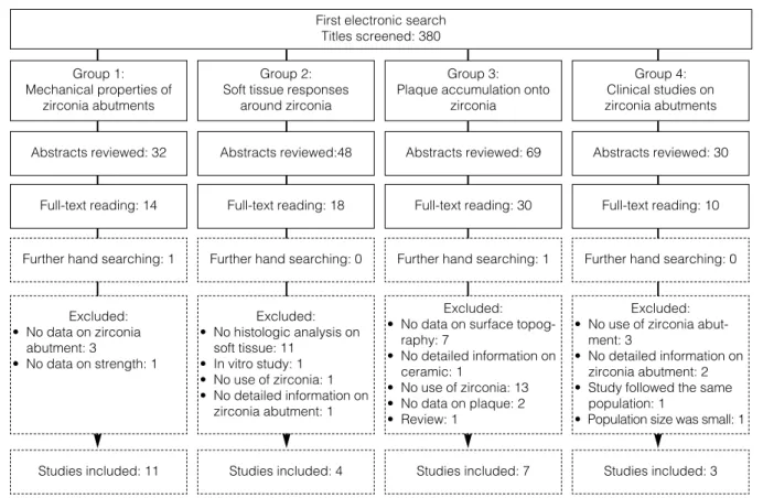
The initial PubMed search resulted in 380 papers. After screening and taking into consideration the inclusion criteria for groups 1 to 4, 11, 4, 7, and 3 articles re-mained, respectively (Fig 1). Unfortunately, a direct comparison of the various results was difficult to ob-tain since the study designs of the reviewed articles in each category varied.
Group 1
From the 11 studies reviewed in this category,30–40two types of zirconia abutments were identified (Table 1). One type was composed of all-zirconia30,32–34,36,40and the other was reinforced with metal (titanium) at the implant-abutment interface (zirconia abutment com-plexes).31,35,37 All reviewed literature in this category, except for 1 study,35 evaluated the strength of the zirconia abutment combined with implants or implant replicas and crowns. Of these 10 studies, 4 evaluated the strength of the zirconia abutment after thermomechan -ical fatigue31–33or after cyclic loading,34 whereas the
remaining 6 studies evaluated strength using static loading only.30,36–40In the experiment without cyclic loading of zirconia abutments, Yildirim et al30reported a mean fracture load of the zirconia abutments of 737 N. This finding has also been confirmed by other re-searchers.37,38The fracture strength against cyclic load-ing or thermomechanical fatigue was, however, reduced significantly.31–34Gehrke et al34reported a decreased strength of zirconia from 672 N without cyclic loading to less than 405 N after cyclic loading using loads be-tween 100 and 450 N for up to 5,000,000 loading cycles. Thermomechanical fatigue studies on zirconia31–33at loads of less than 50 N for 1,200,000 loading cycles showed decreased strength (between 457 and 281 N) compared to the results of Yildirim et al.30
Three studies30,32,33compared the strength of zirco-nia abutments with alumina abutments. Two of them showed that zirconia abutments had significantly higher strength than alumina abutments,30,32 whereas one failed to show any significant difference between them.33 Although it is not possible to compare fracture strength values between various studies because of differences in study design, the reviewed articles demonstrated that zirconia abutments could be used in the anterior region of the dentition safely, where the physiologic maximal First electronic search
Titles screened: 380
Group 1: Mechanical properties of
zirconia abutments
Group 2: Soft tissue responses
around zirconia
Group 3: Plaque accumulation onto
zirconia
Group 4: Clinical studies on zirconia abutments
Abstracts reviewed: 32 Abstracts reviewed:48 Abstracts reviewed: 69 Abstracts reviewed: 30
Full-text reading: 14 Full-text reading: 18 Full-text reading: 30 Full-text reading: 10
Further hand searching: 1 Further hand searching: 0 Further hand searching: 1 Further hand searching: 0
Studies included: 11 Studies included: 4 Studies included: 7 Studies included: 3 Excluded: • No data on zirconia abutment: 3 • No data on strength: 1 Excluded: • No histologic analysis on soft tissue: 11 • In vitro study: 1 • No use of zirconia: 1 • No detailed information on zirconia abutment: 1 Excluded: • No data on surface
topog-raphy: 7 • No detailed information on ceramic: 1 • No use of zirconia: 13 • No data on plaque: 2 • Review: 1 Excluded: • No use of zirconia
abut-ment: 3
• No detailed information on zirconia abutment: 2 • Study followed the same
population: 1
• Population size was small: 1
occlusal forces reach approximately 300 N.41,42 Concerning mechanical strength, zirconia abutments work as well as alumina abutments.
Group 2
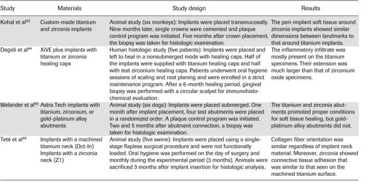
Four papers were found to fulfill the inclusion criteria in this group (Table 2).43–46These studies indicated that zirconia was a suitable abutment material compared to titanium concerning tissue responses. Furthermore, Table 1 In Vitro Studies on Mechanical Properties of Zirconia Abutments
Study Materials Study design Results
Yildirim et al30 Alumina abutments (n = 10) Implant: Brånemark Mean fracture load: All-zirconia abutments (partially Crown: glass ceramic Al = 280.1 N, Zr = 737.6 N stabilized, n = 10) Cyclic loading: none (only static loading) Significant difference: Al vs Zr
Angle of force application: 30 degrees to vertical
Butz et al31 Zirconia abutment complexes (n = 16) Implant: Osseotite During the cyclic loading, one Alumina abutments (n = 16) Crown: metal crowns (nonprecious alloy) alumina abutment fractured. Titanium abutments (n = 16) Cyclic loading: 1.2 million cycles of thermo- Mean fracture load: Zr = 281 N,
mechanical fatigue with a force of 30 N Al = 253 N, Ti = 305 N
Angle of force application: 50 degrees to vertical Significant difference: Zr vs Al, Ti vs Al Att et al32 Titanium abutments (n = 16) Implant: Replace Select All specimens survived 1.2 million
Alumina abutments (n = 16) Crown: all-ceramic with zirconia copings cycles of dynamic loading. Zirconia abutments (n = 16) Cyclic loading: 1.2 million cycles of thermo- Median fracture resistance:
mechanical fatigue with a force of 49 N Ti = 1,251 N, Al = 457 N, Zr = 241 N Angle of force application: 45 degrees to vertical Significant difference: Ti vs Al and
Zr, Al vs Zr
Att et al33 Titanium abutments (n = 16) Implant: Replace Select All specimens survived 1.2 million Alumina abutments (n = 16) Crown: all-ceramic with alumina copings cycles of dynamic loading. Zirconia abutments (n = 16) Cyclic loading: 1.2 million cycles of thermo- Median fracture resistance:
mechanical fatigue with a force of 49 N Ti = 1,454 N, Al = 422 N, Zr = 443 N Angle of force application: 45 degrees to vertical Significant difference: Ti vs Al and Zr Gehrke et al34 All-zirconia abutments (n = 7) Implant: XiVE During static loading: 672 N
Crown: spherical caps (no description in detail) After 811,023 to 5 million cycles: Cyclic loading: at most 5 million cyclic loading 268.8 N
with a force of 100 to 450 N After 10,000 cycles: 403.2 N Angle of force application: 30 degrees to vertical No description of statistics Canullo et al35 Zirconia abutment complexes (n = 20) Implant: none Mean maximum load: 436 N
Crown: none
Cyclic loading: none (only static loading) Angle of force application: 30 degrees to vertical
Sundh and Mg-PSZ abutments (n = 10) Implant: Straumann (titanium with stainless All combinations of ceramic Sjögren36 HIPed Y-TZP abutments (n = 10) steel analog) abutments and implants exceeded
Titanium abutment with zirconia Crown: none but zirconia copies 300 N (individual values not shown). coping (n = 10) Cyclic loading: none (only static loading) Significant difference: Ti vs Mg-PSZ
Angle of force application: 90 degrees to vertical (Ti < Mg-PSZ)
Aramouni et al37 Zirconia abutment complexes (n = 20) Implant: 3i Certain or SLA Straumann Mean fracture strength: ZiReal = 792.7 Zirconia abutment (n = 20) Crown: all-ceramic made of Empress 2 N, synOcta Ceramic Blank = 604.0 N, Noble metal abutment (n = 20) Cyclic loading: none (only static loading) UCLA = 793.6 N
Angle of force application: 45 degrees to vertical Significant difference: synOcta vs ZiReal and UCLA
Kerstein et al38 All-zirconia abutment Implant: Nobel Biocare Mean fracture strength:
(Atlantis zirconia, n = 29) Crown: none Atlantis = 831 N, AllZirkon = 740 N All-zirconia abutment (Procera Cyclic loading: none (only static loading) Significant difference: Atlantis vs AllZirken, n = 29) Angle of force application: 40 degrees to vertical AllZirkon
Kim et al39 Pressable metal ceramic custom Implant: implant analog (Replace Select) Mean fracture strength: pressable implant abutments (n = 10) Crown: all-ceramic lithium disilicate pressable metal ceramic = 901.67 N, All-zirconia abutments (n = 10) ceramic crowns (IPS e.max Press) zirconia = 480.01 N
Cyclic loading: none (only static loading) Angle of force application: 30 degrees to vertical
Adatia et al40 Y-TZP ceramic abutment Implant: implant analog (stainless steel) Mean fracture strength:
Group 1 (n = 10): no modification Crown: none Group 1 = 429 N, Group 2 = 576 N, Group 2 (n = 10) and group 3 (n = 10): Cyclic loading: none (only static loading) Group 3 = 547 N
chamfer preparation with occlusal Angle of force application: 30 degrees to vertical Significant difference: none reduction using a high-speed dental
handpiece
Zr = zirconia; Al = alumina; Ti = titanium; Mg-PSZ = magnesia partially stabilized zirconia; HIPed = hot isostatic pressed; Y-TZP = yttrium oxide partially stabilized zirconia; SLA = sandblasted, large-grit, acid-etched.
peri-implant soft tissues around zirconia exhibit the po-tential to heal faster than when in contact with titanium. Kohal et al43evaluated and compared the conditions of soft and hard tissues in contact with zirconia and ti-tanium implants in monkeys. The authors concluded that bone and soft tissues appeared to integrate with zirconia as well as titanium. This finding was also con-firmed by other researchers.45,46Welander et al45 com-pared abutments made of titanium, zirconia, and gold-platinum alloy in dogs. The soft tissue barrier formed around titanium and zirconia abutments dis-played equal and stable conditions following 2 and 5 months of healing. On the other hand, gold-platinum abutment sites demonstrated an apical shift of the barrier epithelium and the level of marginal bone over the same time period.
Degidi et al44conducted a human histologic study to compare peri-implant soft tissues in contact with ti-tanium and zirconium oxide healing caps. Although no clinically visible plaque accumulation or bleeding on probing was recorded in either group, the inflamma-tory infiltrate was observed more frequently in the peri-implant soft tissues around titanium healing caps compared to the zirconia healing caps.
Group 3
Seven papers were found and reviewed in this category (Table 3).47–53Generally, lower plaque formation was recorded on zirconia specimens compared to other evaluated materials. Nakazato et al47compared plaque
formation in vivo on six different materials, including alumina, titanium, and zirconia. They hypothesized that the implant surface properties might play important roles in bacterial adherence during the early stages of plaque formation after being affected to a greater ex-tent by the material’s surface roughness than by its sur-face free energy. Since the materials used in their study had different surface roughness levels, it could not be determined if and how the various surface factors af-fected the adherence of oral bacteria. Rimondini et al,49however, were able to evaluate the role of bacte-rial adhesion on zirconia and titanium specimens with equivalent average surface roughness (Ra) values both in vivo and in vitro. The various strains of oral bacteria studied included Streptococcus sanguis, Actinomyces, Porphyromonas gingivalis, and Streptococcus mutans. S mutans exclusively displayed significantly increased attachment to zirconia specimens (Ra = 0.18 µm) com-pared to titanium specimens (Ra = 0.22 µm) after an incubation period of 36 hours in vitro. In vivo, however, zirconia specimens accumulated significantly fewer bacteria than titanium specimens after 24 hours. Scarano et al50confirmed the latter finding in vivo by comparing zirconia and titanium specimens with sur-face roughness values of 0.76 µm and 0.73 µm, re-spectively. The percentage of disk surface covered by bacteria was significantly lower on zirconia than on ti-tanium after 24 hours. However, in the studies where early bacterial adhesion (within 2.5 hours) to zirconia and other dental ceramic materials were compared, no differences were reported.52,53
Table 2 In Vivo Studies on Peri-Implant Soft Tissue Responses Around Zirconia Abutments or Implants
Study Materials Study design Results
Kohal et al43 Custom-made titanium Animal study (six monkeys): Implants were placed transmucosally. The peri-implant soft tissue around and zirconia implants Nine months later, single crowns were cemented and plaque zirconia implants showed similar
control program was initiated. Five months after crown placement, dimensions between landmarks to the biopsy was taken for histologic examination. that around titanium implants. Degidi et al44 XiVE plus implants with Human histologic study (five patients): Implants were placed and The inflammatory infiltrate was
titanium or zirconia left to heal in a nonsubmerged mode with healing caps. Half of mostly present on the titanium healing caps the implants were supplied with titanium healing caps and half specimens. Their extension was
with test zirconium healing caps. Patients underwent oral hygiene much larger than that of zirconium sessions of scaling and root planing and were enrolled in a strict oxide specimens.
maintenance program. After a 6-month healing period, gingival biopsy was performed with a circular scalpel for immunohisto-chemical evaluation.
Welander et al45Astra Tech implants with Animal study (six dogs): Implants were placed submerged. One The titanium and zirconia abut-titanium, zirconium, or month after implant placement, four test abutments were placed ments promoted proper conditions gold-platinum alloy in a randomized order. A plaque control program was initiated. for soft tissue healing, but gold-abutments Two and 5 months after abutment connection, a biopsy was platinum alloy abutments did not.
taken for histologic examination.
Teté et al46 Implants with a machined Animal study (five swine): Implants were placed using a single- Collagen fiber orientation was titanium neck (Oct-In) stage flapless surgical procedure and were not functionally similar regardless of implant neck Implants with a zirconia loaded. Oral hygiene was performed on the day of surgery and material. Moreover, zirconia showed neck (Z1) monthly during the experimental period (3 months). Animals were connective tissue adhesion that
sacrificed 3 months after implant insertion for histologic analysis. was similar to that seen on the machined titanium surface.
The long-term effect of plaque accumulation on zir-conia and titanium abutments was investigated by Bollen et al.48Abutments of the two materials were placed in-traorally in six patients receiving implant-supported over-dentures. Clinical and microbiologic examinations after 12 months failed to reveal any major differences quan-titatively or qualitatively between supra- and subgingi-val plaque from the abutment material surfaces. Conclusions from these findings suggest that zirconia might reduce early bacterial adhesion (< 24 hours) com-pared to titanium. However, it is still unclear whether this characteristic of zirconia is of any clinical benefit.
Group 4
Only three papers in the fourth group were found to ful-fill the criteria for the present review (Table 4).54–56 Two of them were prospective clinical trials54,55and the remaining was a randomized controlled trial (RCT).56 Although another RCT was found,57it was excluded be-cause the same population was studied and reported in another paper.56The results of the two prospective studies showed good clinical performance in the an-terior and premolar regions for zirconia abutments without fracture and peri-implant lesion during the Table 3 Studies on Plaque Accumulation or Bacterial Adhesion onto Zirconia
Study Materials Study design Results
Nakazato et al47Single-crystal alumina (Ra = 0.090 µm) In vivo study (3 subjects): Disks of each material were At the 4-hour setting time, Polycrystal alumina (Ra = 0.854 µm) set on the gingiva of the subjects using removable poly crystal alumina and Partially stabilized polycrystal zirconia devices. Tooth brushing was not permitted. The disks hydroxyapatite had a higher (Ra = 0.369 µm) were removed after 4 and 48 hours. The disks were concentration of bacteria. Hydroxyapatite (Ra = 0.518 µm) subjected to SEM observations and microbiologic However, after 48 hours,
Pure titanium (Ra = 0.142 µm) examination. the material surfaces were
Heat-polymerized acrylic resin uniformly covered. No apparent
(Ra = 0.109 µm) differences were observed.
Bollen et al48 CP titanium abutments (Ra = 0.21 µm) In vivo study (6 subjects with implant-supported There were no major differences Zirconia (Polyzir) abutments overdentures): The two test abutments were delivered both quantitatively and qualita-(Ra = 0.06 µm) using the split-mouth technique. After 3 and 23 tively between the supra- and
months, both clinical and microbiologic examinations subgingival plaque of the two test
were repeated. abutment surfaces.
Rimondini et al49Titanium disks (Ra = 0.22 µm) In vitro study (7 specimens of each material): Test In vitro test: Polished zirconia Polished zirconia disks (Ra = 0.18 µm) specimens were anaerobically incubated for 36 hours disks showed significantly more High-polished zirconia disks with bacteria precultured in broth. adherent S mutansthan high-(Ra = 0.04 µm) In vivo study (10 subjects): Removable devices were polished and titanium.
produced and three specimens (one of each type) In vivo test: The zirconia disks were fixed mechanically into the silicone stent. The accumulated significantly fewer device was fixed onto the buccal region of the molar bacteria than titanium disks. or premolar of each volunteer. Subjects suspended
any oral hygiene procedures for 24 hours. Disks were then removed and observed with SEM.
Scarano et al50 Titanium disks (Ra = 0.73 µm) In vivo study (10 subjects): Removable devices having The percentage of the disk Zirconia disks (Ra = 0.76 µm) test disks were adapted to the molar-premolar region. surface covered by bacteria on
Neither cleaning procedures nor agents for chemical the zirconia specimens was plaque control were applied to the disks for 24 hours. significantly lower than that of Disks were then removed and observed with SEM. titaniumspecimens (12.1% and
19.3%, respectively). Scotti et al51 Polished zirconia disk (no Ra available) In vivo study (2 subjects): Samples were fixed on No significant difference was
Glazed zirconia disks (no Ra available) buccal and palatal surfaces of individual oral found in bacteria presence appliances made of light-cured resin. The presence or between glazed and polished absence of bacteria on the surfaces was recorded zirconia samples
using SEM at 20 minutes and 1 and 6 hours.
Meier et al52 Glass (control; Ra = 0.24 µm) In vitro study (5 specimens of each material): The materials' properties, surface Feldspathic ceramic (Ra = 0.26 µm) Before adhesion, specimens were exposed to roughness, and glass content Glass-infiltrated alumina (Ra = 1.33 µm) sterile human saliva for 15 minutes. Adhesion was had only a weak influence on Zirconia-reinforced glass-infiltrated performed using four different streptococci and a streptococci adhesion. alumina (Ra = 1.34 µm) flow chamber for 1 hour.
Tetragonal stabilized zirconia (Ra = 0.26 µm)
Rosentritt et al53Zircona:
Cercon Base (Ra = 0.22 µm) In vitro study (15 specimens of each material): A low adhesion of several Digizon (Ra = 0.08 µm) After 2 hours of incubation with artificial saliva, strep tococci to glass ceramic as Inceram Y-TZP (Ra = 0.22 µm) specimens were incubated with various well as to zirconia was found. Veneering glass ceramic: streptococcal suspension for 2.5 hours. There were only little differences
Cercon Ceram S (Ra = 0.22 µm) between zirconia and glass
Omega 900 (Ra = 0.57 µm) ceramic with regard to
GC Zirconia (Ra = 0.10 µm) streptococci adhesion.
Glass (control)
observation periods (40 and 48 months, respectively). In addition, the RCT showed that zirconia abutments could also function well in the molar region without technical problems, such as abutment fracture, screw loosening, and loss of crown retention.56
Glauser et al54evaluated both peri-implant hard and soft tissue reactions to experimental zirconia abutments made by an individualization process of densely sintered yttrium-stabilized zirconia ingots, supporting single crowns in the esthetic zone, and technical problems re-lated to the abutment materials. Twenty-seven patients (16 women, mean age: 42 years; 11 men, mean age: 46 years) received 54 implants with experimental zirconia abutments (all-zirconia abutments) and all-ceramic crowns (Empress I, Ivoclar Vivadent). Following 1, 12, and 48 months postinsertion, clinical evaluations including assessment of the condition of the peri-implant mucosa were performed. Over the course of this study, no abut-ment fractures were observed, resulting in a survival rate of 100%. The peri-implant mucosa was judged as healthy with regard to Plaque Index and Gingival Index. However, at the 48-month follow-up, a patient/restora-tion dropout rate of 33% was recorded, which inevitably lowered the impact of the 48-month results.
In studying the clinical efficacy of zirconia abutments cemented with a composite resin cement to a titanium substructure (zirconia abutment complex), Canullo55 evaluated 25 patients (14 women, 11 men; mean age: 52 years) requiring 30 zirconia implants provided with zirconia abutments and single implant-supported all-ceramic crowns. No detailed information about the type of all-ceramic crowns used was given. The crowns replaced anterior teeth (eight in the maxilla and eight in the mandible) and premolars (eight in the maxilla and two in the mandible) but only a few molars (two in the maxilla and two in the mandible). The patients were fol-lowed for a mean observation period of 40 months. No abutment fracture or screw loosening was reported during clinical loading, resulting in a cumulative survival
rate of 100%. Since no information was disclosed on patient/restoration dropouts, the reported survival rate must be evaluated cautiously. The Plaque Index and the Gingival Index indicated that the soft tissues around the abutments were considered healthy.
In the RCT,5622 patients (14 women, 8 men; mean age: 41.3 years) who were in need of implant-supported single crowns (n = 40) in the canine, pre-molar, and molar regions were included. At abutment connection, 20 customized all-zirconia abutments (Procera, Nobel Biocare) or 20 customized titanium abutments (Procera, Nobel Biocare) were assigned randomly. All-ceramic crowns were either fabricated out of glass ceramic or out of high-strength ceramic (alumina or zirconia) for the zirconia abutments, while metal-ceramic crowns were fabricated for the tita-nium abutments. During the follow-up period, no tech-nical problems were observed despite the fact that 27% of zirconia abutments supported crowns in the molar region. Hence, the abutment survival rate was 100%. Furthermore, no biologic complications were found at zirconia abutments as well as titanium abutments. Thus, custom-made zirconia abutments might be use-ful for anterior and premolar implant-supported fixed prostheses regardless of whether they are all-zirconia abutments or zirconia abutment complexes. In addition, zirconia abutments might also function well in molar regions.
Discussion
Systematic reviews are often useful in the evaluation of various materials and treatments since they extract the best evidence from the scientific literature.58 Concerning zirconia abutments, no systematic review has been performed thus far. The reason for this may be that zirconia abutments have been used for only a short period of time and data are still limited. Still, the interest in zirconia abutments is increasing due to their Table 4 Clinical Studies on Stability of Zirconia Abutments
Survival
Follow-Study Materials Prosthesis type rate up Results
Glauser et al54 27 patients with 54 experimental Implant-supported single-tooth 100% 48 mo No abutment fractures were all-zirconia abutments restorations in the incisor, observed and the peri-implant No control group canine, and premolar regions mucosa was healthy. Canullo55 25 patients with 30 zirconia Implant-supported single-tooth 100% 40 mo Neither abutment fracture nor
abutment complexes restorations in the incisor, canine, (mean) peri-implant soft tissue lesion No control group premolar, and molar regions was reported during clinical
loading.
Zembic et al56 22 patients with 20 zirconia and Implant-supported single-tooth 100% 36 mo At 3 years, zirconia and titanium 20 titanium abutments (RCT) restorations in the canine, premolar, (mean) abutments exhibited the same
and molar regions survival and technical, biologic, and esthetic outcomes.
favorable mechanical and esthetic properties. In that respect, this systematic review was performed as an at-tempt to evaluate the available data on zirconia abut-ments. Difficulties were experienced in that only a few studies focused on zirconia abutments. Only one RCT was identified. RCTs are regarded as the highest level of evidence. Since few RCTs have been performed on zirconia abutments to date, it should be recognized that the results of the studies reviewed in this article do have limited evidence and clinical relevance.59,60In this re-view, in vitro studies and in vivo animal studies were included because they are well accepted for supplying basic scientific knowledge, although their clinical rel-evance may be questionable.61
Two main factors might cause failure in implant therapy, one of which is related to mechanical failure and the other to biologic complications. The former in-cludes implant fracture, abutment fracture, and frac-ture of the superstrucfrac-ture, whereas the latter includes failure and loss of osseointegration.62–64In addition, the esthetic requirements for implant abutments are or will be more strict in the esthetic zone.
In vitro, the mechanical flexural strength of zirconia (disks or bars) has been recorded from 900 to 1,200 MPa, which is approximately twice that of alumina.17 Concerning zirconia abutments, it was also confirmed that the fracture load of zirconia was more than twice that of alumina.30However, it was observed that the strength of the zirconia abutment decreased after cyclic loading. This could be attributed to the aging process. Still, it remains to be determined whether cyclic load-ing is responsible for the decline in strength and what clinical consequences will result.
The mechanical strength of zirconia abutments can also be affected by the method of fabrication.65–67 Furthermore, zirconia abutments can be adjusted chair-side using a high-speed dental handpiece with a dia-mond bur.40Although these factors must be taken into account for the strength of the abutment, they were dif-ficult to assess in the present review due to lack of valu-able information in a majority of the articles. Although chairside adjustment of zirconia abutments might be possible,40further studies on the subject are needed.
It was apparent from the reviewed literature that even if the mechanical strength of zirconia was reduced by cyclic loading, the zirconia abutment would probably have enough mechanical strength clinically. The all-zirconia abutment and the all-zirconia abutment complex were found to be comparable regarding mechanical strength.
The peri-implant soft tissues around titanium abut-ments have been well documented.1,2,68–72According to histologic studies, periimplant soft tissues and peri -odontal tissues are composed of junctional epithelium and connective tissue attachment and act as barriers
between the oral environment and the internal struc-tures of the body. Although the dimensions of the soft tissue barrier around implants and teeth are similar, the connective tissue attachment is different.68,73,74 Four histologic studies included in the present systematic re-view demonstrated that the peri-implant soft tissues around zirconia are similar to those around the titanium abutments. Furthermore, human histologic analysis44 indicated that peri-implant soft tissues around zirconia might heal faster than when in contact with titanium.
According to the articles reviewed, zirconia appeared to be superior to titanium with less initial plaque ac-cumulation when there were no differences in surface topography. Examination of only the surface roughness value was performed despite other surface parameters existing, such as the skewness factor, which offers valuable information on details of the topography. Due to lack of information, such as the surface free energy value and the surface elemental composition, it is dif-ficult to draw any valid conclusions from the referred articles. The complex evaluation of the importance of plaque in relation to solid abutment surfaces calls for thorough analyses of which bacterial strains constitute the plaque. Also, the importance of the surface free en-ergy as a causative factor for bacterial adhesion on zir-conia abutments warrants further investigation.
It has been reported that although slightly less plaque accumulation on zirconia rather than titanium abut-ments offers no general clinical advantage, plaque ad-herence and plaque removal might be influenced. In a series of studies, Bollen et al48and Quirynen et al75,76 determined the threshold surface roughness value of plaque accumulation on titanium. The threshold value was proposed to be Ra = 0.2 µm, below which no or only minor influence of the surface topography occurred on plaque accumulation. However, Wennerberg et al77failed to find any clinical or histologic differences between surface roughness of titanium abutments and soft tissue inflammatory responses after an evaluation period of 4 weeks. It remains to be determined whether there might be a relationship between surface factors, plaque accumulation, and soft tissue inflammatory responses. In the prospective clinical studies, it was observed that zirconia abutments would not cause technical or biologic problems, at least over short or intermediate observation periods (40 to 48 months).54,55 This was confirmed by the RCT at the 36-month follow-up.56A prospective study on alumina abutments supporting short-span FPDs displayed a cumulative success rate of 98.1% over 5 years.12Based on the reviewed stud-ies, it can be hypothesized that zirconia abutments, with superior mechanical properties compared to alumina, will function as abutments for anterior FPDs with a suc-cess rate that corresponds to or is better than alumina. Additionally, the possibility of zirconia abutments for
restorations in the molar region has been shown.55,56 Still, the small sample size and the relatively short ob-servation period (36 to 40 months) justify further stud-ies for supporting a decision to possibly expand the indications for zirconia abutments.
It is difficult to recommend an appropriate design of zirconia abutments (eg, dimension) because of a lack of data. Only one clinical study reviewed in the present paper mentioned a minimum thickness of zirconia abutments (0.5 mm).54When computer-aided design/ computer-assisted manufacture is used to mill a presintered zirconia block for the fabrication of restora-tions, it is recommended to not reduce the thickness below 0.5 mm.20Therefore, it is reasonable to assume a minimum thickness of zirconia abutments to be 0.5 mm or more to withstand a functional load. Concerning the height of the abutment and the collar, it apparently varies from patient to patient, and further studies may be needed.
Albeit only a few clinical studies reported the out-come of zirconia abutments, zirconia appears suitable as an abutment material. For future improvements to ceramic abutments, however, two issues require focus: color and the long-term stability of zirconia.27The color of zirconia is too white compared to natural teeth. This might cause another problem in esthetic dentistry, at least in the esthetic zone. Therefore, a tooth-colored zir-conia abutment that matches the cervical portion of the natural teeth has been developed recently.78,79
Regarding long-term stability, the aging of zirconia has become an issue for implants used in orthope-dics.80–82Aging is suggested to be a progressive trans-formation of the metastable tetragonal phase into the monoclinic phase, causing degradation of the me-chanical properties.80,83Aging of zirconia might also be a critical problem in the field of dentistry. Further stud-ies should verify if the aging process causes critical damage to zirconia abutments. It is expected that this will be addressed in the future.
Conclusions
On the basis of the available data, the following con-clusions can be drawn:
• Mechanical properties of zirconia abutments: Based on in vitro studies, zirconia abutments are applicable to the anterior region equally as much as alumina abutments. Additionally, evidence from clinical stud-ies has shown that zirconia abutments functioned up to 4 years without mechanical problems. However, it is still necessary to prove that zirconia abutments will be safe for posterior restorations.
• Peri-implant soft tissue responses around zirconia abutments or implants: From the animal and human
histologic studies reviewed, it can be concluded that zirconia is as suitable a material for dental implant abutments as titanium concerning biocompatibility. This has also been confirmed in clinical studies ad-dressing the maintenance of healthy peri-implant soft tissues.
• Plaque accumulation onto zirconia: Further research is warranted before the clinical relevance concerning the differences in plaque formation on titanium and zir-conia abutment surfaces can be concluded. Zirzir-conia appears to have a lower tendency for surface-bound bacterial plaque at early stages.
• Clinical results on zirconia abutments: Since only three studies addressing the clinical outcome of zir-conia abutments passed the inclusion criteria, the conclusions based on those studies should be inter-preted with caution. The two prospective studies and the RCT indicated that zirconia abutments function without fracture and peri-implant lesions for up to 4 years. From this review, it is suggested that more RCTs comparing zirconia with titanium abutments using a large population should be conducted to evaluate the benefits of zirconia abutments.
Due to the limited number of well-performed scien-tific studies published to date, this systematic review concludes that at present, zirconia abutments should be used with caution for single implant–supported restorations in the esthetic zone. Concerning me-chanical and biologic properties, zirconia abutments seem to be as applicable as titanium or alumina. It re-mains to be determined whether this assumption will hold true for follow-up periods over 5 years in prospec-tive randomized controlled clinical trials. To optimize es-thetics further, development of tooth-colored zirconia is necessary. To expand on the data on zirconia restora-tions in the future, it is of crucial importance to eluci-date the influence of the aging process on zirconia.
References
1. Lindhe J, Berglundh T. The interface between the mucosa and the implant. Periodontol 2000 1998;17:47–54.
2. Linkevicius T, Apse P. Influence of abutment material on stability of peri-implant tissues: A systematic review. Int J Oral Maxillofac Implants 2008;23:449–456.
3. Lekholm U, Gunne J, Henry P, et al. Survival of the Brånemark im-plant in partially edentulous jaws: A 10-year prospective multi-center study. Int J Oral Maxillofac Implants 1999;14:639–645. 4. Ekelund JA, Lindquist LW, Carlsson GE, Jemt T. Implant treatment
in the edentulous mandible: A prospective study on Brånemark system implants over more than 20 years. Int J Prosthodont 2003; 16:602–608.
5. Prestipino V, Ingber A. Esthetic high-strength implant abutments. Part I. J Esthet Dent 1993;5:29–36.
6. Prestipino V, Ingber A. Esthetic high-strength implant abutments. Part II. J Esthet Dent 1993;5:63–68.
7. McKinney RV Jr, Steflik DE, Koth DL. Evidence for a junctional ep-ithelial attachment to ceramic dental implants. A transmission electron microscopic study. J Periodontol 1985;56:579–591. 8. Arvidson K, Fartash B, Hilliges M, Köndell PA. Histological
char-acteristics of peri-implant mucosa around Brånemark and single-crystal sapphire implants. Clin Oral Implants Res 1996;7:1–10. 9. Fartash B, Tangerud T, Silness J, Arvidson K. Rehabilitation of
mandibular edentulism by single crystal sapphire implants and overdentures: 3-12 year results in 86 patients. A dual center in-ternational study. Clin Oral Implants Res 1996;7:220–229. 10. Abrahamsson I, Berglundh T, Glantz PO, Lindhe J. The mucosal
attachment at different abutments. An experimental study in dogs. J Clin Periodontol 1998;25:721–727.
11. Andersson B, Taylor A, Lang BR, et al. Alumina ceramic implant abutments used for single-tooth replacement: A prospective 1- to 3-year multicenter study. Int J Prosthodont 2001;14:432–438. 12. Andersson B, Glauser R, Maglione M, Taylor A. Ceramic implant
abutments for short-span FPDs: A prospective 5-year multicen-ter study. Int J Prosthodont 2003;16:640–646.
13. Henriksson K, Jemt T. Evaluation of custom-made procera ceramic abutments for single-implant tooth replacement: A prospective 1-year follow-up study. Int J Prosthodont 2003;16:626–630. 14. Holst S, Blatz MB, Hegenbarth E, Wichmann M, Eitner S.
Prosthodontic considerations for predictable single-implant es-thetics in the anterior maxilla. J Oral Maxillofac Surg 2005;63 (suppl 2):89–96.
15. Garvie RC, Hannink RH, Pascoe RT. Ceramic steel? Nature 1975;258:703–704.
16. Scott HG. Phase relationships in the zirconia-yttria system. J Mater Sci 1975;10:1527–1535.
17. Piconi C, Maccauro G. Zirconia as a ceramic biomaterial. Bio -materials 1999;20:1–25.
18. Manicone PF, Rossi Iommetti P, Raffaelli L, et al. Biological con-siderations on the use of zirconia for dental devices. Int J Immunopathol Pharmacol 2007;20(suppl 1):9–12.
19. Manicone PF, Rossi Iommetti P, Raffaelli L. An overview of zirco-nia ceramics: Basic properties and clinical applications. J Dent 2007;35:819–826.
20. Denry I, Kelly JR. State of the art of zirconia for dental applications. Dent Mater 2008;24:299–307.
21. Hisbergues M, Vendeville S, Vendeville P. Zirconia: Established facts and perspectives for a biomaterial in dental implantology. J Biomed Mater Res B Appl Biomater 2009;88:519–529. 22. Sailer I, Fehér A, Filser F, Gauckler LJ, Lüthy H, Hämmerle CH.
Five-year clinical results of zirconia frameworks for posterior fixed partial dentures. Int J Prosthodont 2007;20:383–388.
23. Pilathadka S, Vahalová D, Vosáhlo T. The Zirconia: A new dental ceramic material. An overview. Prague Med Rep 2007;108:5–12. 24. Molin MK, Karlsson SL. Five-year clinical prospective evaluation
of zirconia-based Denzir 3-unit FPDs. Int J Prosthodont 2008;21: 223–227.
25. Springate SD, Winchester LJ. An evaluation of zirconium oxide brackets: A preliminary laboratory and clinical report. Br J Orthod 1991;18:203–209.
26. Wenz HJ, Bartsch J, Wolfart S, Kern M. Osseointegration and clinical success of zirconia dental implants: A systematic review. Int J Prosthodont 2008;21:27–36.
27. Kohal RJ, Att W, Bächle M, Butz F. Ceramic abutments and ceramic oral implants. An update. Periodontol 2000 2008;47:224–243. 28. Yildirim M, Edelhoff D, Hanisch O, Spiekermann H. Ceramic
abut-ments—A new era in achieving optimal esthetics in implant den-tistry. Int J Periodontics Restorative Dent 2000;20:81–91. 29. Brodbeck U. The ZiReal Post: A new ceramic implant abutment.
J Esthet Restor Dent 2003;15:10–23.
30. Yildirim M, Fischer H, Marx R, Edelhoff D. In vivo fracture resis-tance of implant-supported all-ceramic restorations. J Prosthet Dent 2003;90:325–331.
31. Butz F, Heydecke G, Okutan M, Strub JR. Survival rate, fracture strength and failure mode of ceramic implant abutments after chewing simulation. J Oral Rehabil 2005;32:838–843.
32. Att W, Kurun S, Gerds T, Strub JR. Fracture resistance of single-tooth implant-supported all-ceramic restorations after exposure to the artificial mouth. J Oral Rehabil 2006;33:380–386. 33. Att W, Kurun S, Gerds T, Strub JR. Fracture resistance of
single-tooth implant-supported all-ceramic restorations: An in vitro study. J Prosthet Dent 2006;95:111–116.
34. Gehrke P, Dhom G, Brunner J, Wolf D, Degidi M, Piattelli A. Zirconium implant abutments: Fracture strength and influence of cyclic loading on retaining-screw loosening. Quintessence Int 2006;37:19–26.
35. Canullo L, Morgia P, Marinotti F. Preliminary laboratory evaluation of bicomponent customized zirconia abutments. Int J Prosthodont 2007;20:486–488.
36. Sundh A, Sjögren G. A study of the bending resistance of implant-supported reinforced alumina and machined zirconia abutments and copies. Dent Mater 2008;24:611–617.
37. Aramouni P, Zebouni E, Tashkandi E, Dib S, Salameh Z, Almas K. Fracture resistance and failure location of zirconium and metal-lic implant abutments. J Contemp Dent Pract 2008;9:41–48. 38. Kerstein RB, Radke J. A comparison of fabrication precision and
mechanical reliability of 2 zirconia implant abutments. Int J Oral Maxillofac Implants 2008;23:1029–1036.
39. Kim S, Kim HI, Brewer JD, Monaco EA Jr. Comparison of fracture resistance of pressable metal ceramic custom implant abutments with CAD/CAM commercially fabricated zirconia implant abut-ments. J Prosthet Dent 2009;101:226–230.
40. Adatia ND, Bayne SC, Cooper LF, Thompson JY. Fracture resis-tance of yttria-stabilized zirconia dental implant abutments. J Prosthodont 2009;18:17–22.
41. Haraldson T, Carlsson GE, Ingervall B. Functional state, bite force and postural muscle activity in patients with osseointegrated oral implant bridges. Acta Odontol Scand 1979;37:195–206. 42. Kiliaridis S, Kjellberg H, Wenneberg B, Engström C. The
relation-ship between maximal bite force, bite force endurance, and facial morphology during growth. A cross-sectional study. Acta Odontol Scand 1993;51:323–331.
43. Kohal RJ, Weng D, Bächle M, Strub JR. Loaded custom-made zirconia and titanium implants show similar osseointegration: An animal experiment. J Periodontol 2004;75:1262–1268.
44. Degidi M, Artese L, Scarano A, Perrotti V, Gehrke P, Piattelli A. Inflammatory infiltrate, microvessel density, nitric oxide synthase expression, vascular endothelial growth factor expression, and proliferative activity in peri-implant soft tissues around titanium and zirconium oxide healing caps. J Periodontol 2006;77:73–80. 45. Welander M, Abrahamsson I, Berglundh T. The mucosal barrier
at implant abutments of different materials. Clin Oral Implants Res 2008;19:635–641.
46. Teté S, Mastrangelo F, Bianchi A, Zizzari V, Scarano A. Collagen fiber orientation around machined titanium and zirconia dental implant necks: An animal study. Int J Oral Maxillofac Implants 2009;24:52–58.
47. Nakazato G, Tsuchiya H, Sato M, Yamauchi M. In vivo plaque for-mation on implant materials. Int J Oral Maxillofac Implants 1989;4:321–326.
48. Bollen CM, Papaioanno W, Van Eldere J, Schepers E, Quirynen M, van Steenberghe D. The influence of abutment surface roughness on plaque accumulation and peri-implant mucositis. Clin Oral Implants Res 1996;7:201–211.
49. Rimondini L, Cerroni L, Carrassi A, Torricelli P. Bacterial colo-nization of zirconia ceramic surfaces: An in vitro and in vivo study. Int J Oral Maxillofac Implants 2002;17:793–798.
50. Scarano A, Piattelli M, Caputi S, Favero GA, Piattelli A. Bacterial adhesion on commercially pure titanium and zirconium oxide disks: An in vivo human study. J Periodontol 2004;75:292–296. 51. Scotti R, Kantorski KZ, Monaco C, Valandro LF, Ciocca L, Bottino
MA. SEM evaluation of in situ early bacterial colonization on a Y-TZP ceramic: A pilot study. Int J Prosthodont 2007;20:419–422. 52. Meier R, Hauser-Gerspach I, Lüthy H, Meyer J. Adhesion of oral streptococci to all-ceramics dental restorative materials in vitro. J Mater Sci Mater Med 2008;19:3249–3253.
53. Rosentritt M, Behr M, Burgers R, Feilzer AJ, Hahnel S. In vitro ad-herence of oral streptococci to zirconia core and veneering glass-ceramics. J Biomed Mater Res B Appl Biomater 2009;91:257–263. 54. Glauser R, Sailer I, Wohlwend A, Studer S, Schibli M, Schärer P. Experimental zirconia abutments for implant-supported single-tooth restorations in esthetically demanding regions: 4-year results of a prospective clinical study. Int J Prosthodont 2004;17:285–290. 55. Canullo L. Clinical outcome study of customized zirconia abut-ments for single-implant restorations. Int J Prosthodont 2007; 20:489–493.
56. Zembic A, Sailer I, Jung RE, Hämmerle CH. Randomized-controlled clinical trial of customized zirconia and titanium implant abutments for single-tooth implants in canine and posterior regions: 3-year results. Clin Oral Implants Res 2009;20:802–808.
57. Sailer I, Zembic A, Jung RE, Siegenthaler D, Holderegger C, Hämmerle CH. Randomized controlled clinical trial of customized zirconia and titanium implant abutments for canine and posterior single-tooth implant reconstructions: Preliminary results at 1 year of function. Clin Oral Implants Res 2009;20:219–225.
58. Carlsson GE. Changes in the prosthodontic literature 1966 to 2042. J Can Dent Assoc 2005;71:328.
59. Creugers NH, Kreulen CM. Systematic review of 10 years of sys-tematic reviews in prosthodontics. Int J Prosthodont 2003; 16:123–127.
60. Carlsson GE. Critical review of some dogmas in prosthodontics. J Prosthodont Res 2009;53:3–10.
61. Albrektsson T, Wennerberg A. Oral implant surfaces: Part 2— Review focusing on clinical knowledge of different surfaces. Int J Prosthodont 2004;17:544–564.
62. Quirynen M, De Soete M, van Steenberghe D. Infectious risks for oral implants: A review of the literature. Clin Oral Implants Res 2002;13:1–19.
63. Pjetursson BE, Tan K, Lang NP, Brägger U, Egger M, Zwahlen M. A systematic review of the survival and complication rates of fixed partial dentures (FPDs) after an observation period of at least 5 years. Clin Oral Implants Res 2004;15:625–642.
64. van Steenberghe D, Naert I, Jacobs R, Quirynen M. Influence of inflammatory reactions vs. occlusal loading on peri-implant mar-ginal bone level. Adv Dent Res 1999;13:130–135.
65. Kosmac T, Oblak C, Jevnikar P, Funduk N, Marion L. The effect of surface grinding and sandblasting on flexural strength and relia-bility of Y-TZP zirconia ceramic. Dent Mater 1999;15:426–433.
66. Kosmac T, Oblak C, Jevnikar P, Funduk N, Marion L. Strength and reliability of surface treated Y-TZP dental ceramics. J Biomed Mater Res 2000;53:304–313.
67. Luthardt RG, Holzhüter M, Sandkuhl O, et al. Reliability and prop-erties of ground Y-TZP-zirconia ceramics. J Dent Res 2002; 81:487–491.
68. Berglundh T, Lindhe J, Ericsson I, Marinello CP, Liljenberg B, Thomsen P. The soft tissue barrier at implants and teeth. Clin Oral Implants Res 1991;2:81–90.
69. Buser D, Weber HP, Donath K, Fiorellini JP, Paquette DW, Williams RC. Soft tissue reactions to non-submerged unloaded titanium im-plants in beagle dogs. J Periodontol 1992;63:225–235. 70. Abrahamsson I, Berglundh T, Wennström J, Lindhe J. The
peri-im-plant hard and soft tissues at different imperi-im-plant systems. A com-parative study in the dog. Clin Oral Implants Res 1996;7:212–219. 71. Berglundh T, Lindhe J. Dimension of the periimplant mucosa.
Biological width revisited. J Clin Periodontol 1996;23:971–973. 72. Ericsson I, Nilner K, Klinge B, Glantz PO. Radiographical and
his-tological characteristics of submerged and nonsubmerged tita-nium implants. An experimental study in the Labrador dog. Clin Oral Implants Res 1996;7:20–26.
73. Mackenzie IC, Tonetti MS. Formation of normal gingival epithe-lial phenotypes around osseo-integrated oral implants in humans. J Periodontol 1995;66:933–943.
74. Moon IS, Berglundh T, Abrahamsson I, Linder E, Lindhe J. The bar-rier between the keratinized mucosa and the dental implant. An experimental study in the dog. J Clin Periodontol 1999;26:658–663. 75. Quirynen M, van der Mei HC, Bollen CM, et al. An in vivo study of the influence of the surface roughness of implants on the micro-biology of supra- and subgingival plaque. J Dent Res 1993;72: 1304–1309.
76. Quirynen M, Bollen CM, Papaioannou W, Van Eldere J, van Steenberghe D. The influence of titanium abutment surface rough-ness on plaque accumulation and gingivitis: Short-term observa-tions. Int J Oral Maxillofac Implants 1996;11:169–178.
77. Wennerberg A, Sennerby L, Kultje C, Lekholm U. Some soft tis-sue characteristics at implant abutments with different surface topography. A study in humans. J Clin Periodontol 2003;30:88–94. 78. Pittayachawan P, McDonald A, Petrie A, Knowles JC. The biaxial flexural strength and fatigue property of Lava Y-TZP dental ceramic. Dent Mater 2007;23:1018–1029.
79. Shah K, Holloway JA, Denry IL. Effect of coloring with various metal oxides on the microstructure, color, and flexural strength of 3Y-TZP. J Biomed Mater Res B Appl Biomater 2008;87:329–337. 80. Chevalier J. What future for zirconia as a biomaterial? Biomaterials
2006;27:535–543.
81. Deville S, Chevalier J, Gremillard L. Influence of surface finish and residual stresses on the ageing sensitivity of biomedical grade zirconia. Biomaterials 2006;27:2186–2192.
82. Chevalier J, Gremillard L, Deville S. Low-temperature degradation of zirconia and implications for biomedical implants. Ann Rev Mater Res 2007;37:1–32.
83. Chevalier J. Low-temperature aging of Y-TZP ceramics. J Am Ceram Soc 1999;82:2150–2154.