Deep Domain Adaptation Learning
Framework for Associating Image
Features to Tumour Gene Profile
Tian Xia
SID:
311150683
Supervisor: Jinman Kim Associate Supervisor: Ashnil Kumar This thesis is submitted in partial fulfilment of
the requirements for the degree of Master of Philosophy (Engineering and IT)
School of Information Technologies The University of Sydney
Australia 12th/July/2018
ii
Student Plagiarism: Compliance Statement
I certify that:
I have read and understood the University of Sydney Student Plagiarism: Coursework Policy and Procedure;
I understand that failure to comply with the Student Plagiarism: Academic Board Policy: Academic Dishonesty and Plagiarism can lead to the University commencing proceedings against me for potential student misconduct under the 2012 Academic Dishonesty and Plagiarism in Coursework Policy.
This Work is substantially my own, and to the extent that any part of this Work is not my own I have indicated that it is not my own by Acknowledging the Source of that part or those parts of the Work.
Name: Tian Xia
iii
Abstract
Establishment of accurate cancer diagnosis involves a series of complex medical procedures and is rarely straightforward. These diagnoses are reliant upon the results of multiple medical examinations comprising of non-invasive imaging for disease localisation, histopathology analysis of tissue specimens and genetic sequencing to differentiate mutation subtypes. Although medical imaging and general pathology are becoming more routine, genetic sequencing is not always practical as human cancers exhibit strong phenotypic and genetic heterogeneity, which requires multiple site sampling in addition to its high cost and accessibility.
Medical imaging provides non-invasive assessments of phenotypic diversities in a number of cancer types. Medical images have the potential to offer insights into the patterns of disease spread and treatment response. Furthermore, advances in computerised medical image analysis enable the extraction of tumours’ visual representation (features) to facilitate characterising tumour phenotypes. The association between tumour imaging biomarkers and existing genetic foreknowledge lead to the emergence of image-genomics.
iv Image-genomics leverages medical image analysis and genetic information to provide a complementary approach that contributes to a non-invasive and accurate cancer diagnosis. This is achieved through associating tumour imaging traits with clinical data (e.g., the underlying tumour genetic information), where recent image-genomics research has demonstrated its potential to identify imaging surrogates for tumour biomarkers.
However, the majority of image-genomics research has focused on the employment of tumour visual traits that are derived from high-level image object descriptors according to human understanding. In particular, image features that were designed to represent a range of image objects may not be ideal to describe tumour phenotypes. The challenge of image-genomics hence lies in the extraction of imaging representations to quantify tumour phenotypic characteristics. A secondary challenge lies within the image feature extraction technique, as it requires a large amount of disease-specific medical image datasets; such datasets are difficult to acquire, in addition to the need for precise annotations.
In this thesis we propose a deep domain adaptation learning framework for associating image features to tumour genetic information. Our approach exploits the potential of domain adaptation technique for image-genomics to quantify image features based on similar knowledge domains. This is accomplished by facilitating the learning of tumour image representations with larger datasets from similar domains to reduce the reliance on large volumes of disease-specific datasets for image-genomics research.
In addition, our proposed framework enables the extraction of additional tumour visual descriptors to provide abstract image representations for associating
v with gene expressions. It leverages the current state-of-the-art in image object recognition to provide image features which encode subtle variations of tumour phenotypic characteristics. The quantification of such features is facilitated by the employment of domain adaptation techniques.
We evaluated our proposed deep domain adaptation learning framework by comparing with current state-of-the-art in: (i) tumour histopathology image classification and; (ii) the degree of image-genomics associations, compare with human-crafted tumour image descriptors.
Our results demonstrated that the proposed deep domain adaptation learning framework facilitates the learning of image descriptors and improves the accuracy of tumour classification. Our framework has also shown to provide additional image features that encode abstract representations of tumour phenotypic characteristics which exhibit stronger associations to patient-specific genetic information, compared to current the-art image-genomics analysis. This thesis advances the state-of-the-art in image-genomics by proposing a novel deep domain adaptation learning framework to offer additional image features for image-genomics analysis. The work in this thesis indicates the potential of image-genomics research to reveal additional imaging surrogates to genetic biomarker, which can be used to facilitate cancer diagnosis.
vi
Acknowledgement
It’s time to write another acknowledgement! Different to the honours thesis, I had less mental pressure this time. For this, I would like to express my sincerest appreciation towards my supervisor, Associate Professor Jinman Kim for the mental support and exceptional supervision for the duration of the candidature. I’d say Master of Philosophy is a little bit tougher than I thought, and I credit this to my glorious, supreme leader Kim Jinman as well.
I also would like to show my gratitude to my co-supervisor Ashnil Kumar, thanks for the caring and support in every way possible. I’ll try my best to produce a nature comm paper during my PhD to repay you!
To all of my friends and mentors from the Biomedical & Multimedia Information Technology (BMIT) research group, I am grateful for the company, especially to Hoijoon Jung, Euijoon Ahn, Dr Younhyun Jung, Dr Lei Bi and the captain of team slack off: Dr Michael de Ridder.
The special place for special people: Jingshu Zhao I love you! And my parents: I love you too!
In the near future I’ll get to write another thesis for the PhD. This thesis is surely the beginning of something greater!
vii
Contents
ABSTRACT ... III ACKNOWLEDGEMENT ... VI CONTENTS ... VII LIST OF FIGURES ... XI LIST OF TABLES ... XIII THESIS PUBLICATIONS ... XIVCHAPTER 1 ... 1
INTRODUCTION ... 1
1.1 MOTIVATION ... 3
1.2 AIMS AND OBJECTIVES... 6
1.3 CONTRIBUTIONS OF THIS THESIS ... 7
IMAGE CLASSIFICATION AND DOMAIN ADAPTATION ... 7
viii
1.4 THESIS STRUCTURE ... 8
CHAPTER 2 ... 9
BACKGROUND AND RELATED CONCEPTS ... 9
2.1 MEDICAL IMAGING AND IMAGE PROCESSING ... 9
2.1.1 DEFINITIONS ... 9
2.1.2 MEDICAL IMAGING MODALITIES ... 11
2.1.3 MEDICAL IMAGE SEGMENTATION ... 14
2.2 GENE EXPRESSION PROFILING ... 16
2.3 MACHINE LEARNING,DEEP LEARNING AND CONVOLUTIONAL NEURAL NETWORKS ... 17
2.3.1 MACHINE LEARNING ... 18
2.3.2 ARTIFICIAL NEURAL NETWORK ... 19
2.3.3 DEEP LEARNING ... 21
2.3.4 CONVOLUTIONAL NEURAL NETWORKS ... 22
2.4 DOMAIN ADAPTATION FOR TRANSFER LEARNING ... 28
CHAPTER 3 ... 32
IMAGE-GENOMICS ... 32
3.1 OVERVIEW OF IMAGE-GENOMICS ... 32
3.2 IMAGE FEATURE EXTRACTION... 36
3.3 SUMMARY OF GAPS ... 39
3.3.1 MEDICAL IMAGE FEATURE EXTRACTION ... 40
3.3.2 DOMAIN ADAPTATION ... 40
ix
DOMAIN ADAPTATION FACILITATED MEDICAL IMAGE ANALYSIS ... 42
4.1 OVERVIEW ... 42
4.1.1 FRAMEWORK OVERVIEW ... 44
4.2 DATASETS ... 46
4.3 IMAGE PRE-PROCESSING AND PATCH EXTRACTION... 46
4.4 CNNTRAINING AND FINE-TUNING ... 48
4.5 EVALUATION STRATEGY ... 49
4.6 RESULTS ... 50
4.7 DISCUSSIONS AND SUMMARY ... 52
CHAPTER 5 ... 54
DEEP IMAGE-GENOMICS ... 54
5.1 OVERVIEW ... 55
5.2 FRAMEWORK ... 55
5.2.1 DATASET ... 56
5.2.2 TUMOUR IMAGE FEATURE EXTRACTION ... 58
5.3.3 IMAGE-GENOMIC ASSOCIATION AND ENRICHMENT ANALYSIS ... 60
5.3.4 EVALUATION STRATEGY ... 61
5.3 RESULTS ... 62
5.3.1 DEEP FEATURES AS TUMOUR PHENOTYPE DESCRIPTOR ... 62
5.3.2 DEEP FEATURES DERIVED IMAGE-GENOMICS SIGNATURES ... 63
5.4 DISCUSSIONS AND SUMMARY ... 65
CHAPTER 6 ... 71
CONCLUSION AND FUTURE WORK ... 71
x
6.2 FUTURE RESEARCH DIRECTIONS ... 73
REFERENCES... 74
APPENDIX ... 86
HUMAN-CRAFTED IMAGE FEATURES ... 87
FIRST ORDER STATISTICS ... 87
SHAPE AND SIZE BASED FEATURES ... 89
TEXTURAL FEATURES ... 91
xi
List of Figures
2.1: Pixels, voxels and spatial resolutions of medical images………..………....10
2.2: The CT image of the lung field from a NSCLC patient……….12
2.3: The overview and zoom-in view of WSI of sentinel lymph node with presence of breast cancer metastasis………..……13
2.4: The segmented region of interests in different medical image modalities……….14
2.5: Representation of a single model neuron……….……….………20
2.6: An example 2D convolution……….23
2.7: An example CNN with many convolution layers………24
2.8: The GoogLeNet architecture………26
2.9: The building block of ResNet………...28
2.10: Traditional and transfer learning-based approaches for CNN models………....29
2.11: Overview of shallow tuning-based transfer learning approach………...30
3.1: The current image-genomic approach for associating image features with gene expression profiles………..35
4.1: A n o ve rvi e w o f t he pr opos ed fr am e work fo r pat ch -l ev el t um o u r detection………45
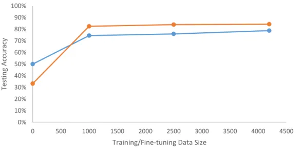
xii 4.3: Accuracy of CNNs with various prostate cancer training size from fine-tuned and scratch-trained strategies……….50 5.1: The proposed deep image-genomics framework………..57 5.2: The 2.5D representation of a tumour physical centroid with axial, sagittal and coronal slices of the image volume………..60 5.3: Association heat map between deep learning derived image features and tumour phenotype………69 5.4: Image-genomics association between deep image features and oncology gene sets using gene-set enrichment analysis (GSEA)………...70
xiii
List of Tables
4.1: Test accuracy of CNNs from both training approaches for cancer classification task with different training size………...…51 4.2: Area under receiver operation characteristics (AUC) ………...51 5.1: Statistical analysis of radiogenomic signatures from human-crafted and deep learning derived image features………...64 5.2: Right-tailed two-sample t-test result summarisation of deep-learnt and human-crafted features………...65 A1. Patients’ characteristics of the NSCLC data set………....73
xiv
Thesis Publications
The following publications have been produced over the course of the candidature:
Accepted paper:
T. Xia, A. Kumar, D. Feng, J. Kim, “Patch-level tumour classification in digital histopathology images with domain adapted deep learning,” submitted to the 40th Internation Conference of the IEEE Engineering in Medicine and Biology Society, accepted, submitted in February, 2018
Paper under preparation
T. Xia, A. Kumar, MJ. Fulham, D. Feng, J. Kim, “Deep radiogenomics: Quantifying Associations between Deep Learning-Based Medical Image Features with Tumour Gene Expression.” For submission to NatureScientific Reports.
1
Chapter 1
Introduction
The current practices for the treatment of human cancers rely upon the establishment of accurate diagnosis, which may involve a series of complex medical procedures comprising of patient screening for symptom evaluation, non-invasive imaging for disease localisation and histopathology analysis of tissue specimens. In recent years, genetic sequencing is becoming an increasingly important addition to the existing diagnostic pipeline. Cancer diagnosis may be the result of analysis of data generated through each medical examinations; the integration of such data contributes towards the understanding of the disease and hence exhibit potentials to offer optimal cancer therapy at individual patient level [1].
Although medical imaging and general pathology are becoming more routine in current cancer diagnosis, genetic sequencing is not always practical. This is due to human cancers exhibits strong phenotypic and genetic heterogeneity, where disease develops at multiple sites with genetic differences [2]. It is not practical or feasible to obtain tissue samples from all sites of disease as invasive biopsies have the potential
2 to induce tumour haemorrhage/proliferation. Further, the cost of genetic examination is high in addition to its limited accessibility.
As an alternative, medical images provide non-invasive assessments of phenotypic diversities in a number of cancer types and contribute to clinical decision-making, e.g., tumour detection, subtype characterisation and treatment responses [3, 4]. Advances in computerised medical image analysis allow the extraction of tumour’s visual representations (features) to facilitate the characterisation of tumour phenotypic differences, which offers insights into the patterns of disease spread and prognosis. The association between tumour imaging features and genetic foreknowledge allows the derivation of imaging surrogates to genetic biomarkers, and ultimately lead to the emerging field of image-genomics [5].
Image-genomics aims to associate the tumour imaging trait and clinical data (e.g., the underlying tumour gene expression) to provide an alternative approach that contributes to a non-invasive and accurate cancer diagnosis [6]. As such, the discovery and extraction of optimised tumour imaging descriptors represent a major challenge in the current image-genomics research.
This thesis address this challenge in the domain of medical image analysis, where our approach offers additional image features with domain adaptation technique to encode extra tumour phenotype representation. This thesis describes research to address the two key hypotheses: can domain adaptation facilitate medical image analysis and processing applications? If so, is domain adaptation capable of deriving tumour visual descriptors that produce stronger associations with genetic information?
3
1.1 Motivation
Precision medicine refers to the approach of disease prevention, treatment and care that takes into account individual variability in genes, environment, and lifestyle [1]. Precision medicine allows the classification of individuals into cohorts who are susceptible to particular diseases and forecasts response to a specific treatment, thereby improving disease prognosis [7]. The combined knowledge of patient’s genetics, bioinformatics, imaging and proteomics ultimately contribute towards the optimisation of therapeutic strategies [8]. One of the near-term focus of precision medicine is with cancer, a disease that is among the leading causes of death incidence worldwide. Traditional cancer therapeutics involve the utilisation of existing knowledge of the clinical outcomes from previous patient populations to draft treatment plans for future patients. Precision medicine has the potential to improve clinical oncology practices by utilising the molecular profile of cancer genetic behaviour, together with patient’s biological information, to optimise treatment plans at individual patient level [9].
However, pathways to cancer diagnosis are complicated and rarely straightforward. Cancer diagnosis may be the result of analysis of data generated by a series of medical procedures including non-invasive medical imaging, invasive tumour biopsies, genetic sequencing or from other evaluation of symptoms [10, 11]. Such procedures are prone to diagnostic errors, time-consuming and expensive in labour. In particular, obtaining tissue samples are not always feasible or practical as human cancers display phenotypic and genetic heterogeneity with multiple sites and genetic differences of the disease.
4 Medical imaging is a fundamental aspect of modern healthcare and plays an important role in cancer diagnosis and post-treatment monitoring. Medical images encode visual features which represent cancer phenotypic characteristics such as cancer location, size, texture and shape, which are useful for disease diagnosis and oncologic research [12]. Furthermore, the growing quantity and quality of medical images allow the creation of big data-driven approaches to computer-aided diagnosis (CAD, where clinicians routinely utilise CAD tools such as segmentation and registration to aid in their interpretation and decision making, as well as automated detection and classification of diseases as the second opinion to improve diagnostic accuracy [13, 14].
Although medical imaging is now a routine for cancer diagnosis, patient-specific genetic information can be valuable for its ability to reveal the underlying tumour biology and mutation types [15, 16]. Unfortunately, most human cancers exhibit strong genotypic heterogeneity, implying that tumours may have different gene expression at various sites and thus limits the usage of surgical operations such as tumour biopsies. In addition, tumour heterogeneity exhibits the potential requirement for multiple tumour biopsies during the diagnosis, the process of which further escalate the expenses along with patients discomfort, and risks the occurrence of undesired side effects such as tumour haemorrhage [17-19].
Imaging surrogates that reflect tumour genetic information have been explored as an alternative approach to invasive biopsies for genetic sequencing. This is achieved by associating tumour imaging descriptor with the tumours’ underlying genetic information which exhibits strong potential to the discovery of tumour biomarkers, and has been a key factor for the emergence of “image-genomics” domain [20, 21].
5 Image-genomics aims to convert tumour imaging data into high-dimensional image features to derive associations with tumour genetic information through the use of quantitative feature extraction techniques [20].
In the medical domain, image-genomics has potential applications in medical image analysis and biomedical research. Recent studies have demonstrated the capability of associating tumour image features with genetic information [22]; and the feasibility for potential non-invasive imaging surrogates to specific tumour prognosis biomarkers through the use of image-genomics [23]. Image-genomics approaches have also been shown to associate image features to a number of biological molecules and processes such as the transcriptome [24]. Furthermore, studies suggested that image-genomics associations are capable of predicting clinical outcomes in hepatocellular carcinoma [25, 26].
Despite the advances in image-genomics for identifying imaging surrogate to genetic biomarkers, there is an inherent reliance on tumour imaging representations for use in the association. The current state-of-the-art image-genomics approach employs image features that are defined based upon the human understanding of a range of objects (we denote this as human-crafted image features). The direct employment of such image object descriptors (by extracting human-crafted image features from medical images) is restricted by its capacity to fully quantify tumour imaging descriptors and therefore unable to reflect specific cancer types, nor variants among the patient cohorts. An alternative approach is to extract additional image representations that describe the subtle variations in tumour images, which are potentially important to specific tumour types [27]. The quantification of such abstract image features offer complementary information to existing image-genomics approach
6 and have the opportunities to: (i) derive stronger associations to gene expressions and; (ii) explore and discover additional surrogate non-invasive imaging biomarkers.
Another challenge that exists in the field of image-genomics is the lack of annotated medical images for rare diseases or different image modalities, where large volumes of annotated datasets are required for the learning of image representations of particular diseases [22, 28]. The demand for such datasets limits the image-genomic applications to specific disease types or image modalities. This restricts the ability to derive associations between image features with tumour underlying gene expressions.
1.2 Aims and Objectives
In this thesis, we address the identified challenges through: (i) the extraction of additional image representation to specific cancer types and; (ii) overcome the reliance of large volumes of annotated datasets with a novel image-genomics framework. Our framework leverages deep learning image analysis and domain adaptation techniques to extract disease-specific abstract image representations for associating tumour gene expressions.
Our framework expands upon traditional approaches in medical image feature extraction by adapting the state-of-the-art deep learning techniques for image object recognition, and hence offering additional tumour image descriptors for genetic associations. The thesis address the following two aims:
1. The development of a medical image analysis scheme with domain adaptation approach. The proposed scheme emphasis on the learning of tumour image
7 representations which leads to the enhanced performance of medical image analysis applications, e.g., tumour image classification.
2. The derivation of an image feature extraction scheme based on deep learning techniques to quantify additional image representation from tumour image through the employment of domain adaptation technique to associate with tumour genetic information.
1.3 Contributions of this thesis
Our contributions can be summarised in the following two major areas:
Image Classification and Domain Adaptation
Chapter 4 presents our contribution towards the domain adaptation facilitated medical image analysis scheme with limited volumes of datasets. In our thesis, the tumour classification system employs the domain adaptation technique from different clinical histopathology images to facilitate the prediction of the presence of abnormalities. Our system overcomes the reliance of large volumes of annotated datasets and offers improved tumour classification accuracy through the utilisation of domain adaptation technique compared to the traditional approach.
Image-Genomics Framework
Detailed in Chapter 5, we present a new image-genomics framework to associate tumour imaging representations to the underlying tumour gene expressions. Our framework utilises the current state-of-the-art in image object recognition to learn the abstract image representations that describe the subtle variations in tumour
8 images through a domain adaptation facilitated data-driven approach. Our framework utilises different mathematical and genetic analysis tools to associate extracted image features to cellular pathways as well as cancer prognostic biomarkers.
1.4 Thesis Structure
The remainder of this thesis is structured as follows:
Chapter 2 and 3 orients the readers by providing theoretical background knowledge of the thesis. Chapter 2 provides an overview of medical imaging and the image processing techniques within this domain. Chapter 3 presents the overview of the state-of-the-art in the image-genomics analysis. In particular, it provides a summary of gaps from recent image-genomics research.
Chapter 4 and 5 provide the detailed contribution of this thesis. We describe the domain adaptation facilitated medical image analysis system in chapter 4. In chapter 5 we highlight the deep domain adaptation learning framework for associating image features to tumour genetic information.
Chapter 6 summarises the contribution of this thesis and indicates directions for future research in the domain of image-genomics analysis.
9
Chapter 2
Background and Related Concepts
2.1 Medical Imaging and Image Processing
Medical imaging is a fundamental component of the modern healthcare system and is essential for the accurate diagnosis and staging of cancers. Medical images provide a fast and non-invasive assessment of phenotypic diversity in many cancers [12] through a variety of different imaging modalities (or techniques). Medical image processing, in turn, allows the extraction of meaningful information which offers insights of different aspects of patients’ conditions. In this chapter, we provide an overview of medical imaging and the theoretical background in image processing techniques that are crucial for cancer diagnosis.
2.1.1 Definitions
Digital images consist of a collection of numerical picture elements, called pixels. The term pixel resolution refers to the number of pixels in an image, which can be
10 represented as a single number or by the number of pixels in each dimension. For example, an image that contains 8,294,400 pixels can also be referred to as having a resolution of 8.3 megapixels or 3840 ×2160 (width × height) pixels.
Many medical imaging techniques consist of sampling 2D images along a third spatial axis to form 3D images, known as volumetric images or image volumes. These 3D volumetric images encode the spatial relationships between 3D pixels, called
voxels, which both 2D and 3D images allow the extraction and interpretation of the encoded information through the use of image processing techniques. Both pixels and
voxels exhibit spatial resolutions, which describe the size of the details captured by individual pixels or voxels. For instance, a spatial resolution of 10.00mm × 10.00mm × 5.00mm means that a voxel depicts a region with a volume of 500.00mm3.
Contrast resolution refers to the range of distinct intensity or a set of intensities for red, green and blue (RGB) channels that can be distinguished in greyscale and coloured images respectively. A relatively low contrast resolution can be interpreted as pixel/voxel intensities that are similar in an image and are difficult to distinguish. Figure 2.1: Pixels, voxels and spatial resolutions of medical images: (a) illustrate a 2D image with pixel resolution of 8 × 8 and a spatial resolution of 1mm × 1mm. (b) illustrate a 3D image volume with voxel resolution of 5 × 5 × 3 where its spatial resolution is 1mm × 1mm × 1mm.
11 Figure 2.1 illustrates an example of a 2D image and a 3D volumetric image. Pixels and voxels are visualised in forms of 2D and 3D arrays of grids respectively.
In medical image processing, a region of interest (ROI) consists collections of pixels in 2D images that represent an area which encodes important information for particular domain or application in cancer diagnosis. The corresponding term volume of interest (VOI) refers to the emphasised anatomical structures that encode important knowledge for clinical uses.
2.1.2 Medical Imaging Modalities
Modern healthcare utilises different medical imaging modalities to capture different aspects of the human body for cancer diagnostic and treatment [29, 30].Medical image modalities can be categorised into two classes based on their technique and process in visualising different aspects of the disease [31]: (i) Anatomical and (ii) Physiological (functional) imaging. Anatomical medical images capture and visualise the anatomical structures of the ROI in the form of 2D or 3D images. Anatomical medical images allow physicians to interpret and evaluate the disease conditions for diagnosis purposes, and can also be used for the monitoring of treatment responses [4]. Functional imaging, on the other hand, captures the metabolic status of the ROI, which allows physicians to assess the physiological status of patients, and to identify structures with abnormalities, such as tumours [32].
Common anatomical medical image modalities include X-ray, computed tomography (CT), magnetic resonance imaging (MRI). Common functional imaging includes single-photon emission computed tomography (SPECT) and positron
12 emission tomography (PET). Such image techniques produce a single type of image or image volumes that are referred as single-modality medical imaging.
Beyond the use of single-modality medical images for cancer diagnosis, the employment of multi-modality imaging types such as PET/CT and SPECT/CT has become one of the fundamental diagnosis protocol in clinical practices and basic medical research [33]. Despite multi-modality medical imaging exhibits strong potential to offer both tumour anatomical and functional properties, the evaluation of our proposed image-genomic framework did not directly involve datasets with multiple modalities. In this subsection, we emphasise on several single modality imaging techniques that are most relevant to our contribution.
2.1.2.1
Computed Tomography (CT)
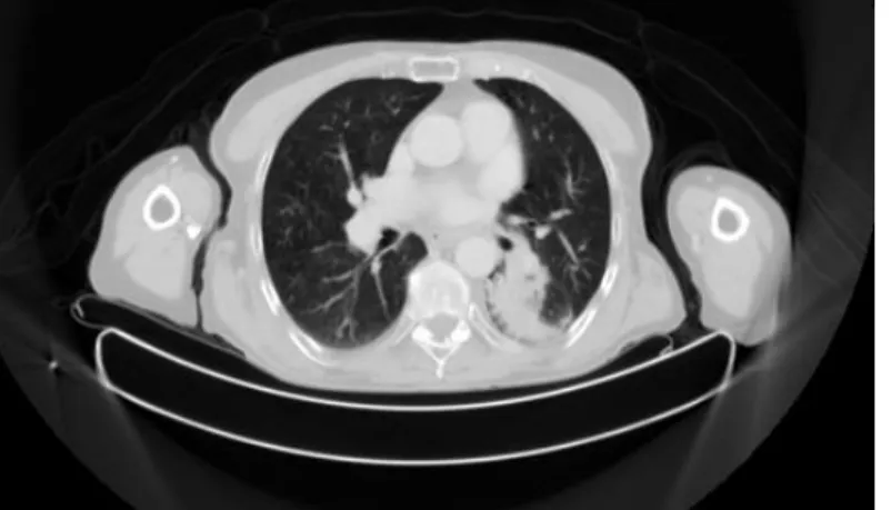
Computed tomography is one of the most utilised imaging modalities in cancer detection, diagnosis and prognosis in modern clinical practices [34, 35]. CT scan makes use of the combination of multiple X-ray measurements to produce tomographic images. Figure 2.2 illustrates an example of CT image of the lung field from a Non-small Cell Lung Cancer Patient (NSCLC). In the coming chapters, we will explore the use of CT image volume with medical image analysis techniques,
Figure 2.2: The CT image of the lung field from a NSCLC patient (data description detailed in Chapter 5).
13 including image feature extraction and its association to tumour phenotypic characteristics, which is the most relevant to our thesis contribution.
2.1.2.2
Digital Histopathology Images
Tumour histopathology is considered as the golden standard for cancer diagnosis. It is produced as the result of tumour tissue biopsies, where specimens of tumour tissue are stained and mounted onto glass slides for visual inspection [36]. Tumour histopathology offers the visualisation of cellular structures of tumour-containing ROIs, providing diagnostic information on mutated cell types, and hence offer therapeutic insights about the disease [36, 37].
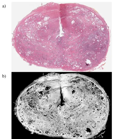
The digitisation of histopathology was introduced through high-resolution whole-slide-imaging (WSI), which have enabled the quantitative analysis of tumour histomorphometry as well as its association with clinical practices for cancer diagnosis
(a) Overview of WSI (b) A zoomed-in view of a WSI region
Figure 2.3: The overview and zoom-in view of WSI of sentinel lymph node with presence of breast cancer metastases. (a) The highest-level view of the entire WSI. (b) The zoom-in view of a region from the same WSI image (data description detailed in Chapter 4)
14 [21, 38]. Similar to CT images, digital WSI enables medical image analysis techniques to be applied to extract image features of abnormal cells. Image traits can then be exploited to correlate to genetic profiles of the tumour. Figure 2.3 illustrates an example of sentinel lymph node WSI with the presence of breast cancer metastases.
2.1.3
Medical Image Segmentation
This subsection describes several approaches for medical image segmentation that are most relevant to our contribution. We describe the current automated image segmentation approaches and compare with manual image segmentation by experienced physicians to reflect their distinct advantages.
Medical image segmentation refers to the process of image partitioning where each image partition contains a collection of pixels that together represent an ROI [39]. Medical image segmentation is commonly used to identify and emphasise ROIs, as it
(a) The region of interest (lung cancer)
in a CT image (b) The region of interest (Breast cancer metastases) in a histopathology image Figure 2.4: The segmented region of interests in different medical image modalities. (a) The dineated presence of lung cancer in an CT image. (b) The segmented regions in a WSI that shows the presence of breast cancer metastases in sentinel lymph node.
15 removes unrelated image regions to reduce the complexity of medical image analysis [40].
Medical image segmentation with manual delineation from experienced physicians is considered as the golden standard in most clinical applications. However, the current procedure of medical image segmentation for cancer patients relies upon the visual inspection of imaging data and the manual delineation of tumour ROI. Figure 2.4 provides examples of ROIs in different medical image modalities that were delineated by experienced physicians. The production of delineated tumour imaging data is time-consuming and prone to diagnostic errors, based on the expertise of the physician [41]. These challenges are exacerbated by the requirement of identifying quantitative morphological parameters of tumours such as shape and size [42].
To address this issue, computerised automated algorithms have been designed to facilitate medical image segmentation tasks. Commonly used medical image segmentation algorithm involves: pixel thresholding [43], region growing [44], edge detection [45] and fuzz clustering [46]. In the recent years, deep learning has also been utilised to perform classification and image segmentation tasks and achieved improved performance [47, 48]. Despite the performance improvement by deep learning-based approaches, few studies have shown superior accuracy to human perception in cancer diagnosis [49, 50]. The contribution of this thesis relies on accurate tumour delineations for domain adaptation purpose; we hence obtained tumour segmentation from experienced physicians as the foundation of our work. We will discuss the principle and applications of deep learning in later sections of this chapter.
16
2.2 Gene Expression Profiling
While the visual inspection of tumour histopathology is considered to provide a definitive diagnosis, it is prone to diagnostic errors in diseases where tumour classification is challenging due to visually indistinguishable morphological properties (e.g. diffuse large B-cell lymphoma and breast cancer) [51, 52]. Gene expression profiling refers to the technique of quantitative measurement of genetic activities that facilitate tumour classification and is also capable of predicting the clinical outcome for cancer patients [53]. In this subsection, we will describe the core concept of genes, gene expressions and its application in clinical applications that are relevant to our contribution.
A gene refers to a specific region of the deoxyribonucleic acid (DNA) strands, which exhibits genetic codes that produce ribonucleic acid (RNA) through transcription. RNAs are capable of synthesising proteins that encode biological functions through translation. However, human genetic codes are prone to mutation, permanent alteration of genetic elements that can be caused by many different factors such as DNA damage from environmental factors. Mutations can result in various types of changes in human DNA and may result in changes in behaviour as well as functions of genes. Clinical studies have shown evidence of genetic mutations in the prognosis of multiple human cancers [54-57]. Gene expression profiling identifies genes with abnormal expressions in tumour samples, thus offering insights into therapeutic options [16]. Gene expression profiling also provides patient-specific genetic information with normal expressions which encodes the genetic variables at the individual level that can ultimately contribute to the application of precision medicine [58].
17 Despite the significance of gene expression profiling in the diagnosis and prognosis of cancer patients, gene expression profiling is limited due to its reliance on invasive surgical procedures. Unfortunately, human cancers exhibit strong phenotypic and genetic heterogeneity within an individual with tumour manifestation at multiple sites [59]. Additionally, the differences in gene expressions across multiple sites may result in different responses to treatments [60]. The sampling and tissue acquisition of tumours from all disease sites is restricted due to patient discomfort and risk of undesired side-effects such as operation induced tumour proliferation. This thesis addresses this issue through the development of a novel image-genomic framework, discussed in detail in the following Chapter 4 and 5 of this thesis.
2.3 Machine
Learning,
Deep
Learning
and
Convolutional Neural Networks
Machine learning techniques for medical image processing are a well-established field. The ability of machine learning to quantify the representation features of the input medical image empowers numerous automated medical image processing algorithms for different clinical applications. However, machine learning systems were limited due to its requirement of domain expertise with careful engineering to be able to learn and transform the input data [61]. Deep learning is a class of machine learning technique which allows learning of data representations with multiple layers of abstraction. Convolutional neural networks (CNNs) are a deep learning technique that extracts image features from imaging data to learn the sophisticated underlying representations with deep networks [62]. In this chapter, we emphasise on the principle of deep learning and CNNs as they represent the current state-of-the-art in image
18 object recognition. We will present the background and CNN architectures that are relevant to our study in the following sections.
2.3.1
Machine Learning
Machine learning is a major field of computer science that has been utilised to serve many aspects of modern healthcare systems. With carefully engineered mathematical models, machine-learning systems have been applied in pattern recognition [63], image classification [64], medical image retrieval [65] and tumour image segmentation[66]. Compare to the rule-based systems, the core of machine learning techniques are based on the development of models from statistical and artificial intelligence approaches. It is essential for the machine learning model to “learn” to recognise the distinguishing characteristics of the patterns within the data to produce meaningful outputs [67]. Generally, the learning approaches for machine learning models are divided into the following: supervised learning, unsupervised learning and reinforcement learning.
Although unsupervised and reinforcement learning exhibit strong potentials in multiple disciplines, our contribution rely on labelled medical imaging data to explore the region-specific genetic association. As the contributions of this thesis involve mainly supervised approaches, we will only cover those approaches here. Supervised training approaches require several different types of database, defined as follows:
Training data: refers to a collection of data that are used to train the machine learning model. The machine learning model learns the representation of the training data and its predictive relationship to the output labels.
19
Validation data: refers to a separate collection of data that are used in addition to the training data to adjust or guide the training process. This process involves the comparison between the predicted output with the training data labels. This provides an indication of the performance of the model on unseen data during the training process, and allows tuning of model parameters.
Test data: refers to a collection of withheld data which is used to evaluate the performance of the model at the after the completion of the training process. Test data indicates the performance of the trained model with new examples. Test data are not involved in the training process.
2.3.1.1
Supervised Learning:
Supervised learning is one of the most common approaches for medical image processing and analysis. It refers to the approach where the model is trained with labelled data sets so that the model learns the internal representation of the input data to make predictions on the labels [68]. The resulting model from supervised learning is typically used to assign class labels with known predictive features for future data sets. Supervised learning is capable of performing classification or regression with training data set with discrete or continuous properties, respectively [69].
2.3.2 Artificial Neural Network
Artificial Neural Networks (ANNs) are a machine learning approach that was inspired by the biological neural networks that constitute human brains [70]. An ANN consists a collection of connected nodes or “artificial neurons” in a directed graph in the form of networks. ANNs are commonly used in machine learning to learn the complex
non-20 linear relationships from the dataset. This is achieved through the ANN’s mechanisms where each neuron receives, processes and transmits a signal from one to another in a similar way to the biological synapse. To achieve the optimised learning outcome, ANN requires the design of an appropriate network structure and learning approach to tune the weights and biases of the network.
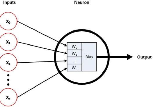
Neurons are the fundamental building blocks of many ANNs. Figure 2.5 illustrates the structure of a single neuron where multiple inputs values are processed in the neuron to produce a single output value, where X is a vector of inputs with n
elements, W is the vector of the weights with a corresponding number of elements, which can be summarised in equation 2.3.1:
L(𝑊, 𝑋) = ∑ 𝑓(𝑥𝑖, 𝑤𝑖) + bias 𝑛
𝑋=0 (2.3.1)
where 𝑓(𝑥𝑖, 𝑤𝑖) is the ANN function that generates the output value.
21 The training of an ANN can be explained by illustrated single neuron model. For each element in the training dataset, its features are extracted and transmitted into the neuron in the form of a feature vector. Assuming that the classification task involves only two classes, the neuron processes the input feature vector by multiplying with its internal weights. A class prediction is based on whether the value of the product is above a threshold. If the predicted class does not match the corresponding label, each weight within the neuron is adjusted individually to refine the prediction process. Additionally, the training dataset is typically divided into collections of batches which allows the weight adjustment after training of each batch. An epoch
refers to the full pass of the entire training dataset; robust weights for classification tasks are the outcome of hundreds of training epochs.
2.3.3 Deep Learning
Deep learning can be categorised as a class of techniques of machine learning [61], which allows the computation models with multiple processing layers to learn the internal representation of the input data with multiple levels of abstraction [71]. Deep learning was proposed to address the limited performance of traditional machine learning approaches to process natural data in their raw form. Deep learning resolves this issue by utilising the multiple processing layers to learn and interpret the low level, abstract representations from the high-level understandings of the input dataset [61]. This is achieved by feeding the raw data through the successive multilayer architecture in a sequential manner, where deeper layers learn the abstract representation from the representation in the previous layers [67].
22 Deep learning technique employs backward propagation of error or “backpropagation” to train the multilayer model for supervised learning [61]. Backpropagation calculates the gradient of the error function of the ANN with respect to its weights and passes the gradient backwards through the neural network. Compared to the traditional approach where the gradient of the error function is calculated for each layer separately, the backflow of error gradient allows more efficient computation of gradience for ANNs [72].
The implementation of deep learning technique utilises specialised GPUs to improve the performance of training process by 10 to 20 times compared to the traditional training approach on standard CPUs [67]. Recent advances in deep learning have lead to the improved state-of-the-art in various domains such as visual object recognition [67], speech recognition [73] and also in medical image analysis [42].
2.3.4 Convolutional Neural Networks
Convolutional Neural Networks (CNNs) is a particular type of ANN and designed to process input data that is in the form of multidimensional arrays, e.g., coloured 2D images which consist of 2D arrays for each RGB (colour) channel. Compared with traditional ANNs with fully connected adjacent layers, CNNs are much easier to train and are more generalised [67]. CNNs are structured in a series of stages where each stage consists of specialised layers with unique functions. The building blocks of a CNN consists three types of specialised layers: convolutional, pooling and activation layers, e.g., rectified linear unit (ReLU) layers [61].
Convolutional layers consist of organised units in the form of feature maps, where each unit is connected to local patches from the previous layer through a set of
23 weights which is referred as filter banks as shown in Figure 2.6. The resultant sum of the local filter bank is typically transmitted through a non-linear activation layer such as ReLU layer, e.g. 𝑓(𝑥) = 𝑚𝑎𝑥(0, 𝑥). Only units within the same feature map share the same filter bank. This design allows the convolutional layers to detect local conjunctions of features from the previous layer, as local values are often highly correlated and are invariant to the location in an image input.
Pooling layers are designed to merge features in spatial proximity which share semantic similarities into one. The principle behind pooling layers is to detect the position of motifs that are typically formed by highly correlated features through a coarse-grained approach. An example pooling layer calculates the maximum of a local patch in one or more feature maps. Pooling layers act to reduce the dimensions of the representations and to create an invariance to small distortions and shifts.
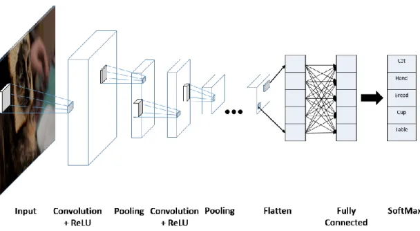
A standard CNN typically involves stacked layers in order of convolution, ReLU and pooling layers, and followed by fully connected layers as shown in Figure 2.7. CNNs also utilise backpropagation to train its filter banks in a similar way compare to regular ANNs.
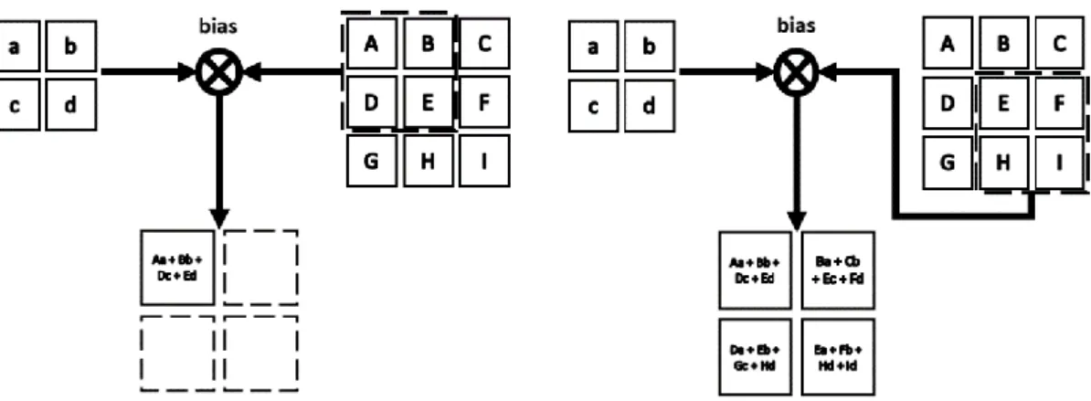
Figure 2.6: An example 2D convolution of a 3 × 3 input using a 2 × 2 filter with the 2 × 2 output feature map.
24 CNNs are inspired by the biological signal transmission through cells with different complexity and functions in visual neuroscience [74]. CNNs exploit the compositional hierarchies in natural signal processing where high-level features are the result of the composition of low-level features, to apply onto images where objects are the results of the composition of edges, motifs and parts.
CNNs have demonstrated large improvement to the state-of-the-art in multiple disciplines. CNNs have been adopted in medical image analysis to perform a range of tasks including medical image segmentation [75], classification [76] and disease detection [50].
In the following subsections, we will explore the two CNN architectures that have shown outstanding performance in image object recognition and that are most relevant to our contributions.
Figure 2.7: An example CNN with many convolution layers. Filters are applied to each input image and the output of each convolutional layer is used as the input to the next layer.
25
2.3.4.1
GoogLeNet (Inception)
GoogLetNet [77], introduced in 2014 as a submission to the ImageNet Large-Scale Visual Recognition Challenge 2014 (ILSVRC14) is a deep CNN architecture consists of 22 layers [78]. GoogLetNet was designed to act as a classifier for natural images with the underlying concept to increase the width and depth of CNN while keeping the computation cost constant. GoogLetNet demonstrated significant improvement to the state-of-the-art at the time by achieving a top-5 error of 6.67% for the classification challenge. A standard GoogLetNet architecture is shown in Figure 2.8.
GoogLetNet employs the “inception modules” concept to exploit the approximation and coverage of optimal local sparse structure by dense data structures. Within each inception model, the input is fed into separated convolution layers with dimensions of 1 × 1, 3 × 3, 5 × 5 and a 3 × 3 max pooling layer. Results from each layer are concatenated into a single output vector to serve as the input for the next stage.
Also, GoogLetNet exploits the “network-in-network” approach [79] by adding a 1 × 1 convolution layer followed by the standard stacked convolutional layers
26 structure for dimension reduction which removes the computation bottleneck and hence increases the representational power of the neural network.
2.3.4.2
Deep Residual Networks (ResNet)
Several studies, including GoogLeNet, have demonstrated that the depth of CNNs is one of the crucial factors to improve the learning outcome for image object classification tasks with ImageNet dataset [80-82]. However, rapid degradation of learning accuracy was observed in CNN architectures that stack many layers together Figure 2.8: The GoogLeNet architecture. Layers labeled with “reduce” represents the additional 1 × 1 reduction layers before each of the 3 × 3 and 5 × 5 convolutions.
27 in a traditional way. The rationale behind the training degradation was not due to overfitting, but an optimisation problem of the mapping of identities for the additional layers.
ResNet [83] was proposed to address the challenge of training accuracy degradation in deep CNN architectures through the use of residual mapping to fit the successive stacked layers. ResNet employed the concept of residual representations [84, 85] and created a shortcut connection [86] with closed gating functions [87, 88] for identity mapping. The residual function and identify mapping by shortcuts are according to:
𝑓(𝑥) = ℋ(𝑥) – 𝑥 (2.3.2)
where 𝑓(𝑥) denotes the residual function, which is the result of removing the input 𝑥 of the first layer of the stacked layers from the underlying mapping ℋ(𝑥). Residual learning drives the weights of the multiple non-linear layers towards zero to approach identity mapping, which is defined as:
𝑦 = 𝑓(𝑥, 𝑊𝑖) + 𝑥 (2.3.3)
where 𝑦 and 𝑥 denote the output and the input of the stacked layers respectively and 𝑓(𝑥, 𝑊𝑖) is the function that represents the residual mapping that needs to be learned.
The shortcut connections in ResNet does not increase the number of parameters of the network nor the computational complexity. A ResNet building block with residual learning and shortcut connection is illustrated in Figure 2.9.
ResNet further improved the state-of-the-art in image object recognition at a top-5 error of 4.49% with a 152-layer ResNet architecture, compared with 6.67% from GoogLeNet. ResNet also demonstrates a good generalisation performance for different
28 recognition tasks [83] and hence has been widely adopted in medical image analysis [89, 90].
2.4 Domain Adaptation for Transfer Learning
Common applications of machine learning-based techniques involve the training of the statistical model to quantify the internal representations of data from the target domain. Such an approach relies on the assumption that the training and real-world (used in a future application) datasets share the same feature space with a similar distribution [91]. The changes of the distribution of feature spaces in the datasets typically require that the trained statistical model is rebuilt to fit the newly collected data. However, the assumption does not always hold, as most machine learning methods require large annotated training datasets that are specific to the target domain. Machine learning applications are thus greatly limited in domains such as medical image analysis where the acquisition of labelled training data is expensive.
Transfer learning is the alternative approach to train the statistical model without the need for the large quantity of domain-specific datasets. Transfer learning refers to the process where the knowledge acquired from one large database is
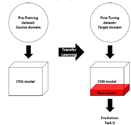
29 transferred to facilitate learning applications on a related but smaller dataset. For neural network applications, transfer learning can be described as an approach to speed up the learning of a specific domain by using and transferring the weights obtained from a network that was trained for a related source task [92]. An illustration of the transfer learning approach is shown in Figure 2.10.
Transfer learning technique allows CNNs to be trained and applied to datasets from different but related domains. The differences between domains can be categorised into two groups: (i) domains with different feature spaces, or (ii) domains with the same feature space but different marginal probability distributions [91]. To evaluate the representation and the similarity between two domains, techniques such as A-distance, have been adopted for transfer learning applications [93, 94].
There are two major approaches to employ domain adapted transfer learning consists for CNNs applications in medical image analysis: (i) using “off-the-shelf CNN” features; and (ii) using domain adaptation with fine-tuning techniques [95]. The Figure 2.10: Traditional and transfer learning-based approaches for CNN models. a) Traditional machine learning. b) Transfer learning-based approach for CNN model.
30 term “off-the-shelf CNN” refers to the procedure of utilising a CNN model which was trained on a larger dataset from scratch as a feature extractor directly on a new dataset with a smaller volume size. Parameters in the convolutional and fully connected layers are not changed. For classification purposes, the extracted features from the “off-the-shelf-CNN” can be used to train a separate classifier, such as support vector machines, random forest classifier or to train only the classification layer of the model to fit the number of classes of the new dataset [95-98].
Fine-tuning refers to the process of updating the weights of a pre-trained CNN model new datasets from the desired domain through the use of backpropagation. Common approaches for fine-tuning involves the training of only the last few convolutional layers, referred as “shallow tuning” or the tuning of all convolutional layers of the network, called “deep tuning” [99]. Deep tuning is typically employed if
Figure 2.11: Overview of shallow tuning-based transfer learning approach.
31 the distance between domains is significant. The tuning of early layers of CNNs involves the learning of low-level image features that are similar to Gabor filters and colour blobs, which are applicable to most vision tasks and hence are not applied to shallow tuning approach [100]. Studies have shown that fine-tuning is as effective as a CNN that is trained from scratch and is more robust to the size of training dataset [99]. Fine-tuning has been applied to a variety of medical image analysis tasks such as ultrasound anatomy identification and medical image modality classification [89, 101]. An illustration of “shallow tuning” is shown in Figure 2.11.
32
Chapter 3
Image-Genomics
This chapter presents the overview of image-genomics research in various clinical contexts. We provide a survey of the current state-of-the-art of image-genomics with a focus on the extraction of genetic-related image features, their application to medical images with different modalities, and identifying the gaps in the existing approaches.
3.1 Overview of Image-Genomics
Cancer treatment relies upon accurate diagnosis, which can benefit from accounting for individual variabilities in genetics, environment and lifestyle [1], but requires a complex combination of information gathered from clinical, histopathology, imaging, and genetic data. Medical imaging is one of the fundamental protocols in modern healthcare for the examination of human cancers [12]. It offers a fast and non-invasive assessment of tumour visual characteristics which can be used as oncologic diagnosis tool as well as providing treatment guidance. Briefly discussed in previous chapters, computerised analysis of medical images has been demonstrated that quantitative
33 image feature analysis is capable of capturing distinct tumour phenotypic characteristics, e.g. in soft tissue sarcomas (STSs) [102], lung adenocarcinoma [22] and colorectal cancers [103], thereby indicating the potential for image features analysis to discover and identify tumour imaging biomarkers. The patient-specific genetic information also offers insights into therapeutic options and hence can be used to optimise patients management and treatment plans, for example, the use of immune-modulating therapies in melanoma and lung cancer [104]. Also, patient-specific geneticdata have been used in medical research to analyse high-dimensional mineable characteristics, which consists detailed characterisation of biological information such as DNA (genomics), RNA (transcriptomics), proteins (proteomics) and metabolites (metabolomics) [105].
Nevertheless, the clinical implementations are restricted by the challenges in acquiring tumour genetic profiles. As previously described in Chapter 1 and 2, human cancers exhibit strong phenotypic and genetic heterogeneity, where different gene behaviours are expressed in various tumour regions. Thus the potential need for multiple biopsies may introduce further risk of patient discomfort and the occurrence of undesired side effects such as tumour haemorrhage. As such, the consequence is that multiple biopsies are rarely performed [17-19].
The emerging field of image-genomics involves the integration of patient-specific clinical information from both imaging and genetics [106]. Image-genomics can be thought of as the identification of surrogate tumour biomarkers through correlating image features with gene expression and has been suggested as a non-invasive alternative to biopsies [5, 107]. It aims to covert clinical images into high dimensional mineable image features using quantitative, high-throughput feature
34 extraction algorithms for correlation to genetic information [12, 108]. Studies have shown promising result in identifying prognostic image-genomics biomarkers for several cancers including lung cancer [109], breast cancer [110], and high-grade primary brain tumours [111].
As such, current image-genomics research requires large volumes of annotated multi-dimensional data. The following clinical data are essential for image-genomics studies:
Segmented medical images with precise annotations
Annotated clinical metadata which provides patient-specific clinical parameters
Labelled patient-specific gene profile or raw expression array data At the current stage, most of the image-genomics research is limited to the publically available datasets that are published in The Cancer Imaging Archive (TCIA) [112] and The Cancer Genome Atlas [113], or data that is privately collected. The limited amount and quality of the dataset make image-genomic research more challenging, for example, the confined feature selection process due to the inadequate amount of clinical metadata and annotations.
In the following subsections, we will explore the use of image features and the techniques for image feature extraction in various contexts. Figure 3.1outlines the crucial procedures of an image-genomic framework. We will focus on the extraction of image features from two of the widely adopted imaging modalities: CT and histopathology and their corresponding application in the field of image-genomics.
35 F ig u re 3.1: The c ur re nt image -g enomi c a pp roa ch for a ssocia ti n g im ag e fe at ure s with g en e e x pre ssi on pr o file s.
36
3.2 Image Feature Extraction
The famous quotation from Bartlett,“A picture is worth ten thousand words”1, illustrates the amount of information encoded within a single image. Similar to the human behaviour, computerised image analysis techniques extract and select information from images to solve problems in various contexts. This is facilitated through the employment of models that were trained on specific domains [114].
Briefly mentioned in Chapter 2, image feature extraction is a process which finds and transforms image encoded information into suitable image representations to be used for various applications [115-117]. In the field of image analysis, extracted image features can be categorised into global, which are calculated from the entire image, or local features, which are extracted from specific ROIs. Image features that are applicable to many vision tasks consist colour, texture, shape and the structural representation of an image.
Texture-based features are one of the most important characteristics to
identify objects and ROIs in an image. Texture is an innate property of surfaces in an image regardless of its modality [118] and contains crucial information about the structural arrangements of surfaces as well as its relationships to the surrounding environment of the ROIs. Texture-based features offer a precise description of the homogeneity of the local spatial variation of pixel intensity [119] and texture analysis serves computerised applications in image segmentation [120], classification [121] and pattern recognition [122]. Commonly adopted texture-based features include the use of Haralick and wavelet features [123, 124]. Haralick features are extracted from
37 the distribution of co-occurring pixel values in the image within a specified spatial neighbourhood, hence providing a statistical summary of the relative distribution of gray tones in the image [118].
The extraction of texture features through the use of wavelets have also contributed to tasks such as content-based image retrieval [125], segmentation [126] and classification [127]. Wavelet features are extracted by decomposing the signal of images at various resolutions based on wavelet orthonormal [128]. The orthogonal multi-resolution representation provides a hierarchical framework to offer image characteristics in a coarse-to-fine strategy that extracts contextual and low-level image features [127, 128].
Colour-based features are one of the most adopted representations of images
due to its robustness to background complications, as well as its independence to the size and orientation fo the image [129]. Colour features also offer both global and local image representations with various applications. Colour representations such as colour indexing, colour descriptors and compact colour moments have been adopted in applications such as image retrieval [130-132] and scene recognition [133]. Colour histograms provide insights on global colour distribution and ranges, and have been used in applications such as face detection [134] and bleeding detection in medical domain [135]. Additionally, the application of colour-based image representations to the three-channel domain offers complementary characteristics of the image compared to single-channel grayscale images [136].
Shape-based features offer descriptions to quantify the shape of ROIs in ways
that agree with human perception for specific tasks [137]. Shape representations capture the geometric details within the ROIs of an image, and are invariant to
38 translation, rotation, scaling and resistant to noise [129]. Shape-based features have been extracted and applied through multiple approaches. For 2D images, shape-based features have been represented as point sets [138], outline curves [139] and shock graphs [140]. Such features have been adopted for image object classifications [141], recognition [142] and content-based image retrieval in various domains [143].
In 3D volumetric images, commonly adopted shape features use spherical harmonics that decompose the 3D object into orientation invariant and descriptive information [144]. Additional approaches for 3D shape-based features extraction involve the analysis of object surface curvature and the correlograms of the objects from various view points [145]. Such 3D shape-based features have been widely used in 3D model search engines [144], classification tasks [146] and content-based retrieval [147].
In addition to the image features that quantify the statistical information of pixels (hereafter referred to as “agnostic features”), yet another set of features have been commonly utilised to represent high-level tumour characteristics based upon human understanding (hereafter referred to as “semantic features”), e.g. tumour shape, size and necrosis [105]. Different from agnostic features, semantic features are commonly used by radiologists to describe tumours, with the foreknowledge of their prognostic value in cancer treatment [148]. The employment of semantic features in image-genomics studies has demonstrated their capability to identify prognostic imaging biomarkers [23] and to predict gene expression patterns in hepatocellular carcinoma [149]. As a collection, agnostic and semantic features quantify the image representation of tumour phenotypic traits that are based on established human knowledge in cancer physiology. To differentiate such a collection of image features
39 from artificial intelligence produced image descriptors, agnostic and semantic features are hereafter referred to as “human-crafted” features.
Human-crafted image features have been an integral component of image-based CAD systems [150] in many clinical applications such as disease detection and classification [151, 152], improving diagnostic performance [153] and ROI segmentations [154].
3.3 Summary of Gaps
A number of image-genomics approaches have been proposed and validated for different image modalities and clinical applications across various cancer types. The multiplicity of image-genomics research has encouraged studies to employ images with multiple modalities and to seek correlations with different genetic information.
Recent image-genomics research stimulated the extraction and quantification of image features that can be significantly correlated to genetic information. Other recent image-genomics studies attempted to derive image features from multi-modality images, e.g., PET/CT, to provide quantification of tumour phenotype as well as its underlying metabolic status to identify imaging surrogate of prognostic biomarkers [23].
Despite the progress and advances in image-genomics, a number of gaps remain unaddressed in the current state-of-the-art. In the following subsections, we provide a detailed explanation of aspects that should be pursued and improved to better seek image-genomics associations.