Fetal ECG Extraction Methods
Raviranjan Gupta
Roll No. 213EC3218
Department of Electronics and Communication Engineering
National Institute of Technology, Rourkela
Fetal ECG Extraction Methods
Thesis submitted in partial fulfillment of the requirements for the degree of
Master of Technology
in
Electronics and Instrumentation
by
Raviranjan Gupta
Roll No. 213EC3218
under the guidance of
Prof. Ajit Kumar Sahoo
Department of Electronics and Communication Engineering
National Institute of Technology, Rourkela
National Institute of Technology
Rourkela
CERTIFICATE
This is to certify that the project work in the thesis entitled”Fetal ECG Extrac-tion Methods” submitted by Raviranjan Gupta is a record of a research work done by him under my supervision and guidance in partial fulfillment for the award of the degree of Master of Technology in Electronics and Communica-tion Engineering with specializaCommunica-tion Electronics and InstrumentaCommunica-tion, NaCommunica-tional Institute of Technology, Rourkela. Neither this thesis nor any part of it, to the best of my knowledge, has been submitted for any degree or academic award elsewhere.
Prof. Ajit Kumar Sahoo
Assistant Professor Department of ECE National Institute of Technology Rourkela
National Institute of Technology
Rourkela
DECLARATION
I declare that the work done in the thesis is original and has been done by myself under the guidance of my supervisor. The work has not been submitted to any other Institute for award of any degree. I have followed the guidelines provided by the Institute in writing the thesis. Whenever I have used materials (data, theoretical analysis, and text) from other sources, I have given due credit to them by citing them in the text of the thesis and giving their details in the references.
Whenever I have quoted written materials from other sources, I have put them under quotation marks and given due credit to the sources by citing them and giving required details in the references.
Acknowledgment
This project work is first achievements of my career. This would not have been possible without the help of my classmates, who have regularly suggest me some ideas regarding my project and I am very thankful to them.
I would like to express my gratitude to my supervisor Prof. Ajit Kumar Sahoo, who has always supervised me for my project work.
I am also very obliged to Prof. K.K. Mahapatra, HOD, Department of Electronics and Communication Engineering for giving such an environment of study. I also want to say thanks to our Faculty Advisor Prof. U.C Pati and Prof. T.K Dan for helping me how to learn.
I might want to thank all faculty members and staff of the ECE Department for their thoughtful participation. I might likewise want to make an exceptional notice of the magnanimous backing and direction I got from my lab-mates and classmates amid my venture work.
When I glance back at my achievements in life, I can see an unmistakable hint of my family’s worries and dedication all around. My dearest mother, whom I owe all that I have accomplished and whatever I have turn into; my cherished father, who constantly trusted in me and propelled me to think be-yond practical boundaries even at the hardest snippets of my life; and my two sisters, who dependably be with me amid all the hardships of this attempt and past.
Abstract
The electrocardiogram signal of fetus, i.e; FECG express very clear infor-mation which helps doctors in making appropriate and timed decision during labor. The profound interest of FECG analysis is in the field of biomedical ap-plications and clinical diagnosis. FECG is extracted from composite abdominal signals using advanced methodologies, and plays a pivotal role in automated fe-tal monitoring systems.
In this thesis we have used various strategies and existing algorithms for FECG detection and analysis to facilitate proficient and detailed understanding of FECG and its role in monitoring of fetus. A comparison has been drawn to show the accuracy and performance of methods used for FECG signal analy-sis.
Additionally, this thesis also throws some light on the hardware implementa-tion for heart rate monitoring of the fetus. This paper clearly opens up a sec-tion for analysts, doctors, and end clients to promoter a superb comprehension of FECG sign and its investigation systems for observing framework for fetal heart rate .
Contents
Certificate iv Declaration v Acknowledgment vi Abstract vii List of Figures x List of Acronyms xi 1 Introduction 2 1.1 Overview . . . 21.2 Characteristics of Fetal Heart . . . 3
1.3 Heart Signal . . . 5 1.3.1 Action Potential . . . 5 1.3.2 Resting Potential . . . 5 1.3.3 Depolarization . . . 6 1.3.4 Repolarisation . . . 6 1.4 Literature Survey . . . 6 1.5 Objective . . . 7 viii
Contents
2 Fetal ECG Signa Extraction 9
2.1 Methodology . . . 9
2.2 FECG Morphology . . . 10
2.3 Techniques for Monitoring FECG . . . 11
2.4 Algorithm . . . 13
2.5 Implementation of the Algorithm . . . 13
2.5.1 ECG signals generation in MATLAB . . . 13
2.5.2 Extraction Methods for FECG . . . 15
3 QRS Complex 20 3.1 Methodology . . . 20
4 Simulation Results 24 5 Conclusion and Future Work 29 5.1 Conclusion . . . 29
5.2 Future work . . . 29
Bibliography 30
List of Figures
1.1 Measurement of Fetal ECG . . . 4
1.2 Fetal Circulation . . . 5
2.1 Comparison of FECG and MECG . . . 10
2.2 Algorithm for FECG Extraction . . . 14
2.3 ANC Block Diagram . . . 16
2.4 ICA Block Diagram . . . 18
3.1 ECG of Normal Sinus Rhythm . . . 21
3.2 Schematic representation of the QRS complex . . . 22
4.1 The Measured Signal . . . 25
4.2 The Reference Signal . . . 25
4.3 Output of the Adaptive Noise Canceller (ANC) . . . 26
4.4 Steady-State error signal . . . 26
4.5 The Filtered Signal . . . 27
4.6 The Reconstructed fetus Signal . . . 27
List of Acronyms
Acronym
Description
AECG
Abdominal Electrocardiogram
ECG
Electrocardiogram
FECG
Fetal Electrocardiogram
BW
Bandwidth
ANC
Adaptive Noise Canceller
ICA
Independent Component Analysis
FHB
Fetal Heart Beat
Chapter 1
Introduction
Extraction Fetal Heart Heart Signal Literature Survey ObjectiveChapter 1
Introduction
1.1
Overview
Cardiac deformalities are manifested with an average of one in every hundred infants conceived in a year. These imperfections happens because of organic disorder, acquired issue or ecological components like exorbitant and abuse of medications. In any case,it is important to routinely screen the heart of the in-fant before the conception of child. Consequently, the role of the Fetal ECG is of prime importance in visualizing the status of the heart of the baby. It helps in discovering irregularities, if any, and prompt medication by the concerned specialists.
Fetal ECG testing is a strategy which is utilized to figure out fetal variations from the norm. It’s a non-obtrusive, multi-functional technique to diagnose various cardiac imperfections. The Fetal ECG (FECG) records the electrical movement of the heart and therefore it gives profitable data about the physio-logical state of the fetus.
The FECG signal can be recorded from mid-region of the body in pregnant ladies. The ECG of the pregnent lady itself is recorded from the mid-section of the body and known as maternal ECG (MECG). In general, FECG is, which is visible as a riding wave over the MECG signal and, hence erratic. The elctrodes terminals are placed on the midriif section of the mother’s body to record the FECG and thus a record is generated which provides minute details in
Chapter 1. Introduction
tion with the fetus developement.
The ECG is a graphical record of the electrical activity of the heart. Typi-cally, an ECG signal is characterized by three type of waves. The peak values of the QRS complexes is used to calculate the heart rate of the AECG, or ab-dominal ECG. Hence it becomes an evident task for the doctors to detect any trace of heart abnormalities before it poses a threat to the fetus or mother.
A big variety of methods viz., wavelet transform, Doppler ultrasound, adap-tive filtering, correlation-based methods, blind source separation etc. are ployed to obtain the FECG. Since FECG is mixed with the MECG, thus de-tection of fetus heart rate from this combined signal is not possible. Moreover, various artifactual factors further make this mixed signal noisy and ambigious. Hence the separation of only FECG signal is compulsorily required to facilitate fetus monitoring.
1.2
Characteristics of Fetal Heart
The fetus is the infant developing within the mother’s uterus. The fetus contains various organs which grows slowly. The growing baby fully dependent on placenta (which is a special organ) for nourishment. The fetal heart is one of the organs of fetus. The variation in the fetal heart with respect to time occurred during initial stage of pregnancy.The heart of the fetus start beating after 22 days of pregnancy.
The FECG can be recorded from 18th-20th week of perception. The skin and the fat is highly conductive in the maternal abdomen. One part of the placenta is linked to the uterus, and the other part is linked to a liquid-filled sac that holds the fetus. The placenta is linked to the fetus by a special cord named as the umbilical cord. The fetus food given by mother’s blood which moves through a dainty layer of cells in the mass of the uterus. Actually the fetus and
Chapter 1. Introduction
Figure 1.1: Measurement of Fetal ECG
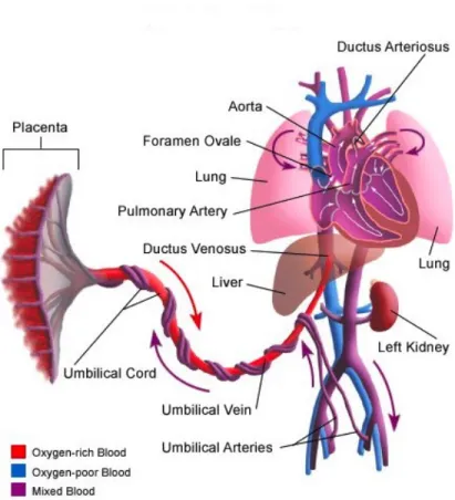
circulatory systems of the mother is not directly connected. The circulatory system of fetus is different from that of a new born baby because fetus does not use its own lungs until birth. The heart of the fetus does not have to pump blood to the lungs to pick up oxygen before birth. i.e; the heart of the fetus does not required a distinct pulmonary artery and aorta, which is blood vessel. These two blood vessels in the fetal heart are joined by a blood vessel termed as the ductus arteriosus. After the birth of baby, the ductus shut down and a distinct left pulmonary artery and aorta form.
The heart of the fetus opens between the upper chambers named as the foramen ovale. It directly circulates blood flow between the right atrium to the left atrium during the fetus development, but after birth it closes . Thus the foramen ovale and the ductus arteriosus are part of the fetal circulatory system before birth but after birth it disappear soon.
A normal heart rate of fetus generally ranges from 120−160 bpm(beats per minute) in the utero period. This heart rate is measured by sonograph from 6
Chapter 1. Introduction
Figure 1.2: Fetal Circulation
weeks and during gestation, the normal range varies which increases to around 170 bpm at10 weeks and it decreases from then to around 130 bpm.
1.3
Heart Signal
1.3.1 Action Potential
The film capability of the cell known as action potential. When an electric current is fortified to a cell at some point then the cell undergoes different mechanical compression and produce a potential known as activity potential. 1.3.2 Resting Potential
The static layer capability of the cell is known as resting potential. When the cells are very still at the point, the layer promptly permits particles likeK+and
Chapter 1. Introduction
Cl− to go into them on the grounds that they are porous layers. The outer part of cell is decidedly charged than within the cell. Subsequently, K+ particles streams into cell to adjust the charge. Consequently, the potential distinction gets grew between within and outer part of the cell at the condition of balance. A resting cell is a spellbound cell.
1.3.3 Depolarization
At first the cell does not permit Na+ particles inside because of larger pen-etrability of the K+ particles. However, when the cell is energized it begins permitting the Na+ particles to move into inner part. This development of Na+ particles contain the ionic streams. Thus, the film boundary lessens to Na+particles. The hurrying of Na+ particles into the phone is called ”Torren-tial slide impact”. Because of higher convergence of the K+ particles in cell theNa+ ions tries to move through outer part of the cell however cannot move as quick as the Na+ particles. Henceforth, within the cell gets to be certain regarding the outside of the cell because of lopsidedness of the K+ particles. This methodology is called depolarization.
1.3.4 Repolarisation
The re-polarization of the cell depends on the time and voltage dependency of the change in permeability of membrane if we compared it for K+ ions with Na+ ions. The Na+ ions moment get slower during depolarisation and hence the re-polarization occurs at the end of depolarization.
1.4
Literature Survey
Seeing an average fetal ECG, the specialist redresses the irregularities in the heart of the fetus. The ECG gadget remove the Fetal ECG signal taken from the AECG flag and reveals to them on the screen. The fetus heart beat get to be more prominent than the heart beat of the mother.
Chapter 1. Introduction
The inspiration driving this venture are difficulties like extracting the low abundancy and high recurrence signal from the high sufficiency and low recur-rence signal. In this proposed work, we have guaranteed extraction of FECG flag however we have not prepared it in a proficient way as did in exploration work by Prasanth K and Baby Paul. We have observed 4000 tests of the Ab-dominal ECG signal with top plentifulness of 3.5 millivolts. And after that the FECG sign rate comes in the reach 120−160 heart pulsates every moment. . Also, consequently the fetal heart thumps quicker than the heart thumps of mother and is extremely touchy to heart anomalies.
Our task guarantees the tops of the FECG is recognized legitimately however not as proficiently did by Butler Pyke, which uses Pan-Tompkins calculation for the identification of the QRS complex. In this venture, we have utilized the separation procedure for the recognition of the R-R crests in a QRS complex. Despite the fact that the system is not all that productive, it issues us the obliged yield as i.e. very nearly exact FECG heart rate.
1.5
Objective
There are two signal in which first one is recorded from the chest of the mother and the second one is recorded from the mother’s abdomen which contains heart beat of both the mother and fetus. The goal of this study is to independent both the FECG and MECG consequently separate the baby pulse and afterward detection of the R-tops to focus the heart beat of the fetus.
1. Extraction of the heart signal of fetus from the abdominal ECG signal.
2. Counting the R-peaks and calculation of the heart beat of the fetus by using the separation of R-R waves.
Chapter 2
Fetal ECG Signal Extraction
Methodology Algorithm Implementation of the AlgorithmChapter 2
Fetal ECG Signa Extraction
2.1
Methodology
The ECG sign of the pregnant women and tyke are recovered from the given data which is inspected at 5 KHz which gives the discrete data utilizing a smoothing filter.The smoothing channel utilized here is a modernized chan-nel called the Savitzky-Golay Filter. The key clarification behind utilizing this channel is to smooth the sign by developing the SNR without reshaping the sign totally. This channel utilizes a convolution system to accomplish the ob-jective. The heart rate of the hatchling is on an exceptionally fundamental level higher than the heart beat that of the mother which is for this situation really around a rate of85pounds every moment while for the embryo it is pretty very nearly 132 throbs every moment. Generally, the hatchling heart rate speedier than that of the mother, which goes from 120 to 160 pounds for each minute. The fetal electrocardiogram sign has an essentially less adequacy when showed up contrastingly in connection to its maternal part. For diagram here the ECG sign of mother has a peak value of 3.5 mv while the ECG sign of fetus has a top of recently 0.25 mv. ECG signs are taken from two exceptional regions of the maternal body, the midsection and the stomach zone. The midsection sign gives the first mother ECG sign however the sign got from the stomach locale is a mix of both mother and hatchling pulse hails commonly summoned by the maternal part reproduced from the midsection opening to the paunch.
Chapter 2. Fetal ECG Signa Extraction
Figure 2.1: Comparison of FECG and MECG
An immediate FIR channel can be utilized with 10 randomized coefficients as a bit of requesting to portray therefore. There may be some extra broad-band impedance joined with both maternal and hatchling standards which can be disposed of by the expansion of a little measure of uncorrelated Gaussian tumult. The try performed by the adaptable channel is to adaptively expel ma-ternal 12area from the creating life pulse signal. To do in that limit it requires a reference signal that is nothing notwithstanding, the sign conveyed from the MECG itself.
2.2
FECG Morphology
Dependable and crucial data about the state of the hatchling amid pregnancy and work is given by FECG, which is only biomedical sign that gives electri-cal representation of Fetus heart beat which is recorded on the maternal belly. The FECG sign is a relatively powerless sign (which is under 20 percent of the MECG) and regularly implanted in AECG and commotion. The FECG lies in the range from 1.3 Hz to 3.5 Hz and here and there it is workable for
Chapter 2. Fetal ECG Signa Extraction
the maternal and a percentage of the FECG signs to be firmly covering. The FECG checking empowers exact estimation of fetal heart execution including transient or perpetual variations from the norm of musicality. For ahead of schedule stage demonstrative of embryo wellbeing and to know its status, at times the FECG is the main data source. The FECG is all that much identified with the mother ECG i.e., MECG, containing the same essential waveforms in-cluding the P wave, the QRS complex, and the T wave . The PQRST intricate as demonstrated in Figure 2.1 is an electric sign created by the compression and unwinding of the embryo’s heart’s muscles. AECG and commotion.
2.3
Techniques for Monitoring FECG
Fetal heart rate examination has turn into a generally acknowledged method for observing fetal status. The most commonplace method for obtaining the fetal heart beat is Doppler ultrasound. Likewise, the FHR checking is addition-ally done by considering fetal magneto cardiogram which employments super-conducting quantum obstruction gadget magnetometers. Aside from this, fetal phonocardiograp permits the heart sounds to be recognized for fetal heart beat checking. The greater part of fetal heart beat examination method is performed utilizing a bedside screen more than a generally short period, with the mother to be in a prostrate position. The greater part of the above methods that are said have been effectively utilized for fetal heart beat checking, in spite of the fact that the beginning decision was which methods among these would be utilized. Clearly, a fetal scalp cathode can’t be utilized stake partum period as there is an extraordinary danger to bring about an imprint or little cut on the fetal head; the instrumentation needed for the obtaining of the FMCG is excessively awkward for wandering utilization; while the FPCG was felt to be excessively defense-less, making it impossible to development curios impacts. Hence, the Doppler ultrasound and the stomach FECG (as it is regularly alluded to) are the most
Chapter 2. Fetal ECG Signa Extraction
feasible alternatives for the checking of fetal heart beat.
As of now, Doppler ultrasound and fetal ECG have turned out to be solid sys-tems for checking fetal heart beat. The FHB observing utilizing the Doppler ultrasound is broadly utilized and suitable be- in light of the fact that an intru-sive test can’t be utilized day by day. The benefit of the Doppler ultrasound strategy is that it can be basically guaranteed that a recording of FHR will be gotten. The drawbacks of such frameworks oblige discontinuous reposition-ing of the transducer and they are suitable for utilization with exceptionally prepared birthing specialists. The ultrasound transducer is hazardous what’s more, uncomfortable while the technique includes dispatching a 2-MHz signal towards the embryo. The utilization of Doppler ultrasound (non-intrusive way) is not suitable for long stretches of FHR checking. This may include skillful position and nonstop re-positioning of the transducer, which would be an ex-treme issue for long haul wandering utilization. It may bring about records of questionable increasing velocities or deceleration and genuine unexpected changes can be misconstrued as clamor. The significant impediment of the Doppler ultrasound system is its affectability to development. The develop-ment of the mother can bring about Doppler-moved reflected waves, which are more grounded than the heart signal. This Doppler ultrasound procedure is wrong for long haul observing of the FHB, because it requires the patients to be rested. Besides, the location of the pulse utilizing Doppler ultrasound de-pends upon an auxiliary impact (the mechanical development of the heart) and is hence not as exact for beat-to-thump investigation as discovery of the QRS complex. United to this disadvantage is the way that most Doppler frameworks depend upon some type of averaging to deliver their FHR information.
Conversely, systems using the stomach electrocardiogram have a more note-worthy prospect for long haul observing of FHB and fetal prosperity utilizing sign handling procedures. The AECG sign can likewise be utilized for
Chapter 2. Fetal ECG Signa Extraction
tum non-intrusive FHR determination through the discovery of little fetal heart possibilities at the surface of the maternal midriff. The AECG can be utilized to create genuine RR interim information, which is suitable for heart rate vari-ability studies if needed. Its point of preference is that it is totally non-obtrusive and subtle, has similarly low power necessities, and can be utilized over broad-ened (e.g., 24 h) periods. The technique moreover permits the maternal heart rate (MHR) to be recorded subsequent to the MECG is likewise identified from the AECG. It is beneficial of utilizing AECG to concentrate FECG with the ex-tra data conex-trasted with utilizing Doppler ulex-trasound. Some new exceedingly exact systems are accounted for observing the FHR.
2.4
Algorithm
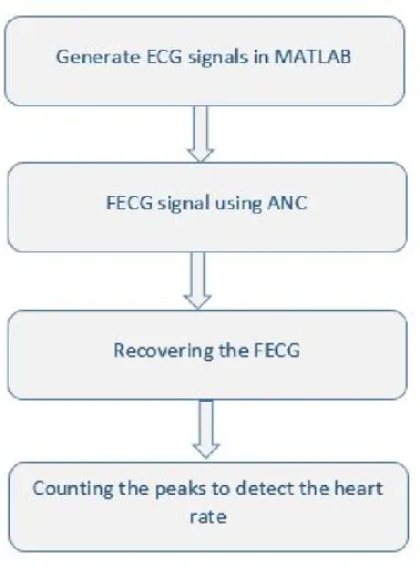
The proposed extraction technique in this proposal is used by MATLAB code which incorporate four general strides as depicted in the flowchart of the pro-posed calculation demonstrated beneath in Fig. 2.2.
2.5
Implementation of the Algorithm
The distinctive strides of the proposed calculation are quickly portrayed under-neath,
2.5.1 ECG signals generation in MATLAB
MATLAB is a simple device which is to a great degree valuable to extract the FECG from AECG. By the use of MATLAB we make the sign on which the proposed algorithm can be performed and executed smoothly. MATLAB con-tains a limit Savitzky-Galoy channel limit and using this request the obliged signs are created.
MATLAB coding is useful to copy the conditions of the both FECG and MECG signals. The MECG sign created from the chest of the mother using
Chapter 2. Fetal ECG Signa Extraction
Figure 2.2: Algorithm for FECG Extraction
the given data as a part of past section is exhibited in Fig. 2.4. The top ade-quacy of this sign is 3.5 mV and the heart beat being 85 thumps each minute. Nevertheless, the infant heart throbs discernibly speedier than heart beat of the mother running between 120−160 beats each minute as communicated pre-viously. The FECG sign made is exhibited in Fig. 2.5 and the planned sign recorded from the midsection of the mother which is commonly directed by the maternal heartbeat sign is demonstrated in Fig. 2.6.
Chapter 2. Fetal ECG Signa Extraction
2.5.2 Extraction Methods for FECG
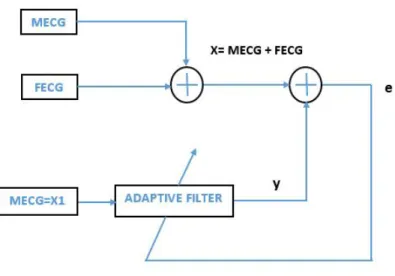
1. Method of Adaptive Noise Canceller (ANC): Two signs are required to empty foundation curios including particular commotions or impedance formally exist in the Fetal ECG movement by using flexible filtering. The principal is the Fetal ECG sign included with MECG sign and the refer-ence signal being the secondary which is moreover the sign to be crossed out to get the Fetal ECG signal. The reference sign is just same as the Maternal ECG signal. Disturbances indicate in the both signs must be very much related i.e. the tumult in the crucial and discretionary signs. In the extraction method of fetal ECG, adaptable channels are used to adap-tively oust a heartbeat of mother from the FECG sign to get the beat sign of the tyke or the hatchling. Right when the workplace is stationary, these channels have reliable shape and presentation for the error execution sur-face. Regardless, for operation in a circumstance that is not stationary, the base of this surface moves reliably nearby the shots of changing pre-sentation and curve. In this manner, for non-stationary inputs, the channel searches for the base of the slip execution surface nearby constantly tailing it down. The square framework for a key Adaptive Noise Canceller struc-ture is shown as indicated in Fig. 2.3. By making accurate estimations of the angle vector at every emphasis and picking the stride size parameter suitably, the tap-weight vector figured would focalize to the ideal Wiener arrangement utilizing the strategy for steepest-plunge. A few recursive calculations can be utilized to change the tap-weight vector after every cycle. Minimum Mean Square (LMS) calculation is one of such calcula-tions that has a certain point of preference as it does excludes any strides with respect to grid reversal. This can be expressed numerically as,
Wk+1=Wk+2µ εkXk
Chapter 2. Fetal ECG Signa Extraction
Figure 2.3: ANC Block Diagram
2. High Frequency Removal Using Digital Filter: The IIR and FIR chan-nels are the two key sorts of chanchan-nels by and large used as a piece of Digital Signal Transforming (DSP). Among those FIR channels can be easily created in straight stage and a rate of the figurings can be blocked by using FIR channels, thusly giving some essential computational viabil-ity. With a particular final objective to check the heart beat of the fetus, the R-highest points of the Fetal ECG sign must be isolated . A FIR channel with fitting channel coefficients is used to empty high frequencies to ac-complish this goal. By then an edge worth is arranged and the finish with higher qualities than the farthest point are distinguished as the R-peaks of the Fetal ECG sign. The amount of R-tops are numbered and heart beat of the fetus is figured from the R-R interval.
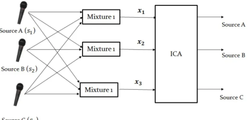
3. Independent Component Analysis(ICA):ICA is a computational system for isolating a multivariate signal into added substance subcomponents. This is finished by expecting that the subcomponents are non-Gaussian signals and that they are measurably free from one another. ICA is a unique instance of visually impaired source partition. A typical case
Chapter 2. Fetal ECG Signa Extraction
plication of ICA is the ”cocktail party problem” of listening in on one individual’s discourse in an uproarious room.
Independent Component Analysis endeavors to break down a multivariate signal into autonomous non-Gaussian signals. As an illustration, sound is generally a flag that is made out of the numerical expansion, at every time t, of signals from a few sources. The inquiry then is whether it is conceivable to independent these contributing sources from the watched aggregate signal. At the point when the factual freedom suspicion is right, dazzle ICA partition of a blended signal gives great results. It is addi-tionally utilized for signals that shouldn’t be created by a blending for investigation purposes. A basic use of ICA is the ”cocktail party prob-lem”, where the hidden discourse signals are isolated from an example information comprising of individuals talking at the same time in a room. Generally the issue is disentangled by accepting no time delays or echoes. A vital note to consider is that if N sources are available, at any rate N perceptions (e.g. receivers) are expected to recoup the first flags. This constitutes the square case (J=D, whereDis the info measurement of the information and J is the measurement of the model). Different instances of underdetermined (J > D) and overdetermined (J < D) have been re-searched. That the ICA partition of blended signals gives great results are in view of two presumptions and three impacts of blending source signals. Two suppositions:
(a) The signals originate from source are not dependent to each other. (b) The signal values of each source have non-Gaussian distributions.
Mixing of source signals has three effects:
(a) Independence: According to suspicion 1, the source signs are free; on the other hand, their sign blends are definitely not. This is on the
Chapter 2. Fetal ECG Signa Extraction
Figure 2.4: ICA Block Diagram
grounds that the sign blends have the same source signals.
(b) Normality: As indicated by the Central Limit Theorem, the dissem-ination of an aggregate of free irregular variables with limited fluctu-ation tends towards a Gaussian dispersion. Freely talking, an entirety of two free arbitrary variables as a rule has a circulation which is closer to Gaussian than any of the two unique variables. Here we consider the estimation of every signal as the irregular variable. (c) Complexity: The worldly unpredictability of any signal blend is more
prominent than that of its easiest constituent source signal.
Chapter 3
QRS Complex
MethodologyChapter 3
QRS Complex
3.1
Methodology
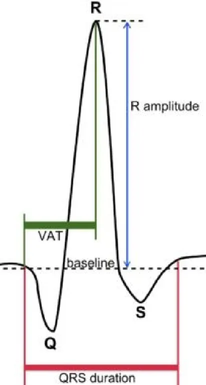
The QRS complex is a mixture of three of the graphical avoidances seen on a normal electrocardiogram (EKG or ECG). It is typically the focal and most outwardly clear piece of the following. It relates to the depolarization of the privilege and left ventricles of the heart. In grown-ups, it typically keeps going 0.06−0.10s; in youngsters and amid physical movement, it might be shorter.
Normally there are five diversions in an ECG, discretionarily named as ”P” to ”T” waves. The Q, R, and S waves happen in quick progression, don’t all show up in all leads, and mirror a solitary occasion, and along these lines are typically viewed as together. The Q waves are any descending avoidance after the P waves. The R wave takes after an upward avoidance, and the S waves are any descending redirection after the R waves. The T wave takes after the S wave, and sometimes an extra U wave takes after the T wave.
Chapter 3. QRS Complex
Figure 3.1: ECG of Normal Sinus Rhythm
Chapter 3. QRS Complex
Figure 3.2: Schematic representation of the QRS complex
Chapter 4
Chapter 4
Simulation Results
Heart beat is estimation of the pounding of the heart. Heart pounds in a settled time compass and the estimation of number of thumps, that is number of R peaks each minute which record the heart beat. Finally the sign is filtered and an edge level is arranged so that any quality above it can be considered as a peak and accordingly the amount of peaks in the sign can be tallied. The heart beat counting is performed by the use of the QRS revelation techniques which is discussed in Chapter 3. The most upgraded method is to count the R peaks of the fetal ECG signal. Counting the R peaks the beat of the Fetus is figured. The heart beat comes to be 135 thumps each minute. This is comes in the degree absolutely. As the fetal heart rate is more unmistakable than the maternal heart rate and it comes in the achieve 120-160 bangs for each minute, this test affirms the same relevant. Finally, all the records shown underneath that illuminates the sign sorts ECG signal of the fetus.Chapter 4. Simulation Results
Figure 4.1: The Measured Signal
Figure 4.2: The Reference Signal
Chapter 4. Simulation Results
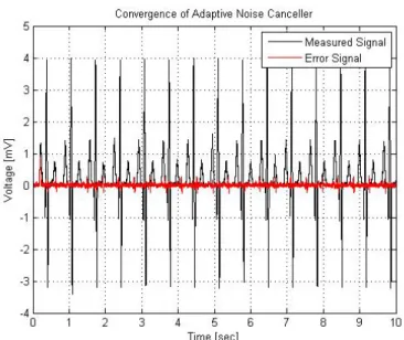
Figure 4.3: Output of the Adaptive Noise Canceller (ANC)
Figure 4.4: Steady-State error signal
Chapter 4. Simulation Results
Figure 4.5: The Filtered Signal
Figure 4.6: The Reconstructed fetus Signal
Chapter 5
Conclusion and Future work
Conclusion Future workChapter 5
Conclusion and Future Work
5.1
Conclusion
The yields of the removed sign were recorded using MATLAB code. At last, the fetal ECG signal is extricated and pulse of the sign is computed. The cal-culation in light of Adaptive Noise Canceller is proposed and actualized ef-fectively. The execution of the calculation has been confirmed effectively on MATLAB and the calculation is discovered to be very productive. It is found after fruitful execution that fetal ECG signal can be effectively separated by utilizing autonomous part investigation. R tops were additionally recognized effectively giving the last heart rate of the signal.
5.2
Future work
The outcomes acquired matches with the real FECG flags fundamentally and will suffice to demonstrate right. The work done here is on-going and it has numerous extensions in future on the grounds that it hasn’t been nitty gritty yet. The future work would incorporate outlining an ECG with in-fabricate programming actualizing the above extraction calculation. At that point the procedure can be utilized generally as a part of item advancement. Yet, the extraction procedures may change with time and henceforth more effective and blunder free procedures will be produced in future. Thusly the undertaking can get up and go in future.
Bibliography
[1] M. Rachid, M. Feham, et al., “Algorithm of remote monitoring ecg us-ing mobile phone: Conception and implementation,” African Journal of Information & Communication Technology, vol. 5, no. 2, p. 11, 2009.[2] T. Ince, S. Kiranyaz, and M. Gabbouj, “A generic and robust system for automated patient-specific classification of ecg signals,”Biomedical Engi-neering, IEEE Transactions on, vol. 56, no. 5, pp. 1415–1426, 2009. [3] K. Prasanth, B. Paul, and A. A. Balakrishnan, “Fetal ecg extraction using
adaptive filters,”International Journal of Advanced Research in Electrical, Electronics and Instrumentation Engineering, vol. 2, no. 4, 2013.
[4] P. P. Kanjilal, S. Palit, and G. Saha, “Fetal ecg extraction from single-channel maternal ecg using singular value decomposition,” Biomedical Engineering, IEEE Transactions on, vol. 44, no. 1, pp. 51–59, 1997.
[5] J. Cardoso, “Multidimensional independent component analysis,” in Acoustics, Speech and Signal Processing, 1998. Proceedings of the 1998 IEEE International Conference on, vol. 4, pp. 1941–1944, IEEE, 1998.
[6] A. Hyv¨arinen and E. Oja, “A fast fixed-point algorithm for independent component analysis,” Neural computation, vol. 9, no. 7, pp. 1483–1492, 1997.
Bibliography
[7] V. Zarzoso and A. K. Nandi, “Noninvasive fetal electrocardiogram ex-traction: blind separation versus adaptive noise cancellation,” Biomedical Engineering, IEEE Transactions on, vol. 48, no. 1, pp. 12–18, 2001.
[8] R. Sameni, C. Jutten, and M. B. Shamsollahi, “Multichannel electrocar-diogram decomposition using periodic component analysis,” Biomedical Engineering, IEEE Transactions on, vol. 55, no. 8, pp. 1935–1940, 2008. [9] M. E. Davies and C. J. James, “Source separation using single channel
ica,”Signal Processing, vol. 87, no. 8, pp. 1819–1832, 2007.
[10] Y. Ye, Z.-L. Zhang, J. Zeng, and L. Peng, “A fast and adaptive ica algo-rithm with its application to fetal electrocardiogram extraction,” Applied Mathematics and Computation, vol. 205, no. 2, pp. 799–806, 2008.
[11] F. S. Najafabadi, E. Zahedi, and M. A. M. Ali, “Fetal heart rate monitor-ing based on independent component analysis,”Computers in biology and Medicine, vol. 36, no. 3, pp. 241–252, 2006.
[12] M. Chawla, H. Verma, and V. Kumar, “Retracted: Artifacts and noise removal in electrocardiograms using independent component analysis,” International journal of cardiology, vol. 129, no. 2, pp. 278–281, 2008.