Susceptibility of Human Iris Stromal Cells to Herpes Simplex Virus 1
Entry
John Baldwin,aPaul J. Park,bBrian Zanotti,aErika Maus,aMichael V. Volin,aDeepak Shukla,b,cVaibhav Tiwaria,c
Department of Microbiology & Immunology, Midwestern University, Downers Grove, Illinois, USAa; Department of Microbiology & Immunologyband Department of
Ophthalmology & Visual Sciences,cUniversity of Illinois at Chicago, Chicago, Illinois, USA
Ocular herpes simplex virus 1 (HSV-1) infection can lead to multiple complications, including iritis, an inflammation of the iris. Here, we use human iris stroma cells as a novelin vitromodel to demonstrate HSV-1 entry and the inflammatory mediators that can damage the iris. The upregulated cytokines observed in this study provide a new understanding of the intrinsic immune mechanisms that can contribute to the onset of iritis.
H
erpes simplex virus 1 (HSV-1) is a significant ocular pathogen affecting multiple regions, including the iris (1). The iris, a specialized eye tissue, is affected in various inflammatory ophthal-mic conditions (2,3). For instance, inflammation of the iris fol-lowing HSV-1 infection may be associated with elevated intraoc-ular pressure, ultimately resulting in glaucoma, although the possibility remains that this may be steroid induced (4–6). The iris has been shown to have histopathologic involvement in HSV-1 infection of the corneal stroma, herpetic stromal keratitis (HSK) (3). Inflammation of the iris is also seen in herpetic anterior uve-itis, a condition that often presents as an inflammation of the iris and ciliary body (iridocyclitis) and is the leading cause of infec-tious anterior uveitis worldwide (2). In addition to iris involve-ment in primary inflammatory conditions of various ocular tis-sues, there had previously been a longstanding question as to whether HSV-induced iritis could occur without concurrent or precedent keratitis. Studies performed since have indicated that HSV may cause iritis that predisposes to glaucoma without nec-essarily causing keratitis (6). Although these results indicate the possible pathogenesis mediated by primary HSV-1 infection of the iris, primary infection of human iris stromal (HIS) cells has not been previously investigated. Likewise, chemokine signaling, which plays a significant role in many inflammatory eye condi-tions (7), has been poorly studied in the context of HSV-1 infec-tion of HIS cells. The current study demonstrates the potential of anin vitroHIS cell model to uncover the molecular basis of viral entry and the identity of the inflammatory mediators involved.HSV-1 starts its infectious journey into cells via interactions of its envelope glycoproteins with their respective host cell receptors (8). The initial attachment involves glycoprotein B (gB) and gly-coprotein C (gC) interactions with host cell surface heparan sul-fate proteoglycans (9,10). HSV-1 glycoprotein D (gD), with its subsequent conformational changes, can bind to any of its host cell receptors, such as nectin-1, herpesvirus entry mediator (HVEM) (11), or 3-O-sulfated heparan sulfate (3-OS HS) (12). This conformational change is important since it allows for the gD-host cell receptor complex to associate with the heterodimer complex of glycoprotein H-glycoprotein L (gH-gL). This allows gH-gL to bind gB, a process that allows the viral and host cell membranes to fuse (12,13).
Following infection by HSV-1, the intrinsic inflammatory re-sponses contribute strongly to subsequent disease manifestations. In cases of HSK, an outburst of cytokine and chemokine activities
by local and infiltrating immune cells can serve as the main con-tributor to the observed scarring of the corneal stromal tissue and vision loss (14,15). Inhibiting the overreactive inflammatory re-sponse elicited by HSV-1 replication may effectively taper the damaging effects on the ocular tissues (16). This possibility has been evidenced with the frequent use of steroids to treat HSK. Here, we demonstrate primary cultures of HIS cells as an excellent model to study and highlight cytokine induction that occurs in the iris in the absence of the other mediators of inflammation, such as neutrophils that are usually recruited to the site upon infection (17).
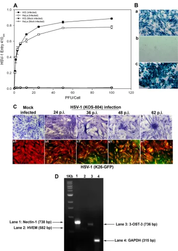
Cultured HIS cells are susceptible to HSV-1 entry and plaque formation.In order to demonstrate susceptibility to HSV-1 infec-tion, HIS cell cultures were prepared in accordance with institu-tional review board-approved protocols and were isolated from anonymously donated human eyes (provided by the Illinois Eye Bank, Chicago, IL) via sterile dissection of the iris and pigmented epithelial layer and subsequent removal with a sterile cotton swab. The tissue was then digested with 0.2% type II collagenase (Sigma-Aldrich, St. Louis, MO) in cell culture medium, MCDB-131 (Sig-ma-Aldrich, St. Louis, MO) at 37°C with gentle stirring for 20 to 30 min. Digested tissues were next centrifuged to remove tissue debris, and HIS cells were cultured in MCDB-131 containing 10% fetal bovine serum (FBS) and antibiotics. To establish an HSV-1 entry model for the cells of the iris, both HIS and HeLa cells were infected with serial dilutions of a recombinant form of HSV-1 (KOS) gL86 that expresses-galactosidase under the HSV-1 im-mediate-early promoter following cell entry (18). Mock infections with PBS and without virus of both cell lines were used as controls. As shown inFig. 1A, compared to the controls, treatment of HIS and HeLa cells demonstrated viral entry in a dose-dependent manner. We further confirmed the above-described result via X-Gal (5-bromo-4-chloro-3-indolyl--D-galactopyranoside) stain-ing after infectstain-ing HIS cells with reporter HSV-1. As shown inFig.
Received20 November 2012Accepted14 January 2013
Published ahead of print23 January 2013
Address correspondence to Vaibhav Tiwari, [email protected].
J.B. and P.P. contributed equally to this article.
Copyright © 2013, American Society for Microbiology. All Rights Reserved.
doi:10.1128/JVI.03235-12
on November 7, 2019 by guest
http://jvi.asm.org/
FIG 1Analysis of herpes simplex virus 1 (HSV-1) entry in primary cultures of HIS cells. (A) Entry of HSV-1 into cultured HIS cells. Cultured HIS cells, along with HeLa cells, were plated in 96-well plates and inoculated with serial dilutions of a recombinant form of HSV-1 (KOS) gL86 that expresses-galactosidase following cell entry at the indicated PFU/cell. After 6 h, the cells were washed, permeabilized, and incubated witho-nitrophenyl--D-galactopyranoside
(ImmunoPure ONPG; Pierce) substrate for quantitation of-galactosidase activity expressed from the input viral genome. The enzymatic activity was measured by spectrophotometer (Molecular Devices) at an optical density (OD) at 410 nm. Values in the figure were plotted as the means from three determinations (⫾standard deviations [SD]). (B) X-Gal staining of HSV-1 entry. Cultured HIS cells inoculated with HSV-1 (KOS) gL86 at an MOI of 0.01 (a) were 100% stained blue, indicating 100% viral infection, whereas uninfected cells (b) did not demonstrate any staining. (c) HeLa cells, with HSV-1 KOS gL86-infected cells stained blue, were used as a positive control. (C) Viral cell-to-cell spread. HIS cells were inoculated with serial dilutions of two recombinant forms of HSV-1, (KOS) 804, a mutant known to form high numbers of syncytia, and green fluorescent protein-tagged HSV-1 (K26) GFP. Confluent monolayer cultures inoculated with HSV-1 (KOS) 804 for 2 h were then washed and incubated with methyl cellulose containing media and stained at the various time points p.i. as indicated (a to e). Cells inoculated with HSV-1 (K26) GFP were subjected to fluorescence imaging (a1 to e1). Confluent monolayer cultures were stained with TRITC-conjugated phalloidin (red color), which binds to F-actin. HSV-1-infected cells showed both green fluorescence from HSV-1 capsid and red fluorescence from F-actin expression (green and red overlapping areas appear yellow). The images were taken with a fluorescence microscope at 20⫻magnification. (D) Standard nonquantitative RT-PCR analysis for entry receptor expression. Total RNA samples were isolated from HIS cells and converted to first-strand cDNAs and analyzed by PCR for the receptors as indicated. A housekeeping gene, GAPDH, was used as a control.
on November 7, 2019 by guest
http://jvi.asm.org/
[image:2.585.113.471.63.566.2]1B, cultured HIS cells that were exposed to HSV-1 (KOS) gL86 were 100% stained blue (Fig. 1B, panel a) compared to uninfected HIS cells (Fig. 1B, panel b). HeLa cells, with HSV-1 KOS gL86-infected cells shown in blue, were used as a positive control
(Fig. 1B, panel c). Next, in order to verify viral replication and
spread to the neighboring cell, we used two recombinant forms of HSV-1 (KOS) 804, a mutant known to form high numbers of syncytia (19) and green fluorescent protein (GFP)-tagged HSV-1, HSV-1 (K26) GFP driven by the UL35 promoter sequences (20). As shown inFig. 1C(panels a to e), more plaques were visualized at greater lengths of time postinfection (p.i.), while no plaques were observed in the mock-infected control. As shown inFig. 1C
(panels a1 to e1), fluorescence imaging was used to observe HSV-1 (K26) GFP spread into HIS cells stained with tetramethyl rhoda-mine isocyanate (TRITC)-conjugated phalloidin binding to F-ac-tin. HSV-1-infected cells demonstrated both green fluorescence due to the HSV-1 capsid and red fluorescence from F-actin ex-pression. Mock-infected cells showed only red fluorescence. Next, using reverse transcriptase PCR (RT-PCR), we focused on dem-onstrating the expression of common HSV-1 entry receptors in HIS cells. RNA samples were isolated from HIS cells and con-verted to first-strand cDNAs using the high-capacity cDNA re-verse transcription kits (Applied Biosystems, Foster City, CA) by following the manufacturer’s protocol. The RNA isolation and the primers were based on our previously published study (21). As shown inFig. 1D, expression of the gD receptor nectin-1 was evident in HIS cells, while levels of HVEM and 3-OST-3 were found to be insignificant, suggesting that it is nectin-1 that is the gD receptor being used. Compared to glyceraldehyde-3-phos-phate dehydrogenase (GAPDH), which was used as the control, the signal for nectin-1 expression was the highest, followed by those of 3-OST-3 and HVEM. A relatively low expression of HVEM has also been reported with other cell types (21–23). Com-bined together, our results demonstrated that cultured HIS cells are highly susceptible to HSV-1 entry and spread. In addition, HIS cells express the required gD receptors that are expressed by other, more studied target cell types, including corneal epithelial cells (21).
Inflammation in HIS cells upon HSV-1 infection.Intrinsic mediators of iritis are poorly understood. Anin vitrocell culture model is the best way to identify the mediators of inflammation that are solely generated by the resident cells of the iris. Using a RayBio cytokine antibody array (RayBio, Norcross, GA) per the manufacturer’s instructions, we assessed inflammatory markers induced as a result of HSV-1 infection of HIS cellsin vitro. We used HSV-1 (KOS) 804 virus strain at a low (0.01) multiplicity of infection (MOI) to infect HIS cells and also mock-infected HIS cells with PBS to monitor inflammation. As shown inFig. 2Aand
B, at 1 day p.i., there were no significant qualitative differences in the signal intensities of the inflammatory markers interleukin 6 (IL-6), IL-8, monocyte chemotactic protein 1 (MCP-1), MCP-2, regulated on activation normal T cell expressed and secreted (RANTES), or TIMP metallopeptidase inhibitor 2 (TIMP-2) be-tween the HSV-1-infected and uninfected cells. However, at 2 days p.i., as noted inFig. 2C andD, there was a greater signal intensity of MCP-2, TIMP-2, IL-6, and IL-8 in the infected cells. In addition, expression of new inflammatory markers, such as gran-ulocyte-macrophage colony-stimulating factor (GM-CSF), I-309, intercellular adhesion molecule 1 (ICAM-1), IL-6 soluble receptor (IL-6sR), interferon-inducible protein 10 (IP-10), macrophage
inflammatory protein 1(MIP-1), and soluble tumor necrosis factor receptor 1 (sTNFR1), was documented in order of decreas-ing qualitative signaldecreas-ing at 2 days p.i. in infected cells, although none are present at 1 day p.i. Although levels of ICAM-1 have been suggested to have only moderate effects on immune response in cases of HSK in a murine model (24), upregulation of other mark-ers at 2 days p.i. may have greater implications in terms of HSV-1-mediated pathology. For instance, GM-CSF has previously been shown to have a role in HSK via a supportive function of the neutrophils infiltrating corneal tissue, an important finding, as neutrophils are believed to contribute greatly to the permanent corneal damage seen in HSK. GM-CSF is also known to induce a maturation stimulus important for antigen-presenting cell (APC) function (25). The fact that this cytokine is first detected 2 days p.i. gives detailed insight into the temporal changes in the inflamma-tory response in infected HIS cells. The upregulation of IL-6 seems critical, too, as previous studies have suggested that IL-6 stimu-lates the secretion of vascular endothelial growth factor (VEGF) in the cornea (26,27), implicating possible roles for initiating neo-vascularization by HSV-1 infection of HIS cells as well. The detec-tion of IL-6sR with upreguladetec-tion of IL-6 is not surprising then, as IL-6sR has been shown to increase the activity levels of IL-6 (28). An antiviral effect against HSV-1 is also documented in IP-10 detection at 2 days p.i., since this is known to directly inhibit HSV-1 replication (29). Upregulation of other inflammatory markers, such as I-309 and sTNFR1, may support viral infection since these chemokines are believed to have antiapoptotic effects (30,31). The fact that these are seen in anin vitromodel suggests that infected cells, independent of the host’s immune response, are an important source of the mediators and cytokines seen in HSV-1 infection.
We next used the cytokine array to evaluate if expression of cytokines is altered during HSV-1-glycoprotein-induced cell fu-sion. As briefly mentioned, virus-mediated cell fusion requires the binding of gD to one of its host cell receptors, HVEM, 3-OS HS, or nectin-1. The subsequent conformational change in gD allows for this gD-host cell receptor complex to interact with gB and gH-gL to initiate the fusion of the viral and host cell membranes. This mechanism initially helps with virus entry and subsequently for the spread of the virus from cell to cell (13). A surrogate form of this assay can be performed by transiently expressing four HSV-1 glycoproteins, gB, gD, gH-gL, in cells and then allowing the cells to fuse with cells expressing a gD receptor such as nectin-1. We hy-pothesized that this biologically relevant assay can improve our understanding of the proinflammatory role of some key glycopro-teins involved in HSV-1 entry. In order to proceed with this assay, we first verified whether HIS cells show the ability to fuse with Chinese hamster ovary (CHO-K1) cells expressing HSV-1 glyco-proteins. CHO-K1 cells were either transfected with an empty vector (pCDNA3.1) or plasmids expressing one or more of the glycoproteins. Data and the schematic in Fig. 3A verify that CHO-K1 cells expressing HSV-1 glycoproteins gB, gD, and gH-gL were able to successfully fuse with HIS cells. Next, the cytokine array was used to determine the changes in the cytokine expres-sion upon cell fuexpres-sion. The assay was performed 2 days after mixing the target HIS cells with glycoprotein-expressing CHO-K1 cells. As shown inFig. 3B, there was overall greater signal intensities detected for IL-6, MCP-1, IL-8, GM-CSF, and RANTES in the test than in the negative control. The variations in cytokines between the test and negative controls are plotted inFig. 3C. These results
HSV-1 Entry and Inflammation
on November 7, 2019 by guest
http://jvi.asm.org/
FIG 2Inflammation in HIS cells upon HSV-1 infection. (A) A RayBio cytokine antibody array (RayBio, Norcross, GA) per the manufacturer’s instructions was used to assess inflammatory markers produced in HSV-1 (KOS) 804 (MOI of 0.01) infection of anin vitroHIS cell culture at 1 day p.i.; (B) signal intensities of various inflammatory markers are plotted in uninfected versus infected cells at 1 day p.i.; (C) inflammatory markers measured at 2 days p.i. utilizing same material and methods as described above; (D) signal intensities of various inflammatory markers are plotted in uninfected versus infected cells at 2 days p.i.
on November 7, 2019 by guest
[image:4.585.133.448.35.652.2]suggest that it is possible that the upregulation of inflammatory cytokines seen in HSV-1 infection of HIS cells as noted earlier may be partly a result of the membrane fusion process that is mediated by the viral glycoproteins. Therefore, targeting some of these
com-mon cytokines may have therapeutic effects against HSV-1 entry as well as viral spread between the cells and an overall reduction in the complications seen with iritis. Further studies would be needed to confirm this possibility.
FIG 3Cytokine response following membrane fusion. (A) HIS cell fusion with HSV-1 glycoprotein-expressing Chinese hamster ovary (CHO-K1) cells. CHO-K1 cells were either transfected with an empty vector (pCDNA3.1) or plasmids expressing one or more of the glycoproteins gB, gD, or gH-gL. A luciferase-based reporter system was used to measure fusion. Relative luciferase activity was measured in relative luciferase units (RLU) (yaxis). Cell fusion was measured in RLU. CHO-K1 cells expressing the empty vector were used as a negative control. The schematic shows both unsuccessful (a) and successful (b) cell fusion between CHO-K1 and HIS cells. (B) A RayBio cytokine antibody array (RayBio, Norcross, GA) per the manufacturer’s instructions was used to assess inflammatory markers produced in HIS cell interactions with CHO-K1 cells in test versus negative-control cases. (C) Signal intensities of various inflammatory markers are plotted in negative-control (no fusion) versus test (fusion) cells.
HSV-1 Entry and Inflammation
on November 7, 2019 by guest
http://jvi.asm.org/
[image:5.585.131.452.64.580.2]Our results provide evidence for the first time that HIS cells are susceptible to HSV-1 entry/spread and verify the essential viral and host receptors involved, thus establishing anin vitromodel to study iris stromal infection and the intrinsic mediators of inflam-mation. The fact that many of the inflammatory mediators are seen in the absence of the host immune response suggests a great importance for a usage of anin vitroHIS cell model. Future studies using this model may further help distinguish between the contri-butions from infected cells and those from the infiltrating im-mune cells during HSV-1 infection. This model may also serve as a platform to test for potential therapeutics that may help against HSV-1-induced iritis. For instance, G2, a peptide isolated against the receptor 3-OS HS, has previously been shown to decrease HSV-1-induced mouse corneal infection (32). With thisin vitro model, we may be able to assess how inhibiting various stages of viral attachment and entry may affect the chemokine induction. Along the same line, this model will also be a new and an easy step to screen for effective drugs that reduce inflammatory conditions in context with HSV-1 infection of the iris stroma.
ACKNOWLEDGMENTS
This work was supported by NIH grants to D.S. (AI057860, AI081869) and V.T. (AI 088429-01A1) and a Midwest Eye Bank research grant to M.V.V. J.B. was supported by a Midwestern University (Downers Grove, IL)-sponsored Kenneth A. Suarez Summer Research Fellowship (10-2014-8172).
REFERENCES
1.Dawson CR, Togni B.1976. Herpes simplex eye infections: clinical man-ifestations, pathogenesis and management. Surv. Ophthalmol.21:121– 135.
2.Rathinam SR, Namperumalsamy P.2007. Global variation and pattern changes in epidemiology of uveitis. Indian J. Ophthalmol.55:173–183. 3.Teitelbaum CS, Streeten BW, Dawson CR. 1987. Histopathology of
herpes simplex virus keratouveitis. Curr. Eye Res.6:189 –194.
4.Sungur GK, Hazirolan D, Yalvac IS, Ozer PA, Aslan BS, Duman S. 2010. Incidence and prognosis of ocular hypertension secondary to viral uveitis. Int. Ophthalmol.30:191–194.
5.Tugal-Tutkun I, O¨ tük-Yasar B, Altinkurt E.2010. Clinical features and prognosis of herpetic anterior uveitis: a retrospective study of 111 cases. Int. Ophthalmol.30:559 –565.
6.Van der Lelij A, Ooijman FM, Kijlstra A, Rothova A.2000. Anterior uveitis with sectoral iris atrophy in the absence of keratitis: a distinct clinical entity among herpetic eye diseases. Ophthalmology107:1164 – 1170.
7.Lundberg P, Cantin E.2003. A potential role for CXCR3 chemokines in the response to ocular HSV infection. Curr. Eye Res.26:137–150. 8.Spear PG, Longnecker R.2003. Herpesvirus entry: an update. J. Virol.
77:10179 –10185.
9.Herold BC, WuDunn D, Soltys N, Spear PG.1991. Glycoprotein C of herpes simplex virus type 1 plays a principal role in the adsorption of virus to cells and in infectivity. J. Virol.65:1090 –1098.
10. Scanlan PM, Tiwari V, Bommireddy S, Shukla D.2005. Spinoculation of heparan sulfate deficient cells enhances HSV-1 entry, but does not abolish the need for essential glycoproteins in viral fusion. J. Virol. Methods128: 104 –112.
11. Krummenacher C, Baribaud F, Ponce de Leon M, Baribaud I, Whitbeck JC, Xu R, Cohen GH, Eisenberg RJ.2004. Comparative usage of herpes-virus entry mediator A and nectin-1 by laboratory strains and clinical isolates of herpes simplex virus. Virology322:286 –299.
12. Shukla D, Liu J, Blaiklock P, Shworak NW, Bai X, Esko JD, Cohen GH, Eisenberg RJ, Rosenberg RD, Spear PG.1999. A novel role for 3-O-sulfated heparan sulfate in herpes simplex virus 1 entry. Cell99:13–22.
13. Eisenberg RJ, Atanasiu D, Cairns TM, Gallagher JR, Krummenacher C, Cohen GH.2012. Herpes virus fusion and entry: a story with many char-acters. Viruses4:800 – 832.
14. Biswas PS, Banerjee K, Kim B, Rouse BT.2004. Mice transgenic for IL-1 receptor antagonist protein are resistant to herpetic stromal keratitis: pos-sible role for IL-1 in herpetic stromal keratitis pathogenesis. J. Immunol. 172:3736 –3744.
15. Zheng M, Deshpande S, Lee S, Ferrara N, Rouse BT.2001. Contribution of vascular endothelial growth factor in the neovascularization process during the pathogenesis of herpetic stromal keratitis. J. Virol.75:9828 – 9835.
16. Carr DJ, Chodosh J, Ash J, Lane TE. 2003. Effect of anti-CXCL10 monoclonal antibody on herpes simplex virus type 1 keratitis and retinal infection. J. Virol.77:10037–10046.
17. Tumpey TM, Cheng H, Cook DN, Smithies O, Oakes JE, Lausch RN. 1998. Absence of macrophage inflammatory protein-1␣prevents the de-velopment of blinding herpes stromal keratitis. J. Virol.72:3705–3710. 18. Montgomery RI, Warner MS, Lum BJ, Spear PG.1996. Herpes simplex
virus-1 entry into cells mediated by a novel member of the TNF/NGF receptor family. Cell87:427– 436.
19. Little SP, Schaffer PAA.1981. Expression of the syncytial (syn) pheno-type in HSV-1, strain KOS: genetic and phenotypic studies of mutants in two syn loci. Virology112:686 –702.
20. Desai P, Person S.1998. Incorporation of the green fluorescent protein into the herpes simplex virus type 1 capsid. J. Virol.72:7563–7568. 21. Shah A, Farooq AV, Tiwari V, Kim MJ, Shukla D.2010. HSV-1 infection
of human corneal epithelial cells: receptor-mediated entry and trends of re-infection. Mol. Vis.16:2476 –2486.
22. Akhtar J, Tiwari V, Oh MJ, Kovacs M, Jani A, Kovacs SK, Valyi-Nagy T, Shukla D.2008. HVEM and nectin-1 are the major mediators of herpes simplex virus 1 (HSV-1) entry into human conjunctival epithelium. In-vest. Ophthalmol. Vis. Sci.49:4026 – 4035.
23. Shukla SY, Singh YK, Shukla D.2009. Role of nectin-1, HVEM, and PILR-␣in HSV-2 entry into human retinal pigment epithelial cells. Invest. Ophthalmol. Vis. Sci.50:2878 –2887.
24. Jung HW, Jung CR, Choi BK, Vinay DS, Hill JM, Gebhardt BM, Kwon BS.2004. Herpesvirus infection of ICAM-1-deficient mice. Curr. Eye Res. 29:201–208.
25. Miyazaki D, Haruki T, Takeda S, Sasaki S, Yakura K, Terasaka Y, Komatsu N, Yamagami S, Touge H, Touge C, Inoue Y.2011. Herpes simplex virus type 1-induced transcriptional networks of corneal endo-thelial cells indicate antigen presentation function. Invest. Ophthalmol. Vis. Sci.52:4282– 4293.
26. Biswas PS, Banerjee K, Kinchington PR, Rouse BT.2006. Involvement of IL-6 in the paracrine production of VEGF in ocular HSV-1 infection. Exp. Eye Res.82:46 –54.
27. Terasaka Y, Miyazaki D, Yakura K, Haruki T, Inoue Y.2010. Induction of IL-6 in transcriptional networks in corneal epithelial cells after herpes simplex virus type 1 infection. Invest. Ophthalmol. Vis. Sci.51:2441–2449. 28. Gaillard JP, Liautard J, Klein B, Brochier J.1997. Major role of the soluble interleukin-6/interleukin-6 receptor complex for the proliferation of interleukin-6-dependent human myeloma cell lines. Eur. J. Immunol. 27:3332–3340.
29. Lokensgard JR, Hu S, Sheng W, vanOijen M, Cox D, Cheeran MC, Peterson PK.2001. Robust expression of TNF-alpha, IL-1beta, RANTES, and IP-10 by human microglial cells during nonproductive infection with herpes simplex virus. J. Neurovirol.7:208 –219.
30. Louahed J, Struyf S, Demoulin JB, Parmentier M, Van Snick J, Van Damme J, Renauld JC.2003. CCR8-dependent activation of the RAS/ MAPK pathway mediates anti-apoptotic activity of I-309/ CCL1 and vMIP-I. Eur. J. Immunol.33:494 –501.
31. Waetzig GH, Rosenstiel P, Arlt A, Till A, Bräutigam K, Schäfer H, Rose-John S, Seegert D, Schreiber S.2005. Soluble tumor necrosis factor (TNF) receptor-1 induces apoptosis via reverse TNF signaling and auto-crine transforming growth factor-1. FASEB J.19:91–93.
32. Tiwari V, Liu J, Valyi-Nagy T, Shukla D.2011. Anti-heparan sulfate peptides that block herpes simplex virus infection in vivo. J. Biol. Chem. 286:25406 –25415.