Evaluation of next-generation sequencing technology in determining infectious causes of fever
222
0
0
Full text
(2) Evaluation of Next-Generation Sequencing Technology in Determining Infectious Causes of Fever. Thesis submitted by. Tri Nugraha Susilawati Bachelor of Medicine, Medical Doctor, Master of Medicine (Infection and Immunity). For the Degree of Doctor of Philosophy in the College of Medicine & Dentistry James Cook University January, 2016. ii.
(3) Statement of Access. I, the undersigned, the author of this thesis, understand that James Cook University Library, by microfilm or by other means, allows access to users in other approved libraries. All users consulting this thesis will have to sign the following statement: ‘In consulting this thesis, I agree not to copy or closely paraphrase it in whole or part without the consent of the author; and to make proper written acknowledgement for any assistance, which I have obtained from it.’ Beyond this I do not wish to place any restriction on access to this thesis.. Signed:. Date: 7 January 2016. iii.
(4) Statement of Sources Declaration. I declare that this thesis is my own work and has not been submitted in any form for another degree or diploma at any university or other institution of tertiary education. Information derived from the published, unpublished or oral work of others has been acknowledged in the text and a list of references is given. Every reasonable effort has been made to gain permission and acknowledge the owners of copyright material. I would be pleased to hear from any copyright owner who has been omitted or incorrectly acknowledged.. Signed:. Date: 7 January 2016. iv.
(5) Statement of Contributions of Others. Nature of assistance. Contribution. Co-contributors. Intellectual The supervisory team provided support assistance within the remit of their roles as described in the policy The Role of the Advisory Panel in Providing Regular Guidance and Support to Research Student. Editorial assistance was provided to the candidate by both the supervisory team and professional editors. Proofreading was restricted to correcting the presentation of the text to conform with standard usage and conventions and the provision of advice in the matters of structure, conventions of grammar and syntax, use of clear language, logical connections between phrases, clauses, sentences, paragraphs and sections, voice and tone and repetition.. Professor John McBride, MBBS, DTM&H, FRACP, FRCPA, PhD College of Medicine & Dentistry, James Cook University, Cairns, Australia Professor Alex Loukas, PhD Centre for Biodiscovery and Molecular Development of Therapeutics, Australian Institute of Tropical Health and Medicine, James Cook University, Cairns, Australia Aaron R. Jex, BSc, PhD Faculty of Veterinary and Agricultural Sciences, The University of Melbourne, Australia Cinzia Cantacessi, PhD Department of Veterinary Medicine, Cambridge Veterinary School, University of Cambridge, UK Elite Editing 213 Greenhill Road, Eastwood, South Australia 5063. Financial support. Project costs Stipend Supplementary academic support (thesis editing and conference presentation). v. James Cook University Far North Queensland Hospital Foundation Australia Awards Scholarships.
(6) Acknowledgements. Study Participants I would like to express my sincere appreciation to the participants of this study. The majority of participating patients were cooperative and happy to contribute. Contact with the study participants, especially with the patients, provided rewarding and unforgettable personal experiences. PhD Supervisors It was Professor John McBride who inspired me to keep up my spirit and persistence throughout this project. As my principal supervisor, he has guided me very well and was always available when I needed his help. He encouraged me to learn many things and try every opportunity. I thank him for his patience and careful supervision throughout the years of my candidature. My appreciation is also extended to Professor Alex Loukas, whose supervision has been always energetic, enthusiastic and thought provoking. I thank him for providing in-kind support and access to his laboratory. The biggest help in the molecular side of my project came from Dr Aaron Jex and Dr Cinzia Cantacessi, whose expertise has encouraged me to learn and learn more about the new fields in my life: molecular technique and genomic data analysis. Lastly, I would like to thank my former supervisor, Dr Jason Mulvenna, who had already made a major contribution in preparation of my proposal and literature review. Funding Bodies The study was made possible by an annual grant from the Far North Queensland Hospital Foundation and from the College of Medicine and Dentistry, James Cook University. I also thank AusAID for the Australia Award Scholarship that covered my tuition fees, living expenses and other necessities. A top-up scholarship from College of Medicine and Dentistry, James Cook University provided an additional stipend during my PhD.. vi.
(7) Cairns Hospital Staff I should highlight the outstanding contributions of the staff from the Clinical Research Unit: Sue Richmond, Sue Dixon, Donna Kreuter and Debra Horold. They provided great help in enrolment of participants and blood collection. The study was conducted with the permission and assistance of Dr Richard Stone (Director of the Emergency Department), Dr Peter Boyd (Director of the Medicine Department), Dr Drew Wenck (Director of the Intensive Care Unit), Mark Porton (Director of the Pathology Department), Peter Hiatt and Deborah Moffat (staff of the Pathology Department). I thank them for their cooperation in helping me obtain the samples I needed. I also thank the staff of the Medical Record Department of Cairns Hospital with their assistance in providing more than a thousand medical records that I requested. Colleagues and Mentors Over the last four and half years, I have received wonderful advice and encouragement from a number of people. They include Intansari Nurjannah, Mercy Rampengan, Lalu Adi Gunawan, Mangalasiri Jayathunge, Sri Warsini, Yvonne Hodder, Lyn Kerr, Dr Cindy Woods, Dr Gregory Maes, A/Prof Malcolm McDonald and Prof Jane Mills. I learnt a great deal from the wonderful mentors I had at the Queensland Tropical Health Alliance laboratory, including Ivana Ferreira, Darren Pickering, Dr Mark Pearson, Dr Severine Navarro, Dr Annette Dougall, Dr Javier Sotillo-Gallego and Dr Paul Giacomin. Thanks so much to all of you who helped me to survive PhD life. Family I sincerely thank my husband, Dr Atik Susianto, for his understanding, continuous support and encouragement during this seemingly never-ending journey. And, to our children, Fatyanaura Arleya, Elena Zedya Renata and Kinza Altair Rashad, thank you so much for giving me a rich life experience as a PhD student with three young children and for keeping my spirits high by simply looking at me with your sparkling eyes and wonderful smiles.. vii.
(8) Abstract. Undifferentiated fever (UDF) is a common complaint in clinical practice, but its aetiology is not always determined due to non-specific symptoms and laboratory findings. While fever of unknown origin (FUO) is a common medical term for fever without obvious cause, this condition is distinguished from acute undifferentiated fever (AUF) in terms of duration, progression of illness and underlying causes. In FUO, fever must exist for more than 3 weeks and can persist for a very long period unless the underlying cause is found and eliminated. In contrast, AUF is more limited in duration and many episodes spontaneously resolve, presumably due to self-limiting infectious diseases. The problem of determining the infectious causes of fever has received considerable attention, particularly in tropical countries. Previous studies in South and Southeast Asia reported high prevalence of infection as the main cause of AUF. This prompted the hypotheses that infection-related AUFs are common in the tropical region of Far North Queensland, Australia, and that a significant proportion of AUFs in this region are undiagnosed. Diagnosing infectious causes of fever is a challenge for clinicians. With hundreds of possible agents and a limited number of specific tests that can be performed, it is very likely that doctors will miss the true cause of fever. Moreover, current diagnostic approaches rely on prior knowledge of the pathogens being sought, thus precluding the detection of unsought or novel pathogens. Thus, the prevalence of undiagnosed undifferentiated fever (UUDF) indicates either that clinicians are failing to order appropriate tests, that current diagnostic methods are inadequate, or that there are causes of fever that are yet to be discovered. This diagnostic challenge necessitates good clinical skills, knowledge of the pattern of signs and symptoms associated with particular infections and a broad diagnostic tool for determining infectious causes of fever. Since there is a wide range of pathogens, the diagnostic tool should be able to distinguish one pathogen from another as well as identify multiple pathogens simultaneously. This can be achieved if the diagnostic tool can ‘read’ unique characteristics of pathogen(s) present in the sample as. viii.
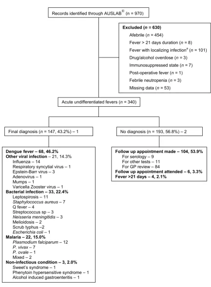
(9) distinguishable nucleic acid sequences so it can facilitate the identification of organism(s). Next-generation sequencing (NGS) technology has the capability to produce large amounts of nucleic acid sequences in a relatively short time. This technology has been applied to the study of biological diversity in environmental samples. The extent of diagnostic problems and the capability of NGS technology to detect the presence of nucleic acids in any environment have brought the theory into clinical application. The potential of NGS as a broad-scale diagnostic tool prompted the hypothesis that NGS is a practical method for investigating pathogens causing fever. There are several NGS platforms available, but little is known about their effectiveness and efficiency with regards to pathogen detection in clinical samples. The primary aim of this study was to assess the capacity, sensitivity, and more importantly, the specificity of the Illumina HiSeq platform for broad-scale characterisation of pathogens associated with fever. Secondary aims included: to describe the epidemiology of AUF and UUDF in Far North Queensland, Australia, and to optimise the preparation procedure for samples that will be subjected to NGS assay. The study was conducted in three stages, comprising two preliminary studies and the main study. The first preliminary study was a retrospective study of fever patients presenting to Cairns Hospital over the three-year period between 1 July 2008 and 30 June 2011. The findings suggest that AUF is common in the population of Far North Queensland, Australia. A robust definition of UUDF is proposed based on the clinical and laboratory characteristics of patients reviewed in this study, including the following criteria: 1) a fever of ≥ 38.0 °C or symptoms suggestive of fever; 2) a duration of fever of ≤ 21 days; 3) a failure to reach a diagnosis after performing clinical evaluation and laboratory investigations, including complete blood count, serum biochemistry, urinalysis, blood culture, chest X-ray; 4) a request by the clinician of specific test for at least one infectious agent and; 5) a failure to make a specific diagnosis. The proportion of UUDF was 56.8% (193/340), indicating the need for a broad diagnostic tool to determine infectious causes of AUF. In general, the findings provide valuable information regarding the feasibility of conducting a fever study using NGS technology at Cairns Hospital. The second preliminary study was conducted to determine the most suitable type of blood specimen for NGS analysis. It was anticipated that there would be small quantities of pathogen nucleic acids present among abundant human nucleic acids ix.
(10) background. Therefore, it was important to minimise the quantity of human nucleic acids in order to increase the sensitivity of detection of the pathogen. Six healthy volunteers participated in this second preliminary study, which aimed to measure levels of double-stranded DNA in plasma and serum. Specimens were taken using different methods of blood collection: with a syringe and with a vacuum system, and with and without applying a tourniquet. DNA concentration in the samples was measured using microplate fluorescence assays using SYBR Green I as the fluorescent dye. This study found that DNA concentration in plasma was significantly lower than that in serum (p < 0.05). However, the method of blood collection did not significantly affect DNA concentration. A main component of this thesis was a prospective study involving the use of a NGS platform to determine infectious causes of AUFs. Isolation of DNA and RNA from plasma/serum samples was performed using QIAamp® DNA Mini Kit (Qiagen) and TRIzol LS reagent (Life Technologies) respectively. Following nucleic acid extraction, amplification of DNA was conducted according to the SeqPlex Enhanced DNA Amplification Kit (SEQXE) protocol (Sigma-Aldrich). DNase treatment, cDNA synthesis and amplification were performed on RNA samples according to SeqPlex RNA Amplification Kit (SEQR) protocol (Sigma-Aldrich). Sequencing was conducted on 22 DNA/cDNA samples that met the standard input determined by the sequencing company. These samples originated from 17 patients, comprising seven positive control samples from patients who had specific diagnoses and 10 samples from patients for whom diagnoses had not been achieved. Data analysis was conducted using the Kraken program and the traditional assembly-alignment pipeline. The study findings demonstrate the limitation and utility of NGS technology in determining the aetiology of AUF. Various viruses and bacteria were found in every sample, so considered selections were made on pathogens for which there were supporting reads consistent with clinical data and pathology findings. Aetiological diagnosis was verified in 85.7% (6/7) of controls. Among the undiagnosed participants, deep sequencing identified some plausible causes of fever in 60% (6/10) of subjects, including Escherichia coli bacteraemia and scrub typus that eluded conventional tests. Further, the NGS technology generated valuable information for studying microbial diversity in human blood. Further work could be directed at optimising sample preparation and improving sequencing efficiency, as well as developing. x.
(11) efficient bioinformatics tools for analysing sequence data. It is hoped that NGS technology can be adopted in clinical practice at more affordable costs and with timely delivery of results.. xi.
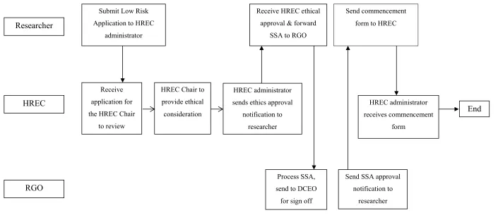
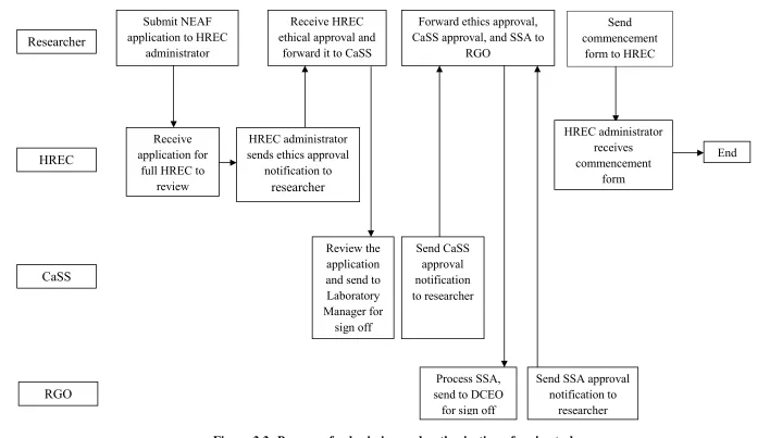
(12) Table of Contents. Statement of Access ...................................................................................................... iii Statement of Sources Declaration ................................................................................iv Statement of Contributions of Others .......................................................................... v Acknowledgements ........................................................................................................vi Abstract ....................................................................................................................... viii Table of Contents..........................................................................................................xii List of Tables ................................................................................................................. xv List of Figures ..............................................................................................................xvi Glossary .......................................................................................................................xvii Abbreviations ............................................................................................................ xviii Chapter 1: Introduction ................................................................................................. 1 1.1 Terminology ........................................................................................................... 1 1.2 Study background ................................................................................................... 3 1.3 Overview of next-generation sequencing ............................................................... 4 1.4 Overview of study design ....................................................................................... 5 1.5 Organisation of thesis ............................................................................................. 6 Chapter 2: Literature Review ....................................................................................... 7 2.1 Overview ................................................................................................................ 7 2.2 Pathogenesis of fever .............................................................................................. 7 2.3 Previous studies on acute undifferentiated fever .................................................. 10 2.4 Identification of new pathogens in historical perspective .................................... 13 2.4.1 Methods for detecting microbes .................................................................... 13 2.4.2 Guidelines for determining disease causation ............................................... 15 2.4.3 Viral discovery: challenges and success stories ............................................ 22 2.5 Next-generation sequencing (NGS) ..................................................................... 26 2.5.1 Development of sequencing technology ....................................................... 26 2.5.2 Principle of NGS ........................................................................................... 30 2.5.3 Application of NGS in metagenomics studies .............................................. 33 2.6 Research questions ............................................................................................... 36 2.7 Chapter summary.................................................................................................. 37 Chapter 3: General Methodology ............................................................................... 38 3.1 Study design and aims .......................................................................................... 38 3.2 Hypotheses ........................................................................................................... 41 3.3 Ethical clearance ................................................................................................... 42 3.3.1 First study: Undifferentiated fever in Cairns (Base) Hospital: a retrospective study ........................................................................................ 42 3.3.2 Second study: Quantitative analysis of nucleic acid in serum and plasma ... 45 3.3.3 Third (main) study: Evaluation of NGS technology in determining infectious causes of human febrile illness..................................................... 45 xii.
(13) 3.4 Consent and enrolment ......................................................................................... 48 3.5 Handling and storage of data ................................................................................ 50 3.6 Returning the results ............................................................................................. 51 3.7 Chapter summary.................................................................................................. 52 Chapter 4: Undiagnosed Undifferentiated Fever (UUDF) in Far North Queensland, Australia .................................................................................................. 53 4.1 Introduction .......................................................................................................... 53 4.2 Methods ................................................................................................................ 55 4.3 Results .................................................................................................................. 56 4.3.1 Dengue infection ........................................................................................... 59 4.3.2 Other viral infection ...................................................................................... 59 4.3.3 Leptospirosis ................................................................................................. 60 4.3.4 Central nervous system (CNS) infection ....................................................... 60 4.3.5 Other bacterial infection ................................................................................ 60 4.3.6 Malaria........................................................................................................... 61 4.3.7 Non-infectious condition ............................................................................... 61 4.3.8 Undiagnosed cases ........................................................................................ 61 4.3.9 Fatal cases ...................................................................................................... 64 4.4 Discussion............................................................................................................. 65 4.4.1 Aetiologies of AUF in Far North Queensland, Australia .............................. 65 4.4.2 Definition of UUDF ...................................................................................... 66 4.4.3 Diagnostic challenges .................................................................................... 66 4.4.4 Study strengths and weaknesses .................................................................... 67 4.4.5 Suggestions for implementation .................................................................... 68 4.5 Chapter summary.................................................................................................. 70 Chapter 5: Quantitative Analysis of Circulating DNA in Plasma and Serum ....... 71 5.1 Introduction .......................................................................................................... 71 5.2 Materials and methods .......................................................................................... 72 5.2.1 Study participants .......................................................................................... 72 5.2.2 Sample collection and processing ................................................................. 73 5.2.3 Extraction of total nucleic acids .................................................................... 74 5.2.4 Quantification of double-stranded DNA (dsDNA) ....................................... 75 5.3 Results .................................................................................................................. 76 5.4 Discussion............................................................................................................. 81 5.5 Chapter summary.................................................................................................. 85 Chapter 6: Fever Investigation Using Deep Sequencing Approach ........................ 87 6.1 Introduction .......................................................................................................... 87 6.2 Materials and methods .......................................................................................... 88 6.2.1 Participants and samples ............................................................................... 88 6.2.2 Sample preparation and sequencing .............................................................. 91 6.2.3 Bioinformatics analysis ................................................................................. 96 6.3 Results .................................................................................................................. 99 6.3.1 Participants and samples ............................................................................... 99 6.3.2 Sample preparation and sequencing ............................................................ 100 6.3.3 Summary of bioinformatics analysis ........................................................... 105 6.3.4 Control subjects ........................................................................................... 109 6.3.5 Undiagnosed subjects .................................................................................. 113 6.4 Discussion........................................................................................................... 122 6.4.1 Participants and samples ............................................................................. 122 xiii.
(14) 6.4.2 Sample preparation ...................................................................................... 122 6.4.3 Bioinformatics analysis ............................................................................... 126 6.4.4 Microbial diversity in human blood ............................................................ 130 6.5 Chapter summary................................................................................................ 138 Chapter 7: General Discussion and Conclusions ..................................................... 140 7.1 The deep sequencing approach to fever investigation ........................................ 140 7.2 Practical challenges in the study......................................................................... 142 7.3 Study strengths and limitations .......................................................................... 145 7.4 Suggestions for implementation ......................................................................... 147 7.5 Revisiting research questions ............................................................................. 152 7.6 Reflections .......................................................................................................... 154 7.7 Conclusions ........................................................................................................ 156 References ................................................................................................................... 157 Appendices. xiv.
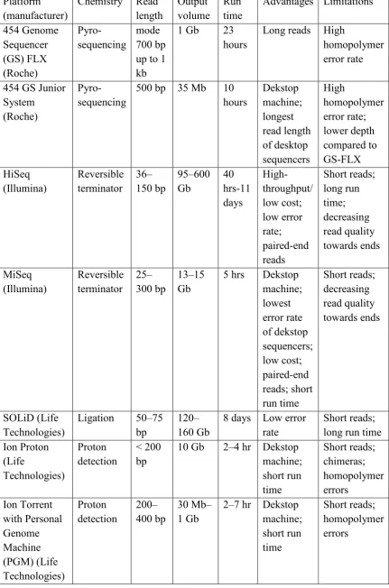
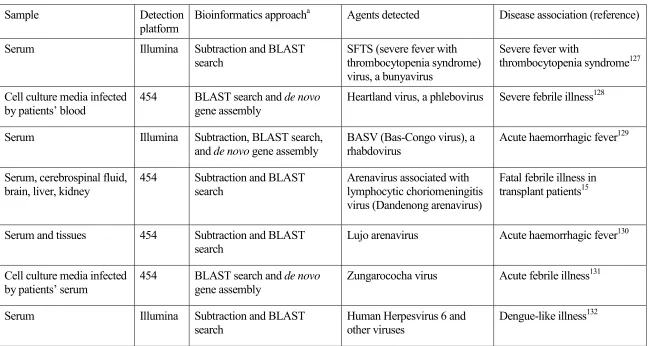
(15) List of Tables. Table 2.1: Next-generation sequencing platforms .......................................................... 29 Table 2.2: Recent applications of next-generation sequencing technology for detecting infectious agents causing fever ...................................................... 35 Table 4.1: Demographic and significant laboratory characteristics of patients with diagnosed and undiagnosed undifferentiated fevera ...................................... 63 Table 4.2: Scoring system to determine the significance of undifferentiated fever ....... 68 Table 5.1: Specimens obtained from each participant.................................................... 74 Table 5.2: Fluorescence intensity (FI) of test samples in duplicates .............................. 77 Table 5.3: Average fluorescence intensity (AFI) after subtraction of ‘blank’ values and the concentration of DNA (ng/µl) in test samples .................................. 79 Table 5.4: Statistical analysisa comparing DNA concentration in various specimens ... 81 Table 6.1: Concentrations of DNA and cDNA in total volume of 20 µl per sample, measured by NanoDrop 2000 spectrophotometer with detection limit of 2 ng/µl.......................................................................................................... 104 Table 6.2: Validation of diagnosis in positive control samples.................................... 108 Table 6.3: Plaussible NGS diagnoses in patients with undiagnosed fevers ................. 115 Table 6.4: Organisms present in all samples, detected by Kraken analysis ................. 132. xv.
(16) List of Figures. Figure 1.1: The outcomes of undifferentiated fever ..................................................................... 3 Figure 2.1: Pathway of fever pathogenesis ................................................................................... 9 Figure 2.2: Schematic workflow in different NGS platforms..................................................... 31 Figure 3.1: Conceptual framework of the study ......................................................................... 41 Figure 3.2: Process of submission and authorisation of low risk research ................................. 44 Figure 3.3: Process of submission and authorisation of main study ........................................... 47 Figure 4.1: Study flow chart ....................................................................................................... 57 Figure 4.2: Seasonal variations of dengue and acute undifferentiated fever (diagnosed and undiagnosed cases) in Cairns Hospital, Far North Queensland, Australia, from 1 July 2008 to 30 June 2011 ................................................................................... 58 Figure 5.1: Standard curve .......................................................................................................... 76 Figure 5.2: dsDNA concentrations (ng/µl) in various blood specimens ..................................... 80 Figure 6.1: Flow chart of patient selection ................................................................................. 90 Figure 6.2: Workflow of sample preparation for sequencing ..................................................... 92 Figure 6.3: SEQXE process workflow ....................................................................................... 94 Figure 6.4: SEQR process workflow .......................................................................................... 95 Figure 6.5: Analysis workflow ................................................................................................... 97 Figure 6.6: Typical quantity and quality of circulating nucleic acids, measured using bioanalyser with detection limit of 200 pg per band ............................................ 101 Figure 6.7: DNA and cDNA samples in 1.5% gel electrophoresis pre-casted with SYBR Green I dye ........................................................................................................... 103 Figure 6.8: Kraken analysis on reads generated by Illumina HiSeq 2000 ................................ 106 Figure 6.9: CLC Genomics Workbench analysis on Kraken unclassified reads ...................... 107 Figure 6.10: Mapping of dengue virus 1 contigs from sample ID# 5c and 17c1 against dengue virus 1 complete genome of 10,735 bp genomic DNA (NCBI Reference Sequence: NC_001477.1) ................................................................... 109 Figure 6.11: Patient ID# 017 with dengue rash over her trunk and extremities ....................... 111 Figure 6.12: Patient ID# 020 with dengue rash on her face...................................................... 111 Figure 6.13: Patient ID# 024 with measles rash on his face, torso and extremities ................. 112 Figure 6.14: Patient ID# 011 with eschar (upper left) and rash on his extremities (bottom left) and trunk (right) ............................................................................................ 120 Figure 6.15: Patient ID# 014 with rash on his body and extremities ........................................ 120 Figure 6.16: Patient ID# 028 with mouth ulcer (left) and tick bite (right) ............................... 121 Figure 6.17: Patient ID# 029 with rash on her face, torso and extremities ............................... 121. xvi.
(17) Glossary. Amplicon. any PCR amplification product. Cloud computing. remote computational resources available via internet. Coverage. the amount by which a genome is over-sampled; the ratio between the cumulative size of the set of reads and the size of the genome. De novo. from the beginning (i.e. without prior information). Errors. percentage of confidently aligned reads that are mapped to the wrong location. E-value. Expect value, a parameter that describes the number of hits one can ‘expect’ to occur by chance when searching a sequence database of a particular size. An E-value of 1 means that it would be expected to find a match with a similar score simply by chance. The lower the E-value, or the closer it is to zero, the more significant the match.. Metagenomics. a novel field that deals with the sequencing and study of entire microbial communities isolated directly from a particular environment. Paired-end reads. DNA sequences from each end of DNA templates. Phage. a virus that infects bacteria. Reads. DNA fragments whose sequence is known. SEQR. SeqPlex RNA Amplification Kit. SEQXE. SeqPlex Enhanced DNA Amplification Kit. SYBR Green I. a fluorescent dye that bind to dsDNA. xvii.
(18) Abbreviations. AFI. Average fluorescence intensity. AGRF. Australian Genome Research Facility. ALT. Alanine aminotransferase. ASCII. American Standard Code for Information Interchange. AST. Aspartate aminotransferase. AUF. Acute undifferentiated fever. BGI. Beijing Genomics Institute. BLAST. Basic Local Alignment Search Tool. CaSS. Clinical and Statewide Service. CMV. Cytomegalovirus. CNA. Circulating nucleic acids. CNS. Central nervous system. CRP. C-reactive protein. CSF. Cerebrospinal fluid. CT. Computed tomography. DNA. Deoxyribonucleic acid. EBV. Epstein-Barr virus. ELISA. Enzyme-linked immunosorbent assay. FI. Fluorescence intensity. FID. Fever of intermediate duration. FUO. Fever of unknown origin. GAS. Group A Streptococcus. GP. General practitioner. HAV. Hepatitis A virus. HBV. Hepatitis B virus. HCV. Hepatitis C virus. HIV. Human immunodefficiency virus. HPV. Human Papilloma virus. HREC. Human Research Ethics Committee. ICD. International Classification of Diseases xviii.
(19) ICU. Intensive care unit. IV. Intravenous. JCU. James Cook University. MFA. Microplate fluorescence assay. NANBH. Non-A, non-B viral hepatitis. NCBI. National Centre for Biotechnology Information. NEAF. National Ethics Application Form. NGS. Next-generation sequencing. NHMRC. National Health and Medical Research Council. PCR. Polymerase chain reaction. PE. Paired-end. PGM. Personal Genome Machine. PUO. Pyrexia of unknown origin. QTHA. Queensland Tropical Health Alliance. RAM. Random-access memory. RGO. Research Governance Officer. RIN. RNA integrity number. RNA. Ribonucleic acid. SD. Standard deviation. SFG. Spotted fever group. SIV. Simian immunodeficiency virus. SPSS. Statistical Package for the Social Sciences. SSA. Site-Specific Assessment. TMV. Tobacco mosaic virus. TTMDV. Torque teno midi virus. TTV. Torque teno virus. UTI. Urinary tract infection. UDF. Undifferentiated fever. UUDF. Undiagnosed undifferentiated fever. WBC. White blood cell. WHO. World Health Organisation. xix.
(20) Chapter 1: Introduction. 1.1 Terminology Fever is a common medical problem in clinical practice with various causes and diverse outcomes. It often poses challenges for clinicians because of its numerous associated diagnostic alternatives. Sometimes the cause of fever is unclear due to nonspecific clinical manifestations and limited information available from the initial laboratory findings. In these cases, the condition is referred to as undifferentiated fever (UDF), and despite the advancement of medical technologies, a considerable proportion of UDF cases go undiagnosed. If the condition lasts for more than three weeks, it is then generally accepted that it meets the criteria for fever or pyrexia of unknown origin (FUO/PUO).1 Reid2 defined PUO as an elevated body temperature of ≥ 38 °C on one occasion or ≥ 37.4 °C on three occasions in a patient over 14 years of age without adequate evidence of local symptoms and signs to be confidently diagnosed after initial examination, chest X-ray, and routine laboratory investigation. This definition did not specify fever duration as a criterion for diagnosing PUO; instead, all fever cases surpassing the temperature threshold were considered as PUO. According to Petersdof and Beeson,3 FUO is defined as a temperature higher than 101 °F (38.3 °C) for more than three weeks without an identified cause after one week of hospital investigation. This conventional definition was modified in 1991 by Durack and Street,4 who differentiated FUO into classical type and three other types: nosocomial, neutropenic and human immunodefficiency virus (HIV)-associated FUO. The classical type of FUO is not associated with prolonged fever acquired during hospital admission (nosocomial). It is also not associated with fever that is often experienced by patients who have an abnormally low level of neutrophils (a type of white blood cell) or those with HIV infection. They also suggested a shorter duration for investigation: three outpatient visits or three days of in-hospital evaluation. The World Health Organisation (WHO) issues the International Classification of Diseases (ICD) as the standard diagnostic tool for epidemiology, health management and clinical purposes (http://www.who.int/classifications/icd/en/). The most current. 1.
(21) version of the ICD, ICD-10 version 2015,5 records ‘fever of other and unknown origin’, in which the aetiology of fever cannot be ascertained. In contrast to FUO, which is clearly defined and widely studied, there is no internationally accepted consensus with regards to the diagnosis of short-term febrile illness with unclear aetiology. This condition is described as ‘acute undifferentiated fever’ (AUF) in this thesis. This term encompasses a variety of causes producing a range of clinical manifestations, with acute fever as a unifying symptom.6 Most clinicians and researchers define acute fever as evidence of raised body temperature to 38 °C or higher for up to three weeks, without detection of systemic disease or the focus of infection or inflammation after initial clinical evaluation and basic laboratory investigations such as complete blood count and urinalysis. In malaria-endemic countries, case definition of AUF usually mandates that malaria is excluded, usually by microscopic examination of a thick blood smear. In addition, a number of researchers refer to fever of intermediate duration (FID) to define fever higher than 38 °C that lasts between one and four weeks without a definite diagnosis after an initial approach.7 The duration of fever defined for FID cases overlaps with that for the case definitions of AUF and FUO, which use a 3-week duration as a cut-off level for distinguishing the two conditions. Therefore, this thesis excludes FID and focuses only on AUF cases. Figure 1.1 depicts the outcomes of UDF and the terminology used in this thesis.8. 2.
(22) Undifferentiated fever (UDF) Acute undifferentiated fever (AUF) ≤ 21 days. Fever/pyrexia of unknown origin (FUO/PUO)> 21 days. Undiagnosed UDF (UUDF). Diagnosed. Resolved. Prolonged. Figure 1.1: The outcomes of undifferentiated fever . Undifferentiated fever (UDF) is any case of fever with unclear aetiology where the results of initial investigations (which include clinical examination, rapidly available pathology and/or radiological investigations) are not conclusive in achieving a diagnosis. Thus, the condition is characterised by a requirement for further investigation to explain the cause of fever and to consider differential diagnoses.. . AUF is a short-term febrile illness lasting up to 21 days without obvious source.. . Undiagnosed undifferentiated fever (UUDF) is a condition of AUF that remains undiagnosed after conducting laboratory and radiology investigation.. . Fever/pyrexia of unknown origin (FUO/PUO) is fever that exists for more than 3 weeks without definite diagnosis.. 1.2 Study background A presumptive diagnosis is frequently made based on focal signs and symptoms accompanying fever. However, sometimes a clear etiology is not found, making treatment problematic. In short-lived episodes, both patient and clinician are usually satisfied that further investigation is not warranted, and an explanation that a patient has a ‘viral infection’ is generally acceptable, although there is a paucity of data to support this common explanation. In many cases, intensive investigative efforts are performed for more prolonged or severe episodes only. This approach is efficient from the health-. 3.
(23) economics point of view, but it is inappropriate in terms of the holistic management of diseases. As the most common feature of infection, AUF requires careful attention because, although it may reflect minor and self-limited infection, it may also be a sign of potentially severe or lethal infection. Some fevers are not easily diagnosed, leading doctors to order several laboratory tests to ascertain an aetiological diagnosis, and the failure to diagnose may represent a failure to test for a pathogen that is already described. Therefore, a broad diagnostic tool that can detect a wide range of pathogens is needed to ascertain the aetiological cause of AUF. Such a broad diagnostic tool is also important for surveillance purposes. Although many recently discovered (‘new’) pathogens may in fact have been present as causes of fever for many years, truly emerging diseases may also present as febrile illnesses. Some factors contributing to the emergence of new infectious diseases include global travel and environmental change. The increased exposure of humans to a habitat that was previously exclusive to animals may introduce a transfer of diseases from animals to humans. Likewise, the risk for the spread of new, emerging and reemerging infections is ever increasing as a result of global travel. It is therefore vital to have methods and a systematic protocol in place that can be applied to new outbreaks of a presumed infectious disease. A method that can provide unbiased characterisation of pathogens may be the key for the rapid screening of infectious diseases, and will certainly reduce the prevalence of UUDF, which eventually will also reduce FUO cases.. 1.3 Overview of next-generation sequencing Recent years have seen an enormous reduction in the cost of high-throughput sequencing technologies, also known as next-generation sequencing (NGS). These technologies provide enormous capacity to produce large genomic sequence datasets over a relatively short period of time at a reasonable cost. The advancement of NGS technology has enabled a 1,000-fold reduction in sequencing costs, from $500 per megabase using Sanger sequencing to < $0.5 per megabase using an Illumina platform.9, 10 It is now possible to sequence a complete human genome within days for < $1,000; this represents a massive advancement, considering that the first human. 4.
(24) genome sequence was completed just over a decade ago after 13 years of work at a total cost of ~$3 billion.11, 12 NGS costs have now reduced to the point where they are feasible technologies for use in the clinical diagnosis of infections. Previous studies have shown that NGS can reliably identify microorganisms, including viruses, at levels beneath the detection sensitivity of conventional microscopy and/or serological tools.13 The technology also facilitates the discovery of genes that serve as biomarkers in various pathological conditions. In fact, the application of NGS in the area of genetic diseases and cancer has facilitated the identification of genes that serve as biomarkers for diagnosing genetic abnormalities and certain types of cancers, as well as for monitoring disease progression.14 In the field of microbiology, NGS provides huge amounts of sequence data for genetic profiling, the study of biodiversity and pathogen discovery. It has been reported that NGS technology has helped to elucidate a fatal infectious disease and revealed an occult Hepatitis B infection in an apparently healthy individual.15, 16 As the technology is still evolving, there is a need to assess the reliability and practicality of NGS as a new tool for pathogen identification in patients with AUF.. 1.4 Overview of study design This thesis describes efforts to identify the infectious causes of AUF when the symptoms do not exceed a three-week period. It presents three stages of research in which the ultimate objective is pathogen identification using a NGS platform. The first stage of the research was to study the epidemiology of AUF in Far North Queensland, Australia, and to identify the characteristics of patients who would subsequently be included in the main study employing the NGS method. The next phase of the study involved laboratory experiments to optimise the sample preparation technique for the NGS study. The proportion of nucleic acids within a clinical sample that belongs to a pathogen are miniscule, so it was essential to measure the levels of human deoxyribonucleic acid (DNA) in various blood specimens to determine which specimen contained the smallest amount of human DNA contaminant. The last and the main component of the research was a prospective study recruiting patients with AUF. Blood samples were collected from two groups of patients: those who were diagnosed with specific infectious disease and those who were undiagnosed. The samples were subjected to a NGS platform to obtain sequencing data for each individual. Analysis 5.
(25) was directed to identify the pathogen causing febrile illness by examining non-human nucleic acids sequences.. 1.5 Organisation of thesis The thesis is presented in seven chapters. It begins with an introduction (Chapter 1) followed by a review of the relevant literature (Chapter 2). Pathogenesis of fever, case definitions and the scope of undiagnosed AUF are described according to previous reports in the literature. In addition, the development of microbiology and diagnostic tools are discussed to formulate research questions and hypotheses. This is followed by a description of the study design and the three stages of the research (Chapter 3). Chapter 4 presents the first study, which describes the prevalence and characteristics of AUF in Far North Queensland, Australia. Chapter 5 covers the second study, which aimed to optimise the sample collection procedure for subsequent study using the NGS. Chapter 6 presents the main study, which investigates pathogen identification using a NGS platform. The chapter provides details on the study subjects, procedure for sample preparation, sequencing details and bioinformatic analysis. This is followed by interpretation of the results and a discussion of the findings of the main study. Chapter 7 offers general discussion unifying the themes and results from the previous chapters. This final chapter concludes the thesis and makes recommendations for future research.. 6.
(26) Chapter 2: Literature Review. 2.1 Overview This chapter provides a comprehensive review of previous studies and available literature to highlight essential issues around AUF and investigations into the cause of this condition. Few articles have explored the topic of AUF and the use of NGS for its investigation. This lack of research on AUF is surprising given that it is a common presentation, though the use of NGS in this investigation is quite recent. This chapter outlines current understanding on the problem of AUF, identifies gaps in knowledge and provides context for this current study. Initially, this chapter discusses the pathogenesis of fever and how infection can induce it. This is followed by a review of previous studies on AUF and a discussion around several issues related to AUF, including its epidemiology, investigations and diagnostic challenges. A review article about AUF in Asia was published as part of this section. The section is followed by a discussion of methods for pathogen detection and discovery and how causal links are established between microbes and diseases. An indepth discussion of NGS technology is then presented, including the development of sequencing platforms, the principle of NGS and the implementation of NGS in various metagenomics studies (i.e. studies that apply DNA sequencing directly on a sample, bypassing the culture and clonal selection steps that are required in earlier sequencing techniques). Finally, the research questions proposed for this PhD are articulated.. 2.2 Pathogenesis of fever Fever is defined as a rise in body temperature above what is considered ‘normal’ (37 °C ± 1 °C), and it is a common symptom experienced by human beings as an adaptive response to various immune challenges of infectious or non-infectious origin. This response is regulated by the central nervous system (CNS) and involves endocrine, neurological, immunological and behavioural mechanisms.1 In general, body temperature can increase as a result of physiological and pathological states. In physiological situations, an elevated body temperature is a reaction to an increase in internal or external temperature, for example, during exercise, pregnancy, hot weather and dehydration. On the other hand, infections and inflammatory diseases are the most 7.
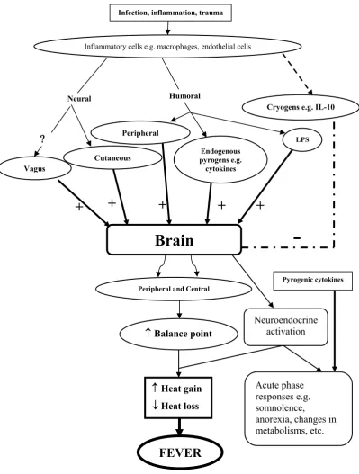
(27) common pathological causes of fever, followed by malignancies and miscellaneous conditions such as medications (e.g., antibiotics and narcotics; drug-induced fevers can be due to adverse reactions or withdrawal), trauma or injury (e.g., heart attack, stroke, burns), autoimmune diseases (e.g., Guillian–Barre syndrome, lupus), hormone disorders (e.g., hyperthyroidism, adrenal insufficiency), embolisms, and various syndromes and diseases (e.g., Caroli’s disease, Castleman’s disease, Kawasaki’s syndrome, Kikuchi’s syndrome).17–20 Fever has been recognised as a major manifestation of inflammation since the sixth century BC.21, 22 A causal relationship between infection and fever became clearer in the late eighteenth century through the works of Louis Pasteur and Robert Koch in the field of microbiology. William H. Welch established that the CNS was involved in regulating body temperature through his experiments, based on which others constructed the modern theory of the pathogenesis of fever. Welch identified the location of the thermoregulatory centre in the CNS and suggested the beneficial effect of fever, either directly by destroying microbes, or indirectly by increasing the host’s resistance to infection.21 At present, it is known that fever is triggered by substances collectively called pyrogens, which may come from sources internal (endogenous) or external (exogenous) to the body. Endogenous pyrogens include cytokines (produced by phagocytic cells) such as interleukin 1 (IL-1), interleukin 6 (IL-6), and tumour necrosis factor-alpha (TNF-α). Lipopolysaccharide (LPS), a cell wall component of Gram-negative bacteria, is an example of an exogenous pyrogen. During infection, exogenous pyrogens cause the release of endogenous pyrogens, which, in turn, activate the arachidonic acid pathway to synthesise prostaglandin E2 (PGE2). The common pathway for fever pathogenesis is through the activation of thermoregulatory cells in the hypothalamus by PGE2, resulting in increased body temperature through various mechanisms, as illustrated in Figure 2.1.. 8.
(28) Infection, inflammation, trauma. Inflammatory cells e.g. macrophages, endothelial cells. Humoral. Neural. Cryogens e.g. IL-10 Peripheral. . LPS. Endogenous pyrogens e.g. cytokines. Cutaneous. Vagus. +. +. +. +. +. -. Brain. Pyrogenic cytokines. Peripheral and Central. Balance point. Heat gain Heat loss. Neuroendocrine activation. Acute phase responses e.g. somnolence, anorexia, changes in metabolisms, etc.. FEVER Figure 2.1: Pathway of fever pathogenesis (Reproduced from1 with permission). 9.
(29) 2.3 Previous studies on acute undifferentiated fever In the first stage of my research, a literature review was conducted in two stages to understand the problem of AUF and to build a study design that would further the current state of knowledge and contribute to the understanding of previously unidentified causes of AUFs. The first stage of the review was performed in January 2012, seeking insight into the global problem of acute fever with unknown infectious cause. The second stage was carried out in September 2012, aiming to identify specific causes of AUF and quantify the proportions of cases that remain undiagnosed. In the first review, the following online databases were used: PubMed, Medline, Web of Science, CINAHL and Cochrane Database of Systematic Reviews. Keywords included (undiagnosed OR undifferentiated OR unknown OR unexplained OR unconfirmed) AND (fever OR febrile) AND acute AND infection. Additional articles were identified from the reference lists of retrieved articles and from articles that had cited the retrieved articles. Studies were included if they were conducted in primary health care, hospital and laboratory settings, and reported a series of laboratory investigations for diagnosing infection as an underlying cause of AUF. The review was not limited to any country or region, but was restricted to articles published in English during the last decade (from 1 January 2001 to 31 December 2011). Although AUF is frequently observed in clinical practice, there is a paucity of published reports on the condition. A total of 25 articles were reviewed. Most of the reviewed AUF studies were conducted in tropical and subtropical areas of the world. Of these, most originated in South and Southeast Asian countries, such as Thailand,23–26 Vietnam,27–29 India,30–33 Nepal,34 Sri Lanka,35 Laos,36 Indonesia37 and Singapore.38 Some AUF studies were conducted in African39–41 and tropical South American countries.42–45 Only two studies were conducted in developed countries in Europe.46, 47 The most commonly reported infections in these studies included malaria,25, dengue,32,. 42, 44. scrub typhus,23,. 31. spotted fever rickettsial infections35,. 41. 30. and. leptospirosis.24, 43, 44 The search strategy did not discover any articles pertaining to AUF in Australia. Fever is not included in the national health priority areas that have been chosen for focused attention by Australian governments.48 Only a small proportion of the Australian population live in areas where tropical infections are commonly found. 10.
(30) Despite this, tropical infections that manifest as acute fever are quite prevalent in some areas in Australia, particularly in areas situated in tropical and subtropical zones. Some Australian cities are major tourist destinations and may serve as points of entry for the spread of tropical infections as a result of increased global travel. Mosquito-borne pathogens such as the malaria parasite (Plasmodium spp.) and the dengue, Ross River and Barmah Forest viruses can be found in Australia, particularly in tropical North Queensland.49–54 It has been reported that malaria and dengue were introduced to this region by travellers,55 while the Ross River and Barmah Forest viruses are endemic in Queensland.50 Other diseases that can present as an AUF, such as melioidosis, leptospirosis and rickettsiosis, are also found in Australia.56, 57 Although previous studies have evaluated the causes of AUF, limited data exists regarding the epidemiology of undiagnosed cases where pathogens other than that already described may be the cause. Those studies have focused on particular pathogens rather than the various causes of AUF. In addition, the proportion of AUF cases that remain undiagnosed was not specifically reported in most studies. In September 2012, the literature review was updated by conducting the review systematically according to PRISMA guidelines (http://www.prisma-statement.org). The second review aimed to determine the case definition, investigations and aetiologies of AUF, and to determine the proportion of AUF cases that remain undiagnosed. It was shown in the first review that AUF studies have predominantly been conducted in Asia, so the second review focused on those studies that were conducted in Asian countries. This second review was limited to articles published in English during the period 1990–2012. This time period was chosen because nucleic acid testing began to be employed as a routine diagnostic tool after 1990. The following terms were used when searching for articles on PubMed database: Fever/etiology (Majr) OR Fever/microbiology (Majr) AND Asia (Mesh) AND Adult (Mesh) AND 1990/01/01 (PDAT): 2012/12/31 (PDAT) AND Journal Article (ptyp) AND English (lang). Literature search was also performed in other databases including Medline, Scopus and Web of Science. From 201 studies retrieved from the online databases, 9 were included in the review. This review was published in the Southeast Asian Journal of Tropical Medicine and Public Health in May 2014.6. 11.
(31) Article: Acute undifferentiated fever in Asia: a review of the literature Declaration of Authorship Publication details. Nature and extent of the intellectual input of each author. Signature. Susilawati, TN & McBride, WJH. Acute undifferentiated fever in Asia: a review of the literature. The Southeast Asian Journal of Tropical Medicine and Public Health. 2014:45(3);719–726. Accepted for publication 12 May 2014 Published May 2014. Developed the initial idea and design the review process, conducted literature search, prepared the manuscript.. Susilawati, TN. Assisted with the article writing and editing.. McBride, WJH. Different case definitions were used for the different studies for AUF; including fever duration and temperature threshold. For example, four studies23, 24, 30, 59 evaluated patients with a duration of fever of less than 14 days whereas another study31 evaluated patients with fever of up to 21 days duration. In terms of the definition of fever, the AUF studies specified a fever cut-off level of ≥ 38 °C, 38.3 °C or 37.5 °C.6 To identify the etiologies of AUF, the studies employed non-specific and specific investigations. Non-specific investigations refer to blood analysis and other laboratory testing to describe the underlying cause of the disease without determining a specific microorganism, such as a complete blood count, serum biochemistry, urinalysis and chest X-ray. Specific investigations refer to laboratory testing investigating specific pathogens, such as malaria films, serological tests, polymerase chain reaction (PCR) assays and bacterial cultures. These methods identified specific infections, such as malaria, dengue fever, leptospirosis and rickettsioses as possible causes of AUF. This review shows that prior studies have often used serological testing (measurement of immunoglobulin M and G [IgM and IgG] levels) as the main diagnostic method for determining infectious causes of AUFs. A drawback of this approach is that collection of convalescent serum is not possible from all patients, and analysis of acute samples only can lead to difficulties with interpretation. If only acute serum samples are collected, results may be falsely negative if collected too early, when antibody titres against a given pathogen are below detectable levels during the first few. 12.
(32) day of illness. Additionally, there are problems associated with cross-reactivity of antibodies of closely related species or organisms, which can result in false positives.58 Direct methods based on pathogen detection by culture, polymerase chain reaction (PCR) or antigen detection provide more reliable results than antibody detection in the acute phase of illness.59 A challenge with antigen detection is that patients may present at a stage of the illness after the antigen or agent causing the illness has been cleared by the immune system. This can result in false negatives and prevent a diagnosis from being established. Given these methodological difficulties, it is not surprising that a large number of AUFs remain undiagnosed. Despite the introduction of PCR as a routine diagnostic test, the identified aetiologies of fever and the proportion of undiagnosed fever cases are similar to those observed in prior serological studies.60, 61 Some conclusions can be drawn from the published article, which forms part of the literature review. First, during the 23-year period from 1 January 1990 to 31 December 2012, only a limited number of reports were published on AUF in Asia. Second, there has been no agreement on the case definition of short-term febrile illnesses with unclear aetiology. Third, the focus of the reviewed articles has largely been on detecting the common aetiologies of AUF, rather than exploring the aetiology of undiagnosed infections. Several gaps in knowledge were identified that underpin the need for further study. The work presented in Chapter 4 goes towards filling the gaps in existing knowledge in several ways. First, the study describes the epidemiology of AUF by examining its prevalence and the proportion of fevers that go undiagnosed in the area of Far North Queensland, Australia. Second, the study compiles clinical and laboratory characteristics of patients to formulate a robust definition of UUDF. Third, the study puts emphasis on the elaboration of information that exists with regards to UUDF and proposes criteria for further investigation with broad diagnostic tools.. 2.4 Identification of new pathogens in historical perspective 2.4.1 Methods for detecting microbes Many tests have been developed for detecting bacterial and viral pathogens. Visualisation was the first method developed, representing an attempt to see the microbial world. The advances in light microscopy achieved by a Dutch botanist, van 13.
(33) Leeuwenhoek (1632–1723), introduced the existence of microbes to humans. The spectrum of known bacterial, fungal and protozoan pathogens has since been expanded with improved staining and culture techniques, in conjunction with the development of more advanced visualisation methods. For example, light microscopy has been refined with the use of immunohistochemical or immunofluorescent stains to detect specific molecules in the host or pathogen. Laser scanning confocal microscopy represents a further quantum improvement in resolution and signal quantification. Most recently, transmission and scanning electron microscopy has revealed microorganisms at very high resolution.62 Traditional methods for diagnosing bacterial infections include staining, culture and biochemical tests. The process of staining is performed by preparing samples on a glass slide. Clinical samples are usually obtained from fluids such as blood, cerebrospinal fluid (CSF), sputum, pus, ear or eye or nasal discharge, and urine. Samples can also be obtained from microbes grown in an artificial medium. Based on the dye used and the purpose of staining, bacterial staining can be categorised into simple staining and differential staining. Simple staining uses only one dye, such as carbol fuchsin, gentian violet or methylene blue. Simple staining is traditionally used to examine the morphology of the bacteria: on microscopic examination, the bacteria are stained more intensely than the background. Differential staining uses more than one dye and is useful for identifying bacteria based on their reaction to the stains. Examples of differential staining include Gram staining and acid-fast staining to differentiate Gram-positive/negative bacteria and to identify acid-resistant bacteria such as Mycobacterium sp. and Nocardia sp. Bacterial and viral cultivation is an important method for detecting microbes. While many bacteria from clinical samples can be grown in artificial media, viruses and some bacteria (such as Chlamydia spp. and Rickettsia spp.) need to be cultivated in living cells. This often hampers the confirmation of a disease caused by those organisms. In clinical laboratories, biochemical testing and sensitivity testing are often performed following bacterial culture for determining the specific diagnosis and for choosing the appropriate antibiotics for treatment. Serological assays are an indirect approach to diagnosis based on the specificity of the immunological response, and enable a clinician to diagnose infection in an individual patient and to study the epidemiology of microbes in host populations. These assays involve the detection of specific antibodies (IgM and IgG) or antigen. 14.
(34) Serological testing is based on the finding that specific antibodies are generated as a reaction to an infectious agent.63,. 64. This serological reaction can be measured. qualitatively or quantitatively. The advantage of performing serological testing is that it can be performed quickly and relatively inexpensively. However, such assays have the potential to yield false positive or negative results, as discussed in the previous section. Arguably, the most revolutionary advance to date in the biomedical sciences was the discovery of nucleic acids as the source of genetic information on the precise characterisation of an organism. The ability to detect pathogens’ nucleic acid and to determine their nucleotide sequences created a powerful diagnostic means in the field of microbiology and infectious diseases.11 Molecular tests, such as PCR, multiplex PCR, quantitative PCR, DNA sequencing and hybridisation techniques, were later developed for the detection of bacterial and viral genetic material. 2.4.2 Guidelines for determining disease causation As a consequence of the technological advances that have facilitated pathogen detection and discovery, the number of putative pathogens reported has increased. At the same time, it is recognised that numerous microbes are normally associated with healthy humans, and indeed are required for our wellbeing.65, 66 This has given rise to the question of how to distinguish between pathogenic microbes and commensal organisms. This issue is discussed further below. In the 1880s, Koch67 postulated the core principles that define the aetiologic role for a potential pathogen, which are known as ‘Koch’s postulates’. According to the postulates, to identify a pathogen as the causative agent of a particular disease, the following conditions must be met: i.. The pathogen must be present in all cases of the disease.. ii.. The pathogen can be isolated from the diseased host and grown in pure culture.. iii.. The pathogen from pure culture must cause the disease when inoculated into a healthy, susceptible host.. iv.. The pathogen must be isolated from the new host and shown to be the same as the originally inoculated pathogen. Koch’s postulates have standardised the discovery of human pathogens by. establishing a causal relationship between a microbe and a disease. In the nineteenth century, microbiologists such as Louis Pasteur, George Miller Sternberg, Robert Koch, Edwin Klebs and Richard Pfeiffer reported newly discovered microbes (e.g., 15.
(35) Streptococcus. pneumoniae,. Mycobacterium. tuberculosis,. Corynebacterium. diphtheriae, Vibrio cholerae and Haemophilus influenzae) after their success in isolating and culturing pathogens. During this era, the most common methods for bacterial identification included Gram staining, culture and biochemical tests. The limitations of Koch’s postulates were immediately identified for some diseases of which the pathogenesis could not be reconciled with the postulates. For instance, the first postulate, that ‘the pathogen must be present in all cases of the disease’, cannot be fulfilled in the case of disease caused by endotoxin. In this instance, the disease occurs without the presence of the pathogen because endotoxin is released by Gram-negative bacteria following the lysis of the bacterial cell wall. The second Koch’s postulate, that ‘the pathogen can be isolated from the diseased host and grown in pure culture’, is violated where an individual can be a healthy carrier of a pathogenic organism. For example, V. cholerae, M. tuberculosis and Neisseria meningitidis can be isolated from patients with diseases and can also be present in healthy subjects.68 This postulate can also be questioned when the disease occurs due to the capability of the microbe to modulate disease-associated genes. For example, S. pneumoniae represents normal flora in the human nose, but can rearrange its genes to cause serious cases of pneumonia;69 thus, this organism can be found in both healthy and diseased people. The most obvious violation of the second Koch’s postulate is when the microbes cannot be grown in pure (lifeless or cell-free) culture; this applies to viruses, chlamydiae and rickettsiae, which require other cells for propagation. In addition, the inability to isolate Plasmodium falciparum and M. leprae from pure cultures also prevents the fulfilment of Koch’s second postulate.68 The third and fourth Koch’s postulates, that ‘the pathogen from pure culture must cause the disease when inoculated into a healthy, susceptible host’ and that ‘the pathogen must be isolated from the new host and shown to be the same as the originally inoculated pathogen’, are violated in cases of limited host availability for a particular microbe. For example, HIV hosts are restricted to humans, and it would be highly unethical to infect and reisolate HIV from an experimental human host. Although simian immunodeficiency virus (SIV), which infects monkeys and chimpanzees, bears a very close resemblance to HIV, these animals are not an ideal model for studying HIV because of the lack of pathogenic consequences despite infection.70, 71 As a consequence of the limitations outlined above, Koch’s postulates clearly need reconsideration. Indeed, as Evans noted: 16.
(36) failure to fulfil the Henle-Koch postulates does not eliminate the putative microbe from playing a causative role in a disease. It did not at the time of Koch’s presentation in 1890 and it certainly does not today. Postulates of causation must change with the technology available to prove them and with our knowledge of the disease.72 The application of new technologies and advances in knowledge in the field of microbiology and infectious disease have indeed led to the revision of Koch’s postulates for defining the causal relationship between a microbe and a disease. In 1937, Rivers73 contended that Koch’s postulates are not satisfied for viral diseases. However, he believed that certain conditions still must be met before the specific relation of a virus to a disease is established. Rivers’ conditions included: i.. a specific virus must be associated with a disease with a degree of regularity;. ii.. the virus must be shown to occur in the sick individual not as an incidental or accidental finding but as the cause of the disease under investigation. Rivers’ postulates differ from Koch’s in that (i) the pathogenic virus does not. need to be present in every case of the disease produced by it; (ii) the existence of the ‘carrier state’ with respect to viral disease is recognised; and (iii) the requirement for viral cultivation is abandoned. Another guideline for establishing a causal link between a virus and a disease was proposed by Huebner in 1957.74 Huebner recognised that some viruses can cause chronic or latent infections in humans and that simultaneous multiple viral infections are extremely common. Therefore, he introduced epidemiological and immunological aspects into the criteria used to judge disease causation. According to Huebner, in order that a virus be regarded as the cause of specific illness, the following factors are essential: i.. The virus should be real; that is, it should be well established on animal or tissue-culture passage.. ii.. The virus must originate in the human specimens, not in the experimental animals, cells or media employed to grow it.. iii.. The agent should evoke antibody response, as shown by an increase in neutralising antibodies or other serological tests.. iv.. The virus should be characterised and compared with known agents, particularly immunological characterisations and comparisons.. v.. There is constant association between the agent and the specific illness.. 17.
(37) vi.. In double-blind studies, the agent should reproduce clinical manifestation(s) consistent with the naturally occurring illness.. vii.. Epidemiological studies are necessary to establish the etiological role of highly prevalent viruses in human disease.. viii.. Prevention of the disease by specific vaccination is one of the best ways to establish the causal link between an agent and a disease. In addition to these factors, Huebner also highlighted that viral research is very. expensive, and thus that financial support is absolutely necessary for proving causal links between virus and disease. In fact, Huebner suggested that this financial factor deserves to be called a postulate. Integrative guidelines considering environmental factors in the development of a disease were proposed by Hill, 1965.75 Hill proposed nine epidemiological criteria for evaluating a possible causal relationship, including: i.. strength of the association between the cause and the disease;. ii.. consistency of the association observed by different persons, in different circumstances and times;. iii.. specificity of the association: that is, that the association is unique to particular types of disease;. iv.. temporality of the association: that is, that exposure precedes the outcome;. v.. biological gradient: that is, evidence of a dose-response relationship;. vi.. plausibility: that is, that the association between the cause and the disease is biologically plausible;. vii.. coherence: that is, that the causal association is compatible and does not conflict with existing knowledge of the disease;. viii.. that experimental or semi-experimental evidence is available to support the causation hypothesis;. ix.. analogy: that is, that the causal relationship conforms to a previously described relationship. When serological assays became widely used for investigating diseases, a causal. link between microbe and disease could be drawn based on the purification of viral antigen and detection of specific antibody. Following this development, Evans proposed his ‘Elements of Immunological Proof of Causation’, which were derived from experience with Epstein-Barr virus (EBV), where the causal relationship between the virus and Burkitt’s tumour was assigned on the basis of the population-based 18.
(38) features of sero-reactivity to EBV antigen.76, 77 Evans’ criteria for establishing causal relationships are as follows: i.. Antibody to the agent is regularly absent prior to the disease and exposure to the agent (i.e., before the incubation period).. ii.. Antibody to the agent regularly appears during illness and includes both IgG and IgM classes.. iii.. The presence of antibody to the agent indicates immunity to the clinical disease associated with primary infection by the agent.. iv.. The absence of antibody to the agent indicates susceptibility to both infection and the disease by the agent.. v.. Antibody to no other agent should be similarly associated with the disease unless it is a cofactor in its production. The discovery of ‘slow virus’ infections of the nervous system, such as the. agents of Kuru and Creutzfeldt–Jakob disease, posed a challenge for establishing disease aetiology using the available guidelines. The agents could not be seen, grown in the laboratory or monitored by serological responses, and thus failed to fulfil Koch’s postulates or to meet many of the criteria for disease causation by viruses suggested by Rivers and Huebner. It is now known that Kuru and Creutzfeldt–Jakob disease are caused by prions, which are infectious, proteinaceous particles. Unlike viruses, prions lack nucleic acids. In order to establish a causal link between prion and disease, one may consider the criteria proposed by Walker in 2006:78 i.. The protein must be invariably present in a disease-specific form and arrangement in the diseased tissue. It is necessary to confirm the presence of protein accumulation (such as in the form of inclusion bodies or extracellular protein deposits) in certain tissues.. ii.. The physicochemical characteristics that confer infectivity on a specific protein must be established. The characterisation of the infectious protein includes the primary amino acid sequence, the secondary, tertiary, and quaternary structure, and post-translational modifications or processing.. iii.. The genetic, biochemical and cellular characteristics that render the host susceptible to infection by a specific proteinaceous agent must be established. The most crucial characteristic to be determined is the amino acid sequence of the host protein.. 19.
Figure


+7

Related documents